Method Article
الكمية التصور والكشف عن سرطان الجلد باستخدام التصوير الحراري الديناميكي
In This Article
Summary
أثبتنا بأن الآفات الخبيثة مصطبغة مع زيادة النشاط الأيضي توليد كميات قابلة للقياس الحرارة وقياس رد فعل عابر الحرارية من الجلد إلى الإثارة التبريد يسمح تحديد كمي لسرطان الجلد وسرطانات الجلد الأخرى (مقابل غير التكاثري حمات) في وقت مبكر مرحلة من مراحل المرض.
Abstract
In 2010 approximately 68,720 melanomas will be diagnosed in the US alone, with around 8,650 resulting in death 1. To date, the only effective treatment for melanoma remains surgical excision, therefore, the key to extended survival is early detection 2,3. Considering the large numbers of patients diagnosed every year and the limitations in accessing specialized care quickly, the development of objective in vivo diagnostic instruments to aid the diagnosis is essential. New techniques to detect skin cancer, especially non-invasive diagnostic tools, are being explored in numerous laboratories. Along with the surgical methods, techniques such as digital photography, dermoscopy, multispectral imaging systems (MelaFind), laser-based systems (confocal scanning laser microscopy, laser doppler perfusion imaging, optical coherence tomography), ultrasound, magnetic resonance imaging, are being tested. Each technique offers unique advantages and disadvantages, many of which pose a compromise between effectiveness and accuracy versus ease of use and cost considerations. Details about these techniques and comparisons are available in the literature 4.
Infrared (IR) imaging was shown to be a useful method to diagnose the signs of certain diseases by measuring the local skin temperature. There is a large body of evidence showing that disease or deviation from normal functioning are accompanied by changes of the temperature of the body, which again affect the temperature of the skin 5,6. Accurate data about the temperature of the human body and skin can provide a wealth of information on the processes responsible for heat generation and thermoregulation, in particular the deviation from normal conditions, often caused by disease. However, IR imaging has not been widely recognized in medicine due to the premature use of the technology 7,8 several decades ago, when temperature measurement accuracy and the spatial resolution were inadequate and sophisticated image processing tools were unavailable. This situation changed dramatically in the late 1990s-2000s. Advances in IR instrumentation, implementation of digital image processing algorithms and dynamic IR imaging, which enables scientists to analyze not only the spatial, but also the temporal thermal behavior of the skin 9, allowed breakthroughs in the field.
In our research, we explore the feasibility of IR imaging, combined with theoretical and experimental studies, as a cost effective, non-invasive, in vivo optical measurement technique for tumor detection, with emphasis on the screening and early detection of melanoma 10-13. In this study, we show data obtained in a patient study in which patients that possess a pigmented lesion with a clinical indication for biopsy are selected for imaging. We compared the difference in thermal responses between healthy and malignant tissue and compared our data with biopsy results. We concluded that the increased metabolic activity of the melanoma lesion can be detected by dynamic infrared imaging.
Protocol
1. الإعداد
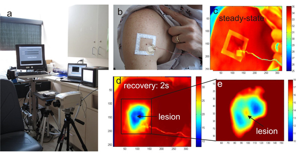
- ويرد درجة حرارة الغرفة امتحان للرقابة الكاميرا مجهزة بالأشعة تحت الحمراء وجهاز الكمبيوتر لالتقاط صور الأشعة تحت الحمراء والتخزين فضلا عن اقتناء بطاقة البيانات المتصلة بجهاز كمبيوتر في Fig.1a.
- ويتم رصد درجة حرارة الغرفة ودرجة حرارة الجلد السطحية التي تعلق على المزدوجات الحرارية بطاقة الحصول على البيانات أثناء الدراسة المريض وقياس البيانات المخزنة على الكمبيوتر.
2. صورة اقتناء
- إذ لا يمكن أن يتم الكشف عن الآفة في صورة حرارية دون تأثير التبريد ، يتم استخدام علامة لاصقة مربع لتوطين آفة مصطبغة المصالح وضواحيها (الشكل 1B).
- علينا الحصول على صورة الضوء الساطع من آفة مصطبغة ، ونافذة لاصقة مع كاميرا رقمية (كانون PowerShot G11) (الشكل 1B).
- ويستخدم dermatoscope متصلة كاميرا رقمية (DermLite صورتي النظام) لالتقاط صورة الضوء المستقطب.
- علينا الحصول على صورة الدولة المستقرة تحت الحمراء مع midwave ميرلين (3-5 ميكرومتر) كاميرا الأشعة تحت الحمراء هو مبين في Fig.1a ، C.
- نحن نطبق تيار من الهواء البارد إلى منطقة جلد المريض تحتوي على الآفة وكذلك المنطقة المحيطة بقطر 50 ملم لمدة دقيقة واحدة.
- بعد دقيقة واحدة ، ونحن إزالة هذا الضغط للسماح للتبريد الجلد لفي درجة حرارة الغرفة في غضون دقائق 3-4 (الحرارية مرحلة الانتعاش) (الشكل 1C - D) إعادة الدافئة.
- خلال مرحلة الانتعاش الحرارية ، ويتم التقاط صور الأشعة تحت الحمراء من آفة مصطبغة كل 2 ثانية (الشكل 1C - D).
- يتم حفظ كل الصور الأشعة تحت الحمراء (بالإضافة إلى الضوء الأبيض والضوء المستقطب الصور) التي اتخذت خلال دراسة وتخزينها باستخدام برنامج LABVIEW.
3. معالجة الصور
- ويتم تحليل هذه الصور باستخدام الأشعة تحت الحمراء رمز مطلب مكرس من أجل الحصول على دقة توزيع الحرارة عابرة على سطح الجلد. لهذا الغرض ، ونحن نقدم العديد من الخطوات معايرة وتحليل صورة النظام المتعدد الوسائط.
- نبدأ بتطبيق خوارزمية الكشف المعالم لصورة الضوء الساطع لتوطين أركان علامة لاصقة. المقبل ، حددنا النقاط المقابلة في صورة الأشعة تحت الحمراء المرجعية.
- من أجل التعويض عن حركة الجسم / أطرافه غير الطوعي للمريض ، ونحن نستخدم هذه النقاط والمعالم في نموذج الحركة التربيعية لمواءمة تسلسل صورة الأشعة تحت الحمراء في مرحلة الانتعاش.
- نستخدم ماشي عشوائي ، وتجزئة الصور خوارزمية التفاعلية حيث يمكن للمستخدم دليل مكانيا للتجزئة عن طريق وضع نقاط البذور ، لخلق صورة قناع ترسيم الآفة.
- بمجرد أن نحدد شكل الآفة ، نحدد المنطقة المقابلة في كل من صور الأشعة تحت الحمراء مسجلة.
- نختار نقطة عشوائية داخل الآفة ، وبعيدا عن الآفة التي تمثل آفة والأنسجة السليمة ، على التوالي.
- قارنا استجابة عابرة الحراري للبشرة صحية واستجابة الآفة.
- نعد جدولا يبين كافة البيانات : الرقمية ، dermoscopy ، مرمزة صور الأشعة تحت الحمراء من الآفة والمنطقة المحيطة بها المسجلة في الظروف المحيطة وبعد 2 ثانية الإثارة التبريد ، واستجابة عابرة الحرارية الآفة والأنسجة السليمة.
4. ممثل النتائج :

الشكل 1. أ) نظام التصوير بالأشعة تحت الحمراء HRIS في غرفة التجارب السريرية ، ب) صورة فوتوغرافية لمنطقة الجسم أكبر السطح مع مجموعة من الآفات الصباغية والإطار قالب بطلبات للحصول على التصوير ، ج) والأشعة تحت الحمراء صورة مرجع في المنطقة عند درجة حرارة الغرفة ، د) تضخيم نفس المنطقة بعد التبريد وه) قسم من آفة الجلد والمناطق المحيطة بها

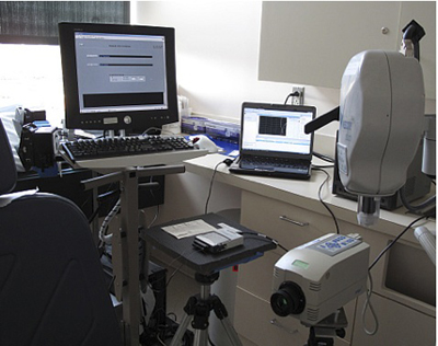
الشكل 2. قاعة الامتحان مع نظامنا التصوير الحراري.

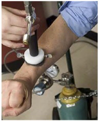
الشكل 3. تبريد الآفة وأنسجة الجلد المحيطة بها تهب تيار من الهواء البارد من أنبوب الدوامة.
Discussion
وتشير النتائج إلى أن الضغط من خلال تطبيق نظام التبريد علينا تعزيز الاختلافات في درجة الحرارة بين الآفة والأنسجة المحيطة صحية. أيضا ، وبسبب حركات صغيرة من المريض خلال التصوير الحراري ، كان لدينا لتطبيق الاقتراح تتبع لتراكب الصور بشكل صحيح لقياس الاختلافات في درجة الحرارة بين الدولة مرجعية وتوزيع درجات الحرارة خلال الانتعاش الحراري. دون أن تتبع الحركة فإننا لم تكن قادرة على كشف وقياس درجة الحرارة الفرق بين الآفة الخبيثة والأنسجة السليمة. واجهت هذه النتائج ، وضرورة تعقب الحركة دقيقة شرح للمحققين صعوبات في الماضي عندما كانت تحاول تشخيص سرطان الجلد باستخدام الأشعة تحت الحمراء والتصوير استنادا إلى معلومات الدولة المستقرة وحدها وبشكل واضح يثبت مزايا التصوير الحراري الديناميكي.
تجدر الإشارة إلى أن القرار المكانية للكاميرا الأشعة تحت الحمراء (عدد البكسل في الصفيف البؤري IR) أمر حاسم عندما آفات صغيرة المميزين. وكان كلا من القرار المكانية وحساسية في درجة الحرارة من كاميرات الأشعة تحت الحمراء في وقت مبكر محدودة ، وهو ما يمثل أيضا بالنسبة للصعوبات في الكشف عن سرطان الجلد في وقت مبكر في المرحلة الماضية. الاختلافات الرئيسية بين نهجنا وقبل محاولات التصوير الحراري -- التي كانت ناجحة نسبيا -- هي تسلسل المعايرة وخطوات معالجة الصور التي تسمح لنا لقياس درجة الحرارة بدقة الخلافات في هذا النظام بالإضافة إلى دينامية عملية التصوير التي تعتمد على التبريد النشطة.
Disclosures
Acknowledgements
وقد تم تمويل هذا البحث من قبل المنحة الوطنية للعلوم رقم 0651981 ومؤسسة الكسندر ستيوارت ومارغريت الثقة على الرغم من أن مركز السرطان التابع لجامعة جونز هوبكنز. فإن الكتاب مثل الاعتراف بمساهمات الدكتور العاني رودا إلى العائد المحلي ودراسة المريض فضلا عن مساعدة ودعم الدكتور كانغ سيوون وزارته خلال دراسة المريض.
Materials
| Name | Company | Catalog Number | Comments |
| اسم المعدات | شركة | فهرس العدد | |
| ميرلين MWIR الكاميرا | FLIR | -- | |
| كانون PowerShot G11 | شريعة | -- | |
| DermLite صورتي النظام | DermLite | -- | |
| دوامة انبوب | Exair | -- | |
| خزانات الهواء | Airgas | -- |
References
- Elder, D. Tumor progression, early diagnosis and prognosis of melanoma. Acta Oncol. 38, 535-547 (1999).
- Wartman, D., Weinstock, M. Are we overemphasizing sun avoidance in protection from melanoma. Cancer Epidemiol Biomarkers Prev. 17, 469-470 (2008).
- Pirtini Cetingul, M. . Using high resolution infrared imaging to detect melanoma and dysplastic nevi [dissertation]. , (2010).
- Jones, B. F. A reappraisal of the use of infrared thermal image analysis in medicine. IEEE Trans. Med. Imaging. 17, 1019-1027 (1998).
- Anbar, M. Clinical thermal imaging today-shifting from phenomenological thermography to pathophysiologically based thermal imaging. IEEE Eng. Med. Biol. Mag. 17, 25-33 (1998).
- Anbar, M., Gratt, B. M., Hong, D. Thermology and facial telethermography. Part I: history and technical review. Dentomaxillofacial Radiology. 27, 61-67 (1998).
- Jones, B. F., Plassmann, P. Digital infrared thermal imaging of human skin. IEEE Eng. Med. Bio. 21, 41-48 (2002).
- Qi, H., Diakides, N. A. . Infrared imaging in Medicine. , (2007).
- Pirtini Cetingul, M., Herman, C. Identification of skin lesions from the transient thermal response using infrared imaging technique. IEEE 5th Int. Symp. on Biomedical Imaging: From Nano to Macro 1-4. , 1219-1222 (2008).
- Cetingul, P. i. r. t. i. n. i., M, ., Herman, C. Quantification of the thermal signature of a melanoma lesion. Int. Journal of Thermal Science. 50, 421-431 (2011).
- Pirtini Cetingul, M., Herman, C. A heat transfer model of skin tissue for the detection of lesions: sensitivity analysis. Physics in Medicine and Biology. 55, 5933-5951 (2010).
- Pirtini Cetingul, M., Herman, C. Quantitative evaluation of skin lesions using transient thermal imaging. Proc. Int. Heat Transfer Conf. , (2010).
Reprints and Permissions
Request permission to reuse the text or figures of this JoVE article
Request PermissionExplore More Articles
This article has been published
Video Coming Soon
Copyright © 2025 MyJoVE Corporation. All rights reserved