
需要订阅 JoVE 才能查看此. 登录或开始免费试用。
Processing Insect Legs for Fluorescence Microscopy: A Method to Preserve Neuromuscular Structures for Imaging
Please note that all translations are automatically generated. Click here for the English version.
Overview
This video describes a method to dissect, fix, and mount the Drosophila adult leg while preserving its neuromusculature intact for imaging analysis.
研究方案
This protocol is an excerpt from Guan et al., Visualize Drosophila Leg Motor Neuron Axons Through the Adult Cuticle, J. Vis. Exp. (2018).
1. Leg Dissection and Fixation
- Take a glass multi-well plate and fill appropriate number of wells with 70% ethanol. Add 15–20 CO2-anesthetized flies (of either sex and any age) to each well and by using a brush, gently dab the flies into the ethanol solution until flies are fully submerged.
NOTE: This step is to remove the hydrophobicity of the cuticle. Do not wash for more than 1 min, because this increases auto-fluorescence of the cuticle. - Rinse the flies 3 times with 0.3% nonionic surfactant detergent solution in 1x phosphate buffered saline (PBS). Keep the flies in this solution for at least 10 min.
NOTE: Legs are better fixed when including detergent, which is likely to increase penetration of the fixative inside the leg. - Place a multi-well plate on the ice along with a tube of 4% paraformaldehyde (PFA).
NOTE: The preparation of fresh 4% PFA from a 16% PFA ethanol free stock solution is critical. - Use forceps to remove the head and abdomen of the flies without damaging the thoracic segment or the legs.
NOTE: Removing the abdomen makes it easier to hold the fly and dissect the legs. - Dissect the legs from the thoracic segment with forceps and place the legs in the wells containing 4% PFA. For this, push gently but firmly at the coxa-thorax junction using the tip of fine forceps until the leg detaches.
- Fix the legs in 4% PFA overnight at 4 °C (approximately 20 h total).
- Wash the legs with 0.3% nonionic surfactant detergent solution in 1x PBS, 5x for 20 min each.
- Replace the washing buffer with mounting medium. Keep the leg in mounting medium for at least a day prior to mounting to allow for its complete penetration into the leg.
NOTE: If the mounting medium is highly viscous, dilute the mounting medium to 80% with 1x PBS because the sudden change in viscosity can cause the cuticle to collapse and damage the overall structure of the leg. Depending on the Gal4 driver, flies can be preserved for 1 to 3 weeks at 4 °C in mounting media until mounting.
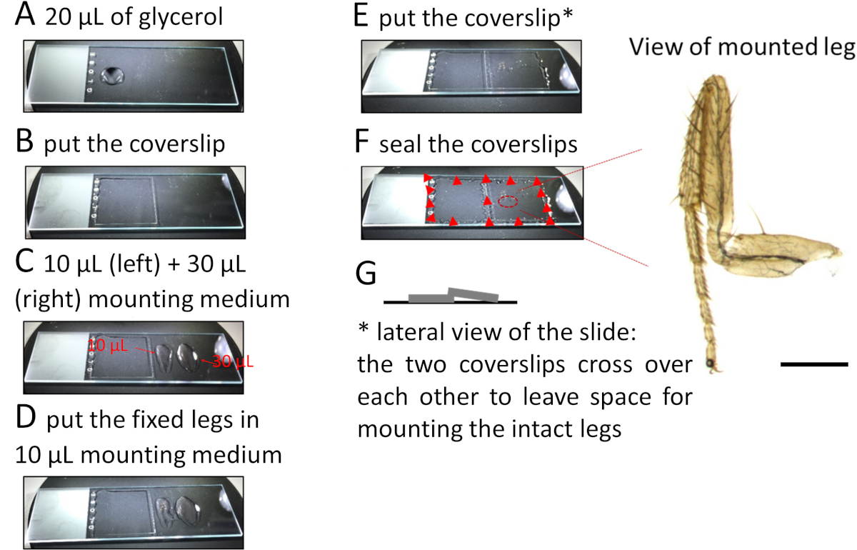
2. Leg Mounting
- Place approximately 20 µL of 70% glycerol on the left-hand side of a microscope slide and cover with a square 22 x 22 mm2 coverslip (Figure 1A,B). Pipette about 10 µL of mounting medium along a line parallel to and at a small distance from the right edge of this coverslip. Add another 30 µL of mounting medium further on the right side of the slide (Figure 1C).
NOTE: When applying a coverslip over the legs (see below) the mounting medium will gently spread under the coverslip and around the legs without displacing them. - Using fine forceps lift the leg from the solution and gently put it on the mounting medium near the left coverslip. Do the same for each leg and align them from top to bottom (Figure 1D).
- Lift and transfer the legs in a drop of medium held between both tips of the forceps. Orient the legs in two ways: external side up or down.
- Once all the legs (up to 6–8 legs can be mounted) are properly aligned, put a second coverslip over the legs such that this coverslip rests slightly on the previously placed coverslip Figure 1E to allow for space between the coverslip and the tissue and to prevent the legs from getting damaged (Figure 1G).
NOTE: Alternatively, use sticker wells or orthodontic wax to create space between the coverslip and the slide. - Use a nail polish to secure the position of the coverslips at each corner (Figure 1F).
NOTE: The tissue is now ready to image.
结果

Figure 1: Procedure to mount legs on microscope slides. Please click here to view a larger version of this figure.
材料
| Name | Company | Catalog Number | Comments |
| Ethanol absolute | Fisher | E/6550DF/17 | Absolute analytical reagent grade |
| nonionic surfactant detergent | Sigma-Aldrich | T8787 | Triton X-100, for molecular biology |
| Fine forceps | Sigma-Aldrich | F6521 | Jewelers forceps, Dumont No. 5 |
| Glass multi-well plate | Electron Microscopy Sciences | 71563-01 | 9 cavity Pyrex, 100 mm x 85 mm |
| PFA | Thermofisher | 28908 | Pierc 16% Formaldehyde (w/v), Methanol-free |
| Glycerol | Fisher BioReagents | BP 229-1 | Glycerol (Molecular Biology) |
| Spacers | Sun Jin Lab Co | IS006 | iSpacer, four wells, around 12 μL working volume per well, 7 mm diameter, 0.18 mm deep |
| Square 22 mm x 22 mm coverslips | Fisher Scientific | FIS#12-541-B | No. 1.5-0.16 to 0.19 mm thick |
| Mounting Medium | Vector Laboratories | H-1000 | Vectashield Antifade Mounting Medium |
This article has been published
Video Coming Soon
Source: Guan, W., et al. Visualize Drosophila Leg Motor Neuron Axons Through the Adult Cuticle. J. Vis. Exp. (2018).
版权所属 © 2025 MyJoVE 公司版权所有,本公司不涉及任何医疗业务和医疗服务。