需要订阅 JoVE 才能查看此. 登录或开始免费试用。
Reverse Phase Protein Arrays Based Protein Expression Analysis: A Procedure for Simultaneous Quantification of Expression of Multiple Proteins from Cell Lysate
Please note that all translations are automatically generated. Click here for the English version.
Overview
This video demonstrates the methodology for performing Reverse Phase Protein Arrays (RPPA) to study protein expression patterns from cell lysate. In RPPA, the lysate containing a mixture of proteins is printed on nitrocellulose slides and proteins of interest are analyzed using fluorescently labeled antibodies.
研究方案
1. RPPA Printing
- Protein lysates were spotted onto nitrocellulose-coated glass slides (Fastslides-Whatman) using a MicroGrid II robotic spotter. The slides used contained 2 pads onto which the samples were spotted. Each pad was spotted with identical samples, in this case, 100 samples. Other available formats include 1, 8, and 16 pad slides. The higher the number of pads the smaller the number of samples which can be spotted onto each.
- A series of five 2-fold dilutions were made from each sample and each was then spotted in triplicate (resulting in a total of 15 spots per sample).
2. RPPA Protein Detection
- Wet slides in excess LiCor Blocking Buffer (diluted 50:50 in PBS). Incubate at RT for 1 hr on a rocking platform.
- Prepare 800 μL of primary antibodies in LiCor Blocking Buffer (diluted 50:50 in PBS) at the desired concentrations and keep on ice.
- Mount the slides in either (i) the single frame Chip Clip or (ii) the 'FastFrame' four-bay slide holder so that a tight seal is formed between the slide and the incubation chamber.
- Remove residual buffer from wells and add 600 μL primary antibody to respective wells.
- Place the slide and chamber into a sealed wet box and incubate on rocking platform overnight at 4 °C.
- Make up 0.1% PBS-Tween20 (PBS-T; 100 μL Tween 20/100 mL PBS).
- Remove slides from the cold room and carefully remove the primary antibodies from each well.
- Add 600 μL of PBS-T and wash slides on rocking platform at RT for 5 min (X3).
- Prepare fluorescently labeled secondary antibodies by diluting in Odyssey Blocking Buffer (diluted 50:50 in PBS) 0.01% SDS at 1:2,000 dilution (1 μL/2 mL) in the first instance.
- Remove buffer from wells and add 600 μL fluorescently labeled secondary antibodies to respective wells. Incubate secondary antibodies at room temperature for 45 min with gentle shaking; it is important to protect the membrane from the light until such time as it has been finally scanned.
- Remove secondary antibodies from the well and briefly wash (x3) in 600 μL PBS-T at room temperature. Remove slide from carrier, transfer to a suitable container and wash in excess PBS-T for 15 min, keeping the membrane in the dark.
- Remove PBS-T and further wash membrane with PBS at room temperature for 15 min to remove residual Tween-20, again keeping the membrane in the dark.
- Dry the Fastslide in 50 °C oven for 10 min and then scan on the Li-Cor Odyssey scanner. Keep the slide in the dark until it has been scanned.
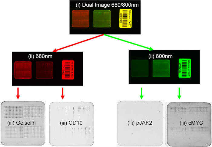
- Scan the slide at 680 nm and/or 800 nm depending on the secondary antibody/antibodies used. For two-color detection always use highly cross-adsorbed secondary antibodies to minimize cross-reactivity. Careful selection of primary and secondary antibodies is necessary for two-color detection. Of primary importance is the selection of different host species (e.g. rabbit and mouse) for the two primary antibodies. This allows discrimination by anti-rabbit and anti-mouse secondary antibodies which are labeled with dye with easily distinguishable emission spectra.
- Image files are saved as .tiff files. Figure 1 depicts the image of scanned files.
结果

Figure 1. Flow diagram of RPPA scanning process.
披露声明
No conflicts of interest declared.
材料
| Name | Company | Catalog Number | Comments |
| Triton X-100 | Triton-X | T8787 | |
| Li-Cor Odyssey Blocking Buffer | Li-Cor | 927-40000 | |
| MicroGrid II robotic spotter | Biorobotics | ||
| FastFrame' four bay slide holder | Whatman | 10486001 | |
| FAST Slide - 2-Pad | Whatman | 10485317 | |
| IRDye 680LT Goat anti-Mouse IgG | Licor | 926-68020 | |
| IRDye 800CW Goat anti-Rabbit IgG | Licor | 926-32211 |
参考文献
This article has been published
Video Coming Soon
Source: O'Mahony, F. C. et al. The Use of Reverse Phase Protein Arrays (RPPA) to Explore Protein Expression Variation within Individual Renal Cell Cancers. J. Vis. Exp. (2013)
版权所属 © 2025 MyJoVE 公司版权所有,本公司不涉及任何医疗业务和医疗服务。