需要订阅 JoVE 才能查看此. 登录或开始免费试用。
Method Article
Erratum: Oxygenation-sensitive Cardiac MRI with Vasoactive Breathing Maneuvers for the Non-invasive Assessment of Coronary Microvascular Dysfunction
Not Published
摘要
An erratum was issued for: Oxygenation-sensitive Cardiac MRI with Vasoactive Breathing Maneuvers for the Non-invasive Assessment of Coronary Microvascular Dysfunction. The Abstract, Protocol, and Representative Results section were updated.
摘要
This corrects the article 10.3791/64149
研究方案
An erratum was issued for: Oxygenation-sensitive Cardiac MRI with Vasoactive Breathing Maneuvers for the Non-invasive Assessment of Coronary Microvascular Dysfunction. The Abstract, Protocol, and Representative Results section were updated.
The second paragraph of the Abstract has been updated from:
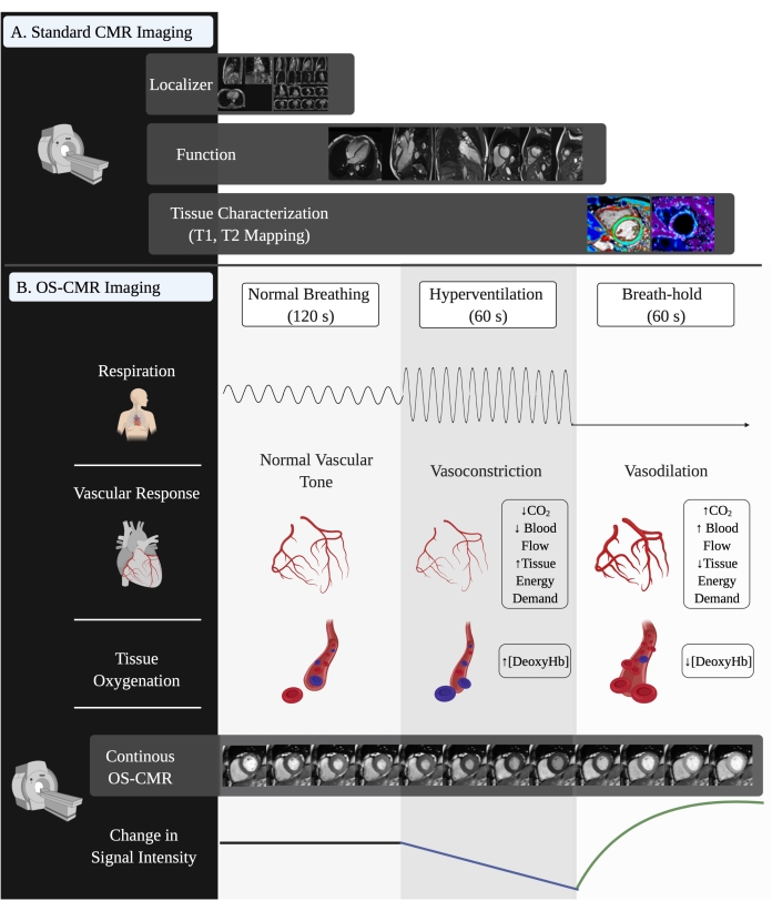
OS-CMR uses the well-known sensitivity of T2*-weighted images to blood oxygenation. Oxygenation-sensitive images can be acquired on any cardiac MRI scanner using a modified standard clinical steady-state free precession (SSFP) cine sequence, making this technique vendor-agnostic and easily implemented. As a vasoactive breathing maneuver, we apply a 4-min breathing protocol of 120 s of free breathing, 60 s of paced hyperventilation, followed by an expiratory breath-hold of at least 30 s. The regional and global response of myocardial tissue oxygenation to this maneuver can be assessed by tracking the signal intensity change. The change over the initial 30 s of the post-hyperventilation breath-hold, referred to as the breathing-induced myocardial oxygenation reserve (B-MORE) has been studied in healthy people and various pathologies. A detailed protocol for performing oxygen-sensitive CMR scans with vasoactive maneuvers is provided.
to:
OS-CMR uses the well-known sensitivity of T2*-weighted images to blood oxygenation. Oxygenation-sensitive images can be acquired on any cardiac MRI scanner using a modified standard clinical steady-state free precession (SSFP) cine sequence, making this technique vendor-agnostic and easily implemented. As a vasoactive breathing maneuver, we apply a 2-min breathing protocol of 120 s of free breathing, 60 s of paced hyperventilation, followed by an expiratory breath-hold of at least 30 s. The regional and global response of myocardial tissue oxygenation to this maneuver can be assessed by tracking the signal intensity change. The change over the initial 30 s of the post-hyperventilation breath-hold, referred to as the breathing-induced myocardial oxygenation reserve (B-MORE) has been studied in healthy people and various pathologies. A detailed protocol for performing oxygen-sensitive CMR scans with vasoactive maneuvers is provided.
Step 1.4.1 of the Protocol has been updated from:
Acquire the second OS-CMR series as a 4 min, continuous acquisition comprised of a short 15 s breath hold and 1 min of paced hyperventilation, followed by a voluntary, maximal breath hold (~45 s). As the continuous acquisition obtains multiple cardiac cycles over 2 min, modify one additional parameter (the number of cardiac cycles acquired by the acquisition) to make this series a repeated-measures acquisition.
NOTE: The minimum required breath-hold length is 30 s, although a breath-hold of 60 s is considered the standard.
to:
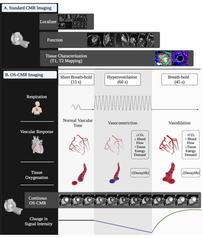
Acquire the second OS-CMR series as a 2 min, continuous acquisition comprised of a short 15 s breath hold and 1 min of paced hyperventilation, followed by a voluntary, maximal breath hold (~45 s). As the continuous acquisition obtains multiple cardiac cycles over 2 min, modify one additional parameter (the number of cardiac cycles acquired by the acquisition) to make this series a repeated-measures acquisition.
NOTE: The minimum required breath-hold length is 30 s, although a breath-hold of 45-60 s is considered the standard.
Step 1.4.3 of the Protocol has been updated from:
Start the breathing maneuver with free breathing, and end the expiration breath hold to acquire one cardiac cycle. Then guide the participant through paced breathing with the use of audible beeps from a metronome at a frequency of 30 breaths/min (one beep indicates breathing in, one beep indicates breathing out). At the 55 s mark of hyperventilation, give a final voice command to "take a deep breath in and then breathe out and hold your breath" to ensure that the breath-hold is performed at an end-expiration level.
to:
Start the breathing maneuver with a short 15 s, and end the expiration breath hold to acquire one cardiac cycle. Then guide the participant through paced breathing with the use of audible beeps from a metronome at a frequency of 30 breaths/min (one beep indicates breathing in, one beep indicates breathing out). At the 55 s mark of hyperventilation, give a final voice command to "take a deep breath in and then breathe out and hold your breath" to ensure that the breath-hold is performed at an end-expiration level.
Step 3.2 of the Protocol has been updated from:
Create two OS series, a baseline (labeled: OS_base) and the continuous acquisition during which the breathing maneuver is performed (labeled: OS_cont_acq). Leave the baseline OS sequence unchanged. In the OS continuous acquisition, increase the repeated measures from 1 to ~25-40 (depending on the scanner type). Increase the number of cardiac cycles (measures) until the acquisition time is ~4.5 min.
to:
Create two OS series, a baseline (labeled: OS_base) and the continuous acquisition during which the breathing maneuver is performed (labeled: OS_cont_acq). Leave the baseline OS sequence unchanged. In the OS continuous acquisition, increase the repeated measures from 1 to ~15-40 (depending on the scanner type). Increase the number of cardiac cycles (measures) until the acquisition time is ~2.5 min.
Step 5.1.1 of the Protocol has been updated from:
If possible, copy slice position and adjust volume from the OS baseline image or duplicate the baseline OS sequence and, in repeated measurements, increase from 1 to ~25-40 (or close to 4.5 min acquisition time).
to:
If possible, copy slice position and adjust volume from the OS baseline image or duplicate the baseline OS sequence and, in repeated measurements, increase from 1 to ~15-40 (or close to 2.5 min acquisition time).
Step 5.5.2 of the Protocol has been updated from:
If manually guiding the participant through the breathing maneuvers, instruct them to breathe in and breathe out, then hold their breath for 10 seconds, and start hyperventilating as soon as they hear the metronome beep.
to:
If manually guiding the participant through the breathing maneuvers, instruct them to breathe in and breathe out, then hold their breath for 15 seconds (for the short breath-hold), and start hyperventilating as soon as they hear the metronome beep.
Step 8.1 of the Protocol has been updated from:
Express B-MORE as a percent change in signal intensity from baseline to vasodilation (see equation 1):
 (1)
(1)
to:
Express B-MORE as a percent change in signal intensity from baseline to vasodilation (see equation 1):
 (1)
(1)
Figure 2 in the Representative Results has been updated from:

Figure 2: Visual representation of a full OS-CMR scan with vasoactive breathing maneuvers. (A) The standard acquisitions of a cardiac magnetic resonance imaging scan, including localizers, short-axis and long-axis cine function images, and tissue characterization images (such as T1 and/or T2 mapping). (B) The performance, physiologic effects, acquisition, and changes in MRI signal intensity throughout the vasoactive breathing maneuver. Abbreviations: OS-CMR = oxygenation-sensitive cardiac magnetic resonance imaging; DeoxyHb = deoxyhemoglobin. Please click here to view a larger version of this figure.
to:

Figure 2: Visual representation of a full OS-CMR scan with vasoactive breathing maneuvers. (A) The standard acquisitions of a cardiac magnetic resonance imaging scan, including localizers, short-axis and long-axis cine function images, and tissue characterization images (such as T1 and/or T2 mapping). (B) The performance, physiologic effects, acquisition, and changes in MRI signal intensity throughout the vasoactive breathing maneuver. Abbreviations: OS-CMR = oxygenation-sensitive cardiac magnetic resonance imaging; DeoxyHb = deoxyhemoglobin. Please click here to view a larger version of this figure.
Table 2 in the Representative Results has been updated from:
| 3T | 1.5T | |||
| bSSFP | mSSFP (OS) | bSSFP | mSSFP (OS) | |
| Repetition Time (TR) | 2.9 ms | 3.5 ms | 31.1 ms | 39 ms |
| Echo Time (TE) | 1.21 ms | 1.73 ms | 1.21 ms | 1.63 ms |
| Flip Angle (FA) | 80 deg | 35 deg | 39 deg | 35 deg |
| Voxel Size | 1.6 mm x 1.6 mm x 6 mm | 2.0 mm x 2.0 mm x 10.0 mm | 1.6 mm x 1.6 mm x 6 mm | 1.6 mm x 1.6 mm x 6 mm |
| Bandwidth (Hertz/ Pixel) | 947 | 1302 | 1313 | 1302 |
Table 2: Parameter differences between balanced SSFP and modified SSFP (BOLD) sequence at 3 Tesla and 1.5 Tesla. Abbreviations: SSFP = steady-state, free precession; bSSFP = balanced SSFP; mSSFP = modified SSFP; OS = oxygen-sensitive; BOLD = blood oxygen level-dependent.
to:
2.1 SIEMENS
| 3T Siemens modified SSFP (OS) | 1.5T Siemens Modified SSFP (OS) | ||
| TR | Repetition Time (ms) | 41.4 | 39 |
| TE | Echo Time (ms) | 1.7 | 1.63 |
| FA | Flip Angle (deg) | 35 | 35 |
| Voxel Size (mm) | 2.0 x 2.0 x 10.0 | 2.0 x 2.0 x 10.0 | |
| Bandwidth (Hz/Px) | 1302 | 1302 |
2.2 GE HEALTHCARE
| 3T GE Healthcare modified SSFP (OS) | 1.5T GE Healthcare modified SSFP (OS) | ||
| TR | Repetition Time (ms) | 2.8 | 3.22 |
| TE | Echo Time (ms) | 0.97 | 1.2 |
| FA | Flip Angle (deg) | 35 | 35 |
| Voxel Size (mm) | 2.0 X 2.0 X 10.0 | 1.6 x 1.6 x 10.0 | |
| Bandwidth (Hz/Px) | 1302 | 1302 |
2.3 PHILIPS
| 3T Philips modified SSFP (OS) | 1.5T Philips modified SSFP (OS) | ||
| TR | Repetition Time (ms) | 3.41 | 3.3 |
| TE | Echo Time (ms) | 1.71 | 1.64 |
| FA | Flip Angle (deg) | 35 | 55 |
| Voxel Size (mm) | 1.98 X 1.98 X 10.0 | 1.19x1.19x10.0 | |
| Bandwidth (Hz/Px) | 571.1 | 571.1 |
Table 2: Parameter differences between of the modified SSFP (BOLD) sequence at 3 Tesla and 1.5 Tesla for 2.1 Siemens, 2.2 General Electric Healthcare, and 2.3 Philips scanners. Abbreviations: GE = General Electric; SSFP = steady-state, free precession; OS = oxygen-sensitive.
Table 3 in the Representative Results has been updated from:
| Modifiable | Non-Modifiable | ||
| Field of View (mm) | 360-400 | Slice Thickness (mm) | 10 |
| Gap (%) | 0-200 | Flip Angle | 35 |
| Acquisition Time (s/measurement) | 8 | Segments | 12 |
| Measurements | 1 (baseline) or 25+ (continuous acquisition) | ECG | Triggered /Prospective |
| Acquisition Window | No set limitations | TE (ms) | 1.7 |
| TR (ms) | 40.68 (3.4) | ||
| Bandwidth (Hertz/Pixel) | 1302 | ||
Table 3: Modifiable and non-modifiable OS-CMR sequence parameters during image acquisition. Abbreviations: OS-CMR = oxygenation-sensitive cardiac magnetic resonance imaging; ECG = electrocardiography; TE = echo time; TR = repetition time.
to:
| Modifiable | Non-Modifiable | ||
| Field of View (mm) | 360-400 | Slice Thickness (mm) | 10 |
| Gap (%) | 0-200 | Flip Angle | 35 |
| Acquisition Time (s/measurement) | 8 | Segments | 12 – 16 |
| Measurements | 1 (baseline) or 15+ (continuous acquisition) | ECG | Triggered /Prospective |
| Acquisition Window | No set limitations | TE (ms) | 1.7 |
| TR (ms) | 40.68 (3.4) | ||
| Bandwidth (Hertz/Pixel) | 1302 | ||
Table 3: Modifiable and non-modifiable OS-CMR sequence parameters during image acquisition. Abbreviations: OS-CMR = oxygenation-sensitive cardiac magnetic resonance imaging; ECG = electrocardiography; TE = echo time; TR = repetition time.
Access restricted. Please log in or start a trial to view this content.
披露声明
参考文献
Access restricted. Please log in or start a trial to view this content.
转载和许可
请求许可使用此 JoVE 文章的文本或图形
请求许可版权所属 © 2025 MyJoVE 公司版权所有,本公司不涉及任何医疗业务和医疗服务。