
Whole Mount Immunofluorescent Staining of the Neonatal Mouse Retina to Investigate Angiogenesis In vivo
July 9th, 2013
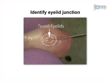
•The neonatal murine retina provides a well characterized physiological model of angiogenesis, which permits investigations of the roles of different genes or drugs that modulate angiogenesis in an in vivo context. Immunofluorescent staining to accurately visualize the vascular plexus is pivotal to the success of these types of studies.
Related Videos

Visualization of the Embryonic Nervous System in Whole-mount Drosophila Embryos

Dissection of a Mouse Eye for a Whole Mount of the Retinal Pigment Epithelium
In vivo Electroporation of Developing Mouse Retina
Whole Mount Immunolabeling of Olfactory Receptor Neurons in the Drosophila Antenna

Assessment of Vascular Regeneration in the CNS Using the Mouse Retina

Whole-mount Imaging of Mouse Embryo Sensory Axon Projections

Whole Mount Dissection and Immunofluorescence of the Adult Mouse Cochlea

Patch Clamp Recording of Starburst Amacrine Cells in a Flat-mount Preparation of Deafferentated Mouse Retina
A Simple One-step Dissection Protocol for Whole-mount Preparation of Adult Drosophila Brains

Transpupillary Two-Photon In Vivo Imaging of the Mouse Retina