Visualizing Cytoskeleton-Dependent Trafficking of Lipid-Containing Organelles in Drosophila Embryos
December 13th, 2021
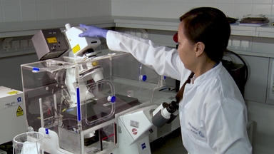
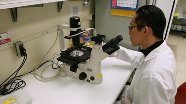
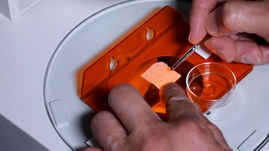
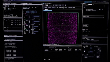
•In the early Drosophila embryo, many organelles are motile. In principle, they can be imaged live via specific fluorescent probes, but the eggshell prevents direct application to the embryo. This protocol describes how to introduce such probes via microinjection, and then analyze bulk organelle motion via particle image velocimetry.
Related Videos

Multi-target Chromogenic Whole-mount In Situ Hybridization for Comparing Gene Expression Domains in Drosophila Embryos

Protocols for Visualizing Steroidogenic Organs and Their Interactive Organs with Immunostaining in the Fruit Fly Drosophila melanogaster

Visualizing the Node and Notochordal Plate In Gastrulating Mouse Embryos Using Scanning Electron Microscopy and Whole Mount Immunofluorescence

Thawing, Culturing, and Cryopreserving Drosophila Cell Lines

Imaging Intranuclear Actin Rods in Live Heat Stressed Drosophila Embryos

Visualizing the Effects of Oxidative Damage on Drosophila Egg Chambers using Live Imaging

Confocal and Super-Resolution Imaging of Polarized Intracellular Trafficking and Secretion of Basement Membrane Proteins During Drosophila Oogenesis

Optogenetic Inhibition of Rho1-Mediated Actomyosin Contractility Coupled with Measurement of Epithelial Tension in Drosophila Embryos (Video) | JoVE

Collection of Drosophila Embryos and Preparing Them for Optogenetic Stimulation (Video) | JoVE

Optogenetic Stimulation, Laser Ablation, and Imaging of Drosophila Embryos (Video) | JoVE