
In Vivo Vascular Injury Readouts in Mouse Retina to Promote Reproducibility
April 21st, 2022
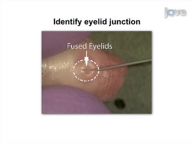
•Here, we present three data analysis protocols for fluorescein angiography (FA) and optical coherence tomography (OCT) images in the study of Retinal Vein Occlusion (RVO).
Related Videos

Transfection of Mouse Retinal Ganglion Cells by in vivo Electroporation
In vivo Electroporation of Developing Mouse Retina

Functional Neuroimaging Using Ultrasonic Blood-brain Barrier Disruption and Manganese-enhanced MRI

Whole Mount Immunofluorescent Staining of the Neonatal Mouse Retina to Investigate Angiogenesis In vivo

Assessment of Vascular Regeneration in the CNS Using the Mouse Retina

Vibratome Sectioning Mouse Retina to Prepare Photoreceptor Cultures

An Ex Vivo Laser-induced Spinal Cord Injury Model to Assess Mechanisms of Axonal Degeneration in Real-time

Simultaneous ex vivo Functional Testing of Two Retinas by in vivo Electroretinogram System

An In Vivo Duo-color Method for Imaging Vascular Dynamics Following Contusive Spinal Cord Injury

Transpupillary Two-Photon In Vivo Imaging of the Mouse Retina