
Method Article
Frühe das Eindringen des Virus-Assays für die Ermittlung und Bewertung von antiviralen Verbindungen
In diesem Artikel
Zusammenfassung
Here, we present a protocol that examines specific steps of the viral entry to identify and evaluate novel antiviral agents.
Zusammenfassung
Cell-based systems are useful for discovering antiviral agents. Dissecting the viral life cycle, particularly the early entry stages, allows a mechanistic approach to identify and evaluate antiviral agents that target specific steps of the viral entry. In this report, the methods of examining viral inactivation, viral attachment, and viral entry/fusion as antiviral assays for such purposes are described, using hepatitis C virus as a model. These assays should be useful for discovering novel antagonists/inhibitors to early viral entry and help expand the scope of candidate antiviral agents for further drug development.
Einleitung
Viral infections are a constant threat to the public health and a significant cause of epidemic diseases, morbidity, and deaths worldwide. Specific modes of control against viral infections include vaccine development and antiviral therapies. While vaccine efforts have proven successful in immunizing against several viruses, many viral pathogens remain without a protective vaccine including dengue virus (DENV), human cytomegalovirus (HCMV), hepatitis C virus (HCV), human immunodeficiency virus (HIV), and respiratory syncytial virus (RSV)1-5. Antivirals, on the other hand, play an important role for the management of these viral infections when prophylactic vaccines are unavailable. However, to date, only few licensed and cost-effective antiviral drugs are available compared to the number of viral pathogens that threatens the public health. More importantly, due to an increase in global travel and rapid urbanization, the situation is aggravated by risks of epidemic outbreaks from emerging/re-emerging viral infections that are being introduced into non-indigenous areas6. Recent outbreaks caused by severe acute respiratory syndrome (SARS) virus, influenza viruses (H1N1, H5N1, H3N2, and H7N9), DENV, West Nile virus (WNV), measles virus (MV), Middle East Respiratory Syndrome (MERS) virus, and Ebola virus6-12 are among the examples reflecting the need for antivirals development when immunization and/or therapeutics are unavailable. In addition, there is always a potential risk of generating drug-resistant infections with currently used antivirals. Thus, the continuous development and expansion of the scope of antivirals to these emerging/re-emerging viral infections are necessary to provide better management strategies and safeguard the public health.
Most antiviral therapies consist of direct acting antivirals (DAAs) which target a specific viral protein or cofactor that mediates important steps in the viral life cycle. For example, the nucleoside analogue Acyclovir inhibits herpesvirus DNA polymerase, protease inhibitors Boceprevir and Telaprevir antagonize the HCV NS3, and Oseltamivir and Zanamivir are neuraminidase inhibitors that block the release of influenza virus particles from infected cells13-15. There are however very few licensed viral entry inhibitors including Enfuvirtide, which targets HIV gp41 to prevent fusion, and Maraviroc, which blocks the HIV co-receptor CCR5, thereby preventing viral entry16. Exploring novel antagonists/inhibitors to viral entry could help provide additional therapeutics for prophylactic or therapeutic use, such as in combination with other antivirals with a different mechanism of action to better manage viral infections17-19.
Identification of antivirals can involve structure-guided drug design and candidate drug screening-based strategy. Methods for assessing antiviral activity of test agents include biochemical assays of enzymatic activity and evaluation by cell-based systems20-23. In cell-based systems, the viral life cycle can be dissected into distinct stages of infection, such as entry events (attachment, fusion, uncoating), the replication phase (viral genome replication and protein translation), and virion egress (assembly, maturation, and release). Since the assays can be adapted to investigate each specific stage using various tools and methods, this approach allows identification/examination of potential candidate antivirals with a specific mechanism of action targeting the distinct stage analyzed. For instance, to analyze drug effect specifically on the free virus particles prior to binding to the host cell, a ‘viral inactivation assay’ can be performed. In this assay, the virus is allowed to incubate with the test drug and then diluted to titrate out the drug before infecting a cell monolayer. Additional steps such as viral attachment and entry/fusion stages can also be analyzed individually, by shifting the temperature during the infection. For many enveloped viruses, viral entry/fusion at the host cell membrane is greatly facilitated at 37 °C, but is typically precluded at 4 °C which does not affect virus binding24-29. Finally, the use of reporter viruses or cell systems could facilitate these studies and permit a high-throughput analysis.
We previously employed the cell-based approach and dissected the early entry of various enveloped viruses for the examination of antiviral compounds that potentially antagonize viral entry30,31. Herein, the various methods used, including viral inactivation, viral attachment, and viral entry/fusion assays, are described.
Protokoll
Hinweis: Stellen Sie sicher, dass alle Verfahren, die Zellkultur und Virusinfektion sind in zertifizierten biologischen Sicherheit Hauben, die für die Sicherheitsstufe der Proben entsprechende gehandhabt werden durchgeführt. Für den Zweck der Beschreibung der Protokolle wird Gaussia-Luciferase-Reporter-markierten HCV als Modellvirus 32 benutzt. Im Zusammenhang mit den repräsentativen Ergebnissen, sind die Verbindungen chebulagic Säure (CHLA) und Punicalagin (PUG) als Kandidaten Virostatika, die virale Glycoprotein Wechselwirkungen mit Zelloberflächen Glycosaminoglycane Target während der frühen viralen Eintritt Stufen 31 verwendet. Heparin, das dafür bekannt ist, mit der Eingabe von vielen Viren 30,31,33,34 eingreifen, wird als positive Kontrolle der Behandlung in diesem Kontext verwendet wird. Für grundlegende Hintergrund Virologie Techniken Vermehrung von Viren, die Bestimmung der Virustiter und die Expression von infektiösen Dosis in plaquebildenden Einheiten (PFU), Fokus-bildenden Einheiten (FFU) oder Multiplizität der Infektion (MOI) ist, worin Re der Leserübertragen auf Bezugs 35. Der vorherigen Beispiele und Viren in den repräsentativen gezeigten Ergebnisse verwendet optimierten Bedingungen wird der Leser auf die in Tabelle 1, 1A und 2A aufgeführten Referenzen 30-32,36-39 sowie Einzelheiten bezeichnet.
1. Zellkultur, Compound Vorbereitung, und die Verbindung Zytotoxizität
- Wachsen die jeweilige Zelllinie für die Virusinfektion zu analysierende System (Tabelle 1). Für HCV, wachsen die Huh-7,5-Zellen in Dulbeccos modifiziertem Eagle-Medium (DMEM) mit 10% fötalem Rinderserum (FBS), 200 U / ml Penicillin G, 200 ug / ml Streptomycin und 0,5 ug / ml Amphotericin B.
- Bereiten Sie die Testverbindungen und Kontrollen unter Verwendung ihres jeweiligen Lösemittel: beispielsweise aufzulösen CHLA und PUG in Dimethylsulfoxid (DMSO); vorzubereiten Heparin in sterilem doppelt destilliertem Wasser. Für alle nachfolgenden Verdünnungen verwenden Kulturmedien.
Hinweis: Die endgültige concentRation von DMSO in der Testverbindung Behandlungen ist weniger als 1% in den Experimenten; 1% DMSO als ein negativer Kontrollbehandlung in den Tests zum Vergleich eingeschlossen. - Bestimmung der Zytotoxizität der Testverbindungen (zB CHLA und PUG) auf die Zellen für die Virusinfektion durch die Verwendung eines Zell-Lebensfähigkeit Bestimmen Reagens wie XTT (2,3-Bis [2-methoxy-4-nitro-5-sulfophenyl] -5-phenylamino) carbonyl] -2H-tetrazolium Hydroxid):
- Für HCV, Samen Huh-7,5-Zellen in einer Platte mit 96 Vertiefungen (1 × 10 4 Zellen pro Vertiefung) und bei 37 ° C in einem 5% CO 2 -Inkubator O / N um eine Monoschicht zu erhalten.
- Gelten DMSO-Kontrolle (1%) oder steigenden Konzentrationen der Testverbindungen CHLA und PUG (ex. 0, 10, 50, 100, und 500 & mgr; M) zu den Kulturvertiefungen in dreifacher Ausführung.
- Bei 37 ° C für 72 Stunden und wirf dann das Medium in der Platte und waschen Sie die Zellen mit 200 ul phosphatgepufferter Kochsalzlösung (PBS) zweimal.
- 100 l assaying Lösung aus dem XTT-basierte in vitro-Toxikologie-Assay-Kit, um jede Vertiefung und inkubieren Sie die Platten bei 37 ° C für weitere 3 h auf XTT ermöglichen Formazan-Produktion.
- Die Optische mit einem Mikroplattenleser bei einer Testwellenlänge von 492 nm und einer Referenzwellenlänge von 690 nm.
- Der prozentuale Anteil der überlebenden Zellen nach der folgenden Formel: Zelllebensfähigkeit (%) = An / Wie × 100%, wobei 'An' und 'Wie' beziehen sich auf die Absorption der Testverbindungen und die Lösungsmittelkontrolle (ex 1% DMSO. ) Behandlungen sind. Bestimmung der Konzentration von 50% zelluläre Zytotoxizität (CC 50) der Testverbindungen aus einer analytischen Software wie GraphPad Prism nach dem Protokoll des Herstellers.
2. Auslesen der Virusinfektion
Hinweis: Das Auslesen der Virusinfektion hängt von der Virus-System verwendet und können Methoden wie Plaque-Assays oder mea beinhaltensuring Reporter Signale von Reporter-markierten Viren. Methode für das Erfassen Reporter-HCV-Infektion auf der Basis der Luciferase-Reporteraktivität wird nachstehend beschrieben.
- Sammeln der Überstände aus den infizierten Vertiefungen und Klärung bei 17.000 xg in einer Mikrozentrifuge für 5 min bei 4 ° C.
- Mix 20 ul Testüberstand wurde in 50 ul Luciferase-Substrat aus der Gaussia-Luciferase-Assay-Kits und direkt mit einem Luminometer gemessen nach den Angaben des Herstellers.
- Auszudrücken HCV Infektiosität als log 10 der relativen Lichteinheiten (RLU) zum Bestimmen virale Inhibition (%) und die Berechnung der 50% effektive Konzentration (EC 50) der Testverbindungen gegen die HCV-Infektion unter Verwendung von Algorithmen von GraphPad Prism Software gemäß dem Protokoll des Herstellers.
3. Vireninaktivierung Assay
Hinweis: Beispiele für Inkubationszeit und Virusdosis für verschiedene Viren einwieder in 1A aufgeführt. Höhere Konzentrationen des Virus kann auch durch Erhöhung der MOI / PFU getestet werden.
- Samen Huh-7,5-Zellen in einer Platte mit 96 Vertiefungen (1 × 10 4 Zellen pro Vertiefung) und bei 37 ° C in einem 5% CO 2 -Inkubator O / N um eine Monoschicht zu erhalten.
- Inkubieren Sie die Testverbindungen oder Kontrollen (Endkonzentrationen: CHLA = 50 & mgr; M; PUG = 50 & mgr; M; Heparin = 1.000 ug / ml; DMSO = 1%) mit den HCV-Partikel bei 37 ° C (1A, "Long-Term ' ) in einem Verhältnis 1: 1. Beispielsweise auf einen 100 & mgr; Virusinokulum enthaltend 1 x 10 4 FFU wird mit 100 ul einer 100 uM CHLA Arbeitsverdünnung; Dies ergibt CHLA Behandlung bei einer Endkonzentration von 50 uM.
- Verdünnt das virus-Wirkstoff-Mischung "sub-therapeutische" (unwirksam) Konzentration der Testverbindungen. Zum Beispiel ist die unwirksame Konzentration CHLA und PUG gegen HCV bei 1 & mgr; M 31; deshalbdies erfordert eine 50-fache Verdünnung des Virus-Wirkstoff-Mischung, die mit 9,8 ml Basalmedium (Zellkulturmedium mit 2% FBS) erreicht werden kann.
Anmerkung: Die Verdünnung auf subtherapeutische Konzentration verhindert erhebliche Wechselwirkung zwischen der Testverbindungen und der Oberfläche der Wirtszelle und ermöglicht die Untersuchung der Wirkung der Behandlung auf den zellfreien Virionen. Beachten Sie, dass diese Verdünnung hängt von der antivirale Dosis-Antwort der Testverbindungen gegen die insbesondere virale Infektion und vor der Durchführung dieses bestimmten Test 31 ermittelt. - Zum Vergleich, mischen das Virus mit den Testverbindungen und sofort zu verdünnen (kein Inkubationszeit), um Unter therapeutische Konzentration vor der Infektion (1A, "Short-Term).
- Zugabe von 100 ul der verdünnten HCV-Wirkstoff-Mischung auf die Huh-7,5 Zellmonolayer (die Virusmenge ist nun auf 1 x 10 2 FFU; final MOI = 0,01) und Inkubation für 3 Stunden bei 37 ° C viral, damitAdsorption.
- Nach der Infektion, entfernen Sie die Inokula verdünnt und vorsichtig waschen die Vertiefungen zweimal mit 200 ul PBS.
Hinweis: Führen Sie die wäscht vorsichtig, um zu vermeiden, heben Sie die Zellen. - Gelten 100 ul Basalmedium in jede Vertiefung und Inkubieren bei 37 ° C für 72 Std.
- Analyse des resultierenden Infektion durch Untersuchung des Überstandes auf Luciferase-Aktivität wie in '2 beschrieben. Auslesen der Virusinfektion.
4. Viral Befestigung Assay
Hinweis: Beispiele für Inkubationszeit und Virusdosis für verschiedene Viren sind in 2A aufgeführt, "Anhang". Höhere Konzentrationen des Virus kann auch durch Erhöhung der MOI / PFU getestet werden.
- Samen Huh-7,5-Zellen in einer Platte mit 96 Vertiefungen (1 × 10 4 Zellen pro Vertiefung) und bei 37 ° C in einem 5% CO 2 -Inkubator O / N um eine Monoschicht zu erhalten.
- Pre-Chill die Zellmonoschichten in Platten bei 4 ° C for 1 Std.
- Co-Behandlung der Zellen mit HCV Inokulum (MOI = 0,01) und die Testverbindungen oder Kontrollen (Endkonzentrationen: CHLA = 50 uM; PUG = 50 uM; Heparin = 1,000 & mgr; g / ml; DMSO = 1%) bei 4 ° C für 3 Std. Zum Beispiel, um eine 90 & mgr; l Virusinokulum mit 1 x 10 2 FFU, fügen Sie 10 ul einer 500 uM CHLA Arbeitsverdünnung; Dies ergibt CHLA Behandlung bei einer Endkonzentration von 50 uM und eine Infektion durch HCV bei MOI = 0,01 auf der Zell-Monoschicht.
Hinweis: Es ist wichtig, für die Durchführung des Experiments bei 4 ° C, da es eine Virusbindung, sondern schließt Eintrag, der am effizientesten bei 37 ° C auftritt. Führen die Zugabe des Virus und die Testverbindungen auf Eis und der anschließenden Inkubation in einem 4ºC Kühlraum, um sicherzustellen, dass die Temperatur auf 4 ° C gehalten. - Entfernen Sie den Überstand und waschen Sie sich leicht die Zellmonoschicht mit 200 ul eiskaltem PBS zweimal.
Hinweis: Führen Sie die wäscht vorsichtig, um zu vermeiden, heben Sie die Zellen <./ li> - Gelten 100 ul Basalmedium in jede Vertiefung und Inkubieren bei 37 ° C für 72 Std.
- Analyse des resultierenden Infektion durch Untersuchung des Überstandes auf Luciferase-Aktivität wie in '2 beschrieben. Auslesen der Virusinfektion.
5. Viral Entry / Fusion Assay
Hinweis: Beispiel für Inkubationszeiten und Virusdosis für verschiedene Viren sind in 2A 'Entry / Fusion "aufgeführt. Höhere Konzentrationen des Virus kann auch durch Erhöhung der MOI / PFU getestet werden.
- Samen Huh-7,5-Zellen in einer Platte mit 96 Vertiefungen (1 × 10 4 Zellen pro Vertiefung) und bei 37 ° C in einem 5% CO 2 -Inkubator O / N um eine Monoschicht zu erhalten.
- Pre-Chill die Zellmonoschichten in Platten bei 4 ° C für 1 Stunde.
- Infizieren die Zellen mit HCV (MOI = 0,01) bei 4 ° C für 3 Stunden. Verwenden Sie beispielsweise eine 100 ul Virusinokulum mit 1 x 10 2 FFU.
Hinweis: Führen Sie die addition des viralen Inokulum auf Eis und der anschließenden Inkubation in einem 4ºC Kühlraum, die Temperatur auf 4 ° C, die die virale Bindung jedoch keine Ausgabe ermöglicht aufrechtzuerhalten. - Entfernen Sie den Überstand und waschen Sie sich leicht die Zellmonoschichten zweimal mit 200 ul eiskaltem PBS.
Hinweis: Führen Sie die wäscht vorsichtig, um zu vermeiden, heben Sie die Zellen. - Behandeln Sie die Vertiefungen mit den Testverbindungen oder Kontrollen (Endkonzentrationen: CHLA = 50 & mgr; M; PUG = 50 & mgr; M; Heparin = 1.000 ug / ml; DMSO = 1%) und bei 37 ° C für 3 Stunden. Fügen Sie beispielsweise 10 & mgr; l einer 500 & mgr; M CHLA Arbeitsverdünnung auf 90 ul Medium, zu mischen und zu behandeln, die Brunnen; Dies ergibt CHLA Behandlung bei einer Endkonzentration von 50 uM.
Hinweis: Der Wechsel von 4 ° C bis 37 ° C ermöglicht nun den Viruseintritt / Fusion Veranstaltung und ermöglicht somit Beurteilung der Testverbindungen "-Effekt an diesem Schritt. - Saugen Sie das Arzneimittel enthaltende Überstand und entfernen Sie nicht internalisiertextrazellulären Viren durch Waschen mit 200 ul Citratpuffer (50 mM Natriumcitrat, 4 mM Kaliumchlorid, pH 3.0) oder PBS. Gelten 100 ul des Grundmediums vor der Inkubation bei 37 ° C für 72 Std.
- Analyse des resultierenden Infektion durch Untersuchung des Überstandes auf Luciferase-Aktivität wie in '2 beschrieben. Auslesen der Virusinfektion.
Ergebnisse
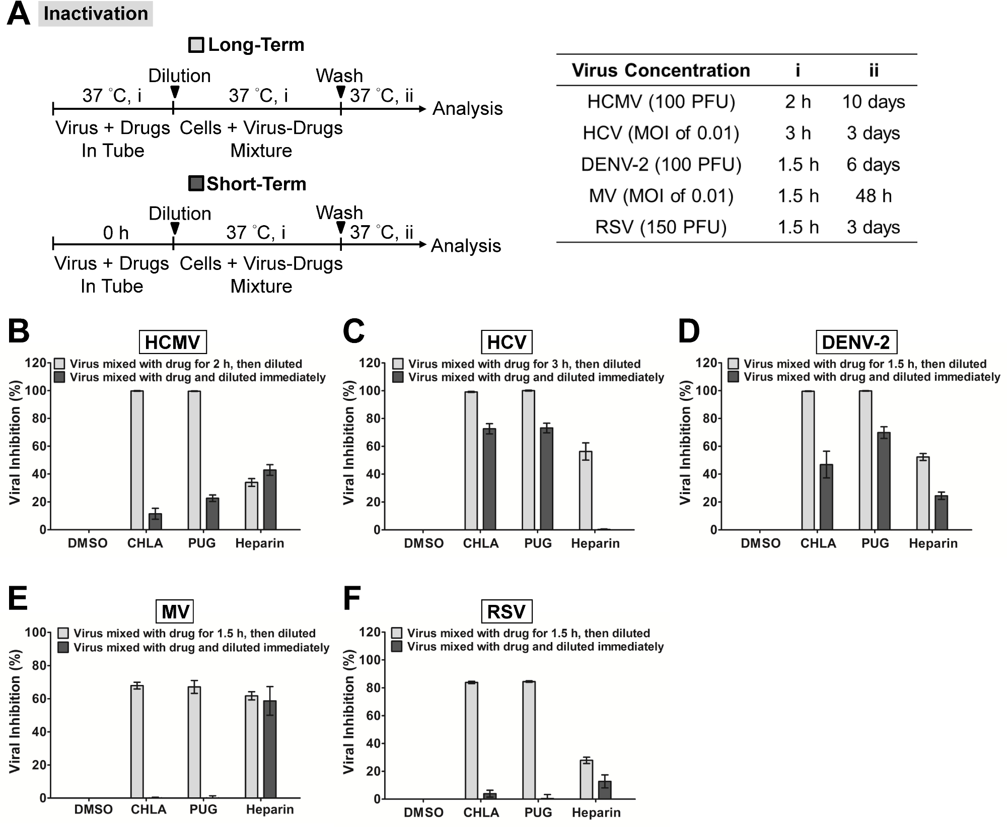
In Abbildung 1 wurde die "Virus-Inaktivierung Test" durchgeführt, um zu prüfen, ob zwei bestimmte natürliche Verbindungen CHLA und PUG könnten die verschiedenen umhüllten Viren in zellfreien Zustand zu inaktivieren und zu verhindern nachfolgende Infektion. Die Zytotoxizität und antivirale Dosis-Antwort dieser Verbindungen vor der Durchführung der mechanistische Studie 31 bestimmt. Die Viren wurden mit den Testverbindungen vorbehandelt und anschließend die Virus-Wirkstoff-Mischungen wurden zu subtherapeutischen Konzentrationen vor der Impfung auf die jeweilige Zellmonoschicht für jede Virus-System verdünnt. Wie in 1 gezeigt, sowohl CHLA und PUG schienen mit den zellfreien Virionen interagieren, was irreversible Effekte, die die Zell-Monoschicht von der nachfolgenden Infektion geschützt. Die beiden Testverbindungen erreicht eine nahezu 100% ige Hemmung gegen HCMV, HCV und DENV-2, während eine 60 bis 80% Block wurde gegen MV und RSV beobachtet. Diese Ergebnisse suggest, dass CHLA und PUG haben direkten Einfluss auf diese freie Viruspartikel durch Inaktivierung von ihnen und zur Neutralisierung ihrer Infektiosität.
In 2 wurden die Befestigung und Ein- / Fusion-Assays durchgeführt, um die Wirkung von CHLA und PUG gegen diesen frühen Viruseintritt bezogene Ereignisse von HCMV, HCV, DENV-2, MV und RSV erkunden. Sowohl CHLA und PUG wirksam verhindert die Bindung der untersuchten Viren auf die jeweilige Wirtszelle, wie durch Hemmung auf die resultierende Virusinfektion (Abbildung 2, "Anhänge": hellgraue Balken) gezeigt. Die inhibitorische Wirkung auf die Virus Befestigung von beiden Verbindungen war ähnlich gegen HCMV (2B), HCV (2C), DENV-2 (2D) und RSV (2F), im Bereich von 90 - 100%. Andererseits erschien PUG wirksamer als CHLA gegen MV Bindung (2E) zu sein, wobei der Hemmungsrate von twO-Verbindungen zwischen 50 - 80%. Die Kontrollbehandlung Heparin, von dem bekannt ist, um den Eintritt von vielen Viren, inhibierte auch die Anbringung von HCMV, DENV-2, RSV, ad MV blockieren, war aber weniger wirksam gegen HCV. Die darauf folgende 'Viruseintritt / Fusionstest "untersucht, ob CHLA und PUG während der Viruseintritt / Schmelzphase behielten ihre Aktivität (Abbildung 2," Entry / Fusion ": dunkelgraue Balken). Auch hier sowohl CHLA und PUG wurden beobachtet, um das Eindringen des Virus / Fusionsschritt der untersuchten Viren (2B - F) wirksam zu beeinträchtigen, was zu einer 50 bis 90% schützende Wirkung auf die jeweilige Zellmonolayer. Heparin auch potent gehemmt Eintrag / Fusion in DENV-2 und RSV-Infektionen, war aber gegen HCMV, HCV, und MV (<40% ige Hemmung im Durchschnitt) weniger wirksam.
| Virus | Zellen-Art |
| HCMV | HEL |
| HCV | Huh-7.5 |
| DENV-2 | Vero |
| MV | CHO-SLAM |
| RSV | HEp-2 |
Tabelle 1:. Wirtszelltyp für die virale Infektion der Zelltyp für jeden der repräsentativen Ergebnisse beschriebenen viralen Infektionssystem verwendet wird, angegeben. Weitere Einzelheiten zu den Zellen in Referenz 31 gefunden werden.

Abbildung 1. Inaktivierung von Virusinfektionen durch die Testverbindungen CHLA und PUG Verschiedene Viren wurden mit den Testverbindungen für einen langen Zeitraum behandelt werden. (1,5 inkubiert - 3 Stunden vor der Titration, hellgraue Balken) oder kurzen Zeitraum (sofort verdünnt; dunkelgrau bar) bei 37 ° C, bevor eine Verdünnung auf subtherapeutischen Konzentrationention und anschließender Analyse der Infektion auf den jeweiligen Wirtszellen. (A) Schematische Darstellung des Experiments (links dargestellt) mit der endgültigen Viruskonzentration (PFU / Vertiefung oder MOI), Langzeit-Virus-Medikament Inkubationszeit (i), und anschließender Inkubation (ii) angegeben für jedes Virus in der Tabelle auf der rechten Seite. Die Analysen für (B) HCMV, (C) HCV, (D) DENV-2, (E) MV, und (F) RSV in jedem zusätzlichen Platte angezeigt. Ergebnisse werden mit dem DMSO negative Kontrolle Behandlung für Virusinfektion und die gezeigten Daten wurden die Mittelwerte ± Standardfehler des Mittelwerts (SEM) aus drei unabhängigen Experimenten aufgetragen sind. Diese Zahl hat sich von Referenz 31 modifiziert worden ist. Bitte klicken Sie hier, um eine größere Version dieser Figur zu sehen.

Abbildung 2. Evaluierung von antiviralen Aktivitäten der Testverbindungen CHLA und PUG Viren Befestigung und Ein- / Fusion. (A) Die experimentelle Vorgehensweise, Viruskonzentration (PFU / Vertiefung oder MOI), und der Zeitpunkt der Zugabe und die Behandlung mit den Testverbindungen (i, ii, iii) für jedes Virus in den Schemata und den zugehörigen Tabellen dargestellt. In Virusanheftung Analyse (hellgraue Balken) Monoschichten aus verschiedenen Zelltypen wurden bei 4 ° C für 1 h vorgekühlt, wobei die jeweiligen Viren und Testverbindungen dann mitbehandelt bei 4 ° C (1,5 - 3 h, i) vor dem Waschen die Inokulate und die Testverbindungen für die anschließende Inkubation (37 ° C, ii) und Prüfung der Virusinfektion. In virus entry / Schmelzanalyse (dunkelgraue Balken), wurden ausgesät Zellmonoschichten vorgekühlte bei 4 ° C für 1 Stunde und dann mit den entsprechenden Viren bei 4 ° C für 1,5 in Frage gestellt - 3 h (i). Zellen wurden danngewaschen und mit den Testverbindungen für eine weitere Inkubationsperiode (ii), während der die Temperatur auf 37 ° C verschoben, um das Eindringen des Virus / Fusionsereignis erleichtern behandelt. Am Ende der Inkubation wurden die extrazellulären Viren entweder durch Citratpuffer (pH 3.0) oder PBS Waschschritten entfernt und die Zellen wurden für weitere Analyse der Virus-Infektion inkubiert (iii). Ergebnisse für (B) HCMV, (C) HCV, (D) DENV-2, (E) MV, und (F) RSV in jedem zusätzlichen Platte angezeigt. Daten werden gegen die DMSO negative Kontrolle Behandlung einer Virusinfektion, aufgetragen und werden als Mittel ± SEM von drei unabhängigen Experimenten. Diese Zahl hat sich von Referenz 31 modifiziert worden ist. Bitte klicken Sie hier, um eine größere Version dieser Figur zu sehen.
Diskussion
In this report the methods to identify and evaluate antiviral compounds based on a mechanistic approach of dissecting the early viral entry events were described. Specifically, the assays allowed us to examine the effect of test compounds on free virus particles, viral attachment, and viral entry/fusion. Critical steps were implemented to distinctly evaluate the drug effect on the specific stage of early viral entry. For instance, in the ‘viral inactivation assay’, the dilution of the virus-drug mixture to sub-therapeutic concentration prevents significant interaction between the test compound and the host cell surface by ‘titrating out’ the drug. This ensures that the inhibitory effect observed on the subsequent infection of the host cell is due to a direct interaction between the test compound and the cell-free virions, rather than an effect from the test compound on host cell membrane or membrane-associated molecules, including viral receptors30. Similarly, the shift in temperature between 4 °C (which allows for virus binding but not entry) and 37 °C (which facilitates virus entry/fusion) in the ‘viral attachment assay’ and ‘viral entry/fusion assay’ are crucial to determine the test compound’s effect on each of these specific events. This is feasible due to the temperature sensitivity of enveloped viruses during these steps in the infection24-29. It is therefore important that the assays are performed at the indicated temperature to ensure the accuracy of the results; for example, by carrying out the experiment on ice to maintain at 4 °C and by placing the sample directly in a 37 °C incubator for the temperature shift. In addition, the use of negative (ex. DMSO solvent for drug preparation) and positive (ex. heparin treatment) controls also help further establish the assays’ accuracy. The utility and applicability of such methods have been demonstrated in many antiviral studies26,30,31,40,41. Note that while heparin is included as a control for all three assays in the context of the representative results, it typically blocks the initial virus binding rather than the ensuing fusion/entry step (as reflected by the data in Figure 2). Additional controls could also be used, such as neutralizing antibodies directed against the virus (for viral inactivation assay), antibodies that mask the cell surface receptors for the virus (for viral attachment assay), and membrane fusion inhibitors (for viral fusion/entry assay).
The assays described in this report, which are specific to the early stages of the viral infection, are useful in terms of application as secondary tests to characterize the mechanism of action of candidate drugs from primary screens which typically target the viral infection more broadly. Alternatively, they could also be incorporated in primary screens if one is specifically looking for inhibitors of early viral entry, including virus inactivating agents, viral attachment antagonists, and inhibitors to viral entry/fusion. In this case, their use allows a more focused and precise screen analysis for the identification of mechanism-specific antiviral candidates, which, in turn, would expedite downstream drug development.
The use of cell-based assays in identifying antiviral agents provides several important advantages compared to biochemical assays, including revealing potential off-target effects (such as cytotoxicity) and adding physiological relevance to the bioactivity of the test agents42. These issues are important considerations for deciding whether a candidate agent is of value for continuation in subsequent phases of drug development. Similarly, the early viral entry-specific assays described in this report allow examination of the drug effect on the distinct viral entry stage at the cellular level, and more specifically in the context of an authentic viral infection in vitro. The results obtained from such assays would therefore help better predict the antiviral efficacy of the test compounds and also identify potentially unwanted off-target effects against the host cell. One potential limitation though, is that an in vitro cell-based assay may not completely reflect the actual in vivo entry step in the context of a natural viral infection. Nonetheless, the assays presented in this protocol do serve as an analytical platform for mechanism-based identification and evaluation of novel antiviral agents.
The development of reporter viruses or reporter cell systems to quantitate the amount of viral infection has greatly facilitated cell-based screening and evaluation of antiviral compounds. Examples include the use of recombinant viruses carrying a reporter gene or by means of recombinant human cell lines containing a reporter gene driven by the specific virus promoter31,43. In this report, the infection from luciferase-tagged HCV can be easily monitored by quantitating the reporter signal, thus facilitating data analysis. By incorporating these useful reporter-based tools, the early viral entry assays described here can essentially be adapted into high-throughput format for mechanism-based screening of small molecule libraries.
In conclusion, a protocol was described for assays dissecting the early viral entry as a means of identifying and evaluating mechanism-specific antiviral compounds. Such assays would be useful for discovering novel antagonists/inhibitors to viral entry and help expand the scope of antiviral agents for development as prophylactic and/or therapeutic treatments.
Offenlegungen
The authors declare that no competing interests exist.
Danksagungen
This study is supported by funding from Taipei Medical University Hospital (102TMU-TMUH-19) and the Ministry of Science and Technology of Taiwan (MOST103-2320-B-038-031-MY3).
Materialien
| Name | Company | Catalog Number | Comments |
| DMEM | GIBCO | 11995-040 | |
| FBS | GIBCO | 26140-079 | |
| Penicillin-Streptomycin | GIBCO | 15070-063 | |
| Amphotericin B | GIBCO | 15290-018 | |
| DMSO | Sigma | D5879 | |
| In vitro toxicology assay kit, XTT-based | Sigma | TOX2 | |
| PBS pH 7.4 | GIBCO | 10010023 | |
| Microplate reader | Thermo Scientific | 89087-320 | |
| Microcentrifuge | Thermo Scientific | 75002420 | |
| BioLux Gaussia luciferase assay kit | New England Biolabs | E3300L | |
| Luminometer | Promega | GloMax-20/20 | |
| Sodium citrate, dihydrate | Sigma | 71402 | |
| Potassium chloride | Sigma | P5405 |
Referenzen
- Munier, C. M., Andersen, C. R., Kelleher, A. D. HIV vaccines: progress to date. Drugs. 71, 387-414 (2011).
- Rothman, A. L. Immunity to dengue virus: a tale of original antigenic sin and tropical cytokine storms. Nat Rev Immunol. 11, 532-543 (2011).
- Sung, H., Schleiss, M. R. Update on the current status of cytomegalovirus vaccines. Expert Rev Vaccines. 9, 1303-1314 (2010).
- Torresi, J., Johnson, D., Wedemeyer, H. Progress in the development of preventive and therapeutic vaccines for hepatitis C virus. J Hepatol. 54, 1273-1285 (2011).
- Wright, M., Piedimonte, G. Respiratory syncytial virus prevention and therapy: past, present, and future. Pediatr Pulmonol. 46, 324-347 (2011).
- Christou, L. The global burden of bacterial and viral zoonotic infections. Clin Microbiol Infect. 17, 326-330 (2011).
- Cascio, A., Bosilkovski, M., Rodriguez-Morales, A. J., Pappas, G. The socio-ecology of zoonotic infections. Clin Microbiol Infect. 17, 336-342 (2011).
- Grais, R. F. Measles vaccination in humanitarian emergencies: a review of recent practice. Confl Health. 5, 21(2011).
- Gautret, P. Emerging viral respiratory tract infections-environmental risk factors and transmission. Lancet Infect Dis. 14, 1113-1122 (2014).
- Sampathkumar, P. Middle East respiratory syndrome: what clinicians need to know. Mayo Clin Proc. 89, 1153-1158 (2014).
- Burd, E. M. Ebola Virus: a Clear and Present Danger. J Clin Microbiol. 53, 4-8 (2015).
- Bishop, B. M. Potential and Emerging Treatment Options for Ebola Virus Disease. Ann Pharmacother. , (2014).
- Arduino, P. G., Porter, S. R. Oral and perioral herpes simplex virus type 1 (HSV-1) infection: review of its management. Oral Dis. 12, 254-270 (2006).
- Mitrasinovic, P. M. Advances in the structure-based design of the influenza A neuraminidase inhibitors. Curr Drug Targets. 11, 315-326 (2010).
- Soriano, V. Directly acting antivirals against hepatitis C virus. J Antimicrob Chemother. 66, 1673-1686 (2011).
- Haqqani, A. A., Tilton, J. C. Entry inhibitors and their use in the treatment of HIV-1 infection. Antiviral Res. 98, 158-170 (2013).
- Melby, T., Westby, M. Inhibitors of viral entry. Handb Exp Pharmacol. , 177-202 (2009).
- Vanderlinden, E., Naesens, L. Emerging antiviral strategies to interfere with influenza virus entry. Med Res Rev. 34, 301-339 (2014).
- Antoine, T. E., Park, P. J., Shukla, D. Glycoprotein targeted therapeutics: a new era of anti-herpes simplex virus-1 therapeutics. Rev Med Virol. 23, 194-208 (2013).
- Pawlotsky, J. M., Chevaliez, S., McHutchison, J. G. The hepatitis C virus life cycle as a target for new antiviral therapies. Gastroenterology. 132, 1979-1998 (2007).
- Beyleveld, G., White, K. M., Ayllon, J., Shaw, M. L. New-generation screening assays for the detection of anti-influenza compounds targeting viral and host functions. Antiviral Res. 100, 120-132 (2013).
- Kilianski, A., Baker, S. C. Cell-based antiviral screening against coronaviruses: developing virus-specific and broad-spectrum inhibitors. Antiviral Res. 101, 105-112 (2014).
- Caillet-Saguy, C., Lim, S. P., Shi, P. Y., Lescar, J., Bressanelli, S. Polymerases of hepatitis C viruses and flaviviruses: structural and mechanistic insights and drug development. Antiviral Res. 105, 8-16 (2014).
- Frey, S. Temperature dependence of cell-cell fusion induced by the envelope glycoprotein of human immunodeficiency virus type 1. J Virol. 69, 1462-1472 (1995).
- Tscherne, D. M. Time- and temperature-dependent activation of hepatitis C virus for low-pH-triggered entry. J Virol. 80, 1734-1741 (2006).
- Madan, R. P. Molecular umbrellas: a novel class of candidate topical microbicides to prevent human immunodeficiency virus and herpes simplex virus infections. J Virol. 81, 7636-7646 (2007).
- Haywood, A. M., Boyer, B. P. Time and temperature dependence of influenza virus membrane fusion at neutral pH. J Gen Virol. 67 (Pt 12), 2813-2817 (1986).
- Haywood, A. M., Boyer, B. P. Sendai virus membrane fusion: time course and effect of temperature, pH, calcium, and receptor concentration). Biochemistry. 21, 6041-6046 (1982).
- Wang, G., Hernandez, R., Weninger, K., Brown, D. T. Infection of cells by Sindbis virus at low temperature. Virology. 362, 461-467 (2007).
- Lin, L. T. Hydrolyzable tannins (chebulagic acid and punicalagin) target viral glycoprotein-glycosaminoglycan interactions to inhibit herpes simplex virus 1 entry and cell-to-cell spread. J Virol. 85, 4386-4398 (2011).
- Lin, L. T. Broad-spectrum antiviral activity of chebulagic acid and punicalagin against viruses that use glycosaminoglycans for entry. BMC Microbiol. 13, 187(2013).
- Marukian, S. Cell culture-produced hepatitis C virus does not infect peripheral blood mononuclear cells. Hepatology. 48, 1843-1850 (2008).
- Baba, M., Snoeck, R., Pauwels, R., de Clercq, E. Sulfated polysaccharides are potent and selective inhibitors of various enveloped viruses, including herpes simplex virus, cytomegalovirus, vesicular stomatitis virus, and human immunodeficiency virus. Antimicrob Agents ChemotheR. 32, 1742-1745 (1988).
- Barth, H. Cellular binding of hepatitis C virus envelope glycoprotein E2 requires cell surface heparan sulfate. J Biol CheM. 278, 41003-41012 (2003).
- Flint, S. J., Enquist, L. W., Racaniello, V. R., Skalka, A. M. Principles of Virology. , 3rd edn, ASM Press. (2008).
- Brown, M. G. Dramatic caspase-dependent apoptosis in antibody-enhanced dengue virus infection of human mast cells. J Leukoc Biol. 85, 71-80 (2009).
- Huang, Y., Cyr, S. L., Burt, D. S., Anderson, R. Murine host responses to respiratory syncytial virus (RSV) following intranasal administration of a Protollin-adjuvanted, epitope-enhanced recombinant G protein vaccine. J Clin Virol. 44, 287-291 (2009).
- Isaacson, M. K., Compton, T. Human cytomegalovirus glycoprotein B is required for virus entry and cell-to-cell spread but not for virion attachment, assembly, or egress. J Virol. 83, 3891-3903 (2009).
- Leonard, V. H., et al. Measles virus blind to its epithelial cell receptor remains virulent in rhesus monkeys but cannot cross the airway epithelium and is not shed. J Clin Invest. 118, 2448-2458 (2009).
- Ciesek, S. The green tea polyphenol, epigallocatechin-3-gallate, inhibits hepatitis C virus entry. Hepatology. 54, 1947-1955 (2011).
- Lin, L. T. Saikosaponin b2 is a naturally occurring terpenoid that efficiently inhibits hepatitis C virus entry. J Hepatol. 62, 541-548 (2015).
- Atkins, C., Evans, C. W., White, E. L., Noah, J. W. Screening methods for influenza antiviral drug discovery. Expert Opin Drug Discov. 7, 429-438 (2012).
- Zhang, J. Identification of novel virus inhibitors by influenza A virus specific reporter cell based screening. Antiviral Res. 93, 48-54 (2012).
Nachdrucke und Genehmigungen
Genehmigung beantragen, um den Text oder die Abbildungen dieses JoVE-Artikels zu verwenden
Genehmigung beantragenThis article has been published
Video Coming Soon
Copyright © 2025 MyJoVE Corporation. Alle Rechte vorbehalten