Y-90 Radioembolization and PD-1 Inhibitor as Neoadjuvant Treatment in Hepatocellular Carcinoma
* These authors contributed equally
In This Article
Summary
This study illustrates the methodological potential of combining Yttrium-90 Trans-Arterial Radioembolization (Y-90 TARE) with an anti-PD-1 monoclonal antibody as an effective neoadjuvant strategy leading to hepatectomy in hepatocellular carcinoma (HCC) patients with a high initial recurrence risk. It emphasizes the safety, feasibility, and step-by-step procedural guidance of this approach.
Abstract
This study showcases a comprehensive treatment protocol for high-risk hepatocellular carcinoma (HCC) patients, focusing on the combined use of Y-90 transarterial radioembolization (TARE) and Programmed Cell Death-1 (PD-1) inhibitors as neoadjuvant therapy. Highlighted through a case report, it offers a step-by-step reference for similar therapeutic interventions. A retrospective analysis was conducted on a patient who underwent hepatectomy following Y-90 TARE and PD-1 inhibitor treatment. Key demographic and clinical details were recorded at admission to guide therapy selection. Y-90 TARE suitability and dosage calculation were based on Technetium-99m (Tc-99m) macroaggregated albumin (MAA) perfusion mapping tests. Lesion coverage by Y-90 microspheres was confirmed through single photon emission computed tomography/computed tomography (SPECT/CT) fusion imaging, and adverse reactions and follow-up outcomes were meticulously documented. The patient, with a 7.2 cm HCC in the right hepatic lobe (T1bN0M0, BCLC A, CNLC Ib) and an initial alpha-fetoprotein (AFP) level of 66,840 ng/mL, opted for Y-90 TARE due to high recurrence risk and initial surgery refusal. The therapy's parameters, including the lung shunting fraction (LSF) and non-tumor ratio (TNR), were within therapeutic limits. A total of 1.36 GBq Y-90 was administered. At 1 month post-therapy, the tumor shrank to 6 cm with partial necrosis, and AFP levels dropped to 21,155 ng/mL, remaining stable for 3 months. After 3 months, PD-1 inhibitor treatment led to further tumor reduction to 4 cm and AFP decrease to 1.84 ng/mL. The patient then underwent hepatectomy; histopathology confirmed complete tumor necrosis. At 12 months post-surgery, no tumor recurrence or metastasis was observed in follow-up sessions. This protocol demonstrates the effective combination of Y-90 TARE and PD-1 inhibitor as a bridging strategy to surgery for HCC patients at high recurrence risk, providing a practical guide for implementing this approach.
Introduction
Hepatocellular carcinoma (HCC) accounts for 85%-90% of primary liver cancer cases worldwide and is a prevalent malignant tumor of the digestive system1 . The problem is even worse in China, where HCC ranks as the 4th most common malignancy and the second leading cause of cancer-related mortality2,3. Compounding this challenge is the high recurrence rate post-hepatectomy, observed in a significant subset of patients within 2 years. These recurrences often evolve into therapeutically resistant and aggressively malignant forms, swiftly leading to fatal outcomes. Consequently, mitigating post-hepatectomy recurrence is crucial for extending survival rates among HCC patients4,5.
Neoadjuvant therapy refers to a comprehensive set of treatments conducted before surgery, aimed at increasing the rate of complete tumor removal (R0 resection), eliminating micrometastatic disease not visible on imaging, reducing the risk of postoperative recurrence, and prolonging the patient's long-term survival6. It is particularly appropriate for HCC patients presenting high-risk factors, including non-anatomical resection, microscopic vascular invasion, elevated serum AFP levels exceeding 32 ng/mL, tumor dimensions greater than 5 cm, multiple tumors, and underlying cirrhosis7. The repertoire of neoadjuvant therapy encompasses various techniques such as transcatheter arterial chemoembolization (TACE), hepatic arterial infusion chemotherapy (HAIC), and selective internal radiation therapy (SIRT). These are often integrated with targeted immunotherapies, applied either singly or in combination8.
Yttrium-90 trans-arterial radioembolization (Y-90 TARE), a specialized form of selective internal radiation therapy (SIRT), stands as a significant treatment option for inoperable primary liver cancers and hepatic metastases. Renowned for its exceptional local control rates, Y-90 TARE excels by delivering targeted high-dose β-radiation directly to the tumor site, while its limited average penetration distance of just 2.5 mm helps protect adjacent healthy tissues9. Yttrium-90 microsphere selective internal radiation therapy (90Y-SIRT) has been clinically utilized in the treatment of liver malignancies for over 50 years since 1970, with large-scale clinical application confirmed for more than two decades10. Its safety and efficacy have been substantiated since its approval in Europe and the United States in the previous century. Yttrium-90 radiotherapy has been conducted internationally for several decades, yielding abundant clinical data11,12,13. Additionally, Yttrium-90, a synthetic radioactive element positioned 39th in the periodic table, emits high-purity beta rays with high energy. It has a short half-life (64.2 hours) and limited tissue penetration distance, eliminating the need for isolation post-surgery14,15,16. Also, the vascular technology, and the decay products are harmless to the human body15,17. Concurrently, anti-PD-1 monoclonal antibodies rejuvenate the cytotoxic potential of immune cells against cancer cells. With more robust immune profiles noted in early-stage HCC patients, PD-1 inhibitors are increasingly being leveraged in neoadjuvant settings. Functionally, these PD-1 monoclonal antibodies enhance the immunogenic cell death induced by Y-90 TARE, boosting the immune system's ability to recognize and eliminate tumor cells. Y-90 TARE's mechanism involves direct tumor cell destruction through β-radiation, which addresses tumor heterogeneity and immune evasion, thus augmenting the impact of PD-1 monoclonal antibodies. However, it is important to note that, as of the present, comprehensive studies on the combined use of Y-90 TARE and PD-1 inhibitors in neoadjuvant therapy are relatively scarce18,19,20.
This case study serves as a practical guide, demonstrating the process, safety, and potential of Y-90 TARE combined with PD-1 inhibitor as a neoadjuvant therapy leading to hepatectomy. By conducting a retrospective evaluation of an HCC patient treated with this neoadjuvant therapy followed by hepatectomy, we detail the therapeutic steps, manage adverse events, and evaluate outcomes. Our findings aim to provide a comprehensive blueprint for clinicians in applying Y-90 TARE and PD-1 inhibitor therapy in managing high-risk HCC patients.
CASE PRESENTATION:
The patient, a 49-year-old male, was 168 cm tall, weighed 62 kg, and had a total liver volume (VOI) of 1236 mL, with a tumor volume of 157 mL and a target tumor perfusion volume of 246 mL. The total lung volume was 2124 mL, with an LSF of 17.17% and a TNR of 8.2. He had a 7.2 cm size HCC in the right hepatic lobe (T1bN0M0, BCLC A, CNLC Ib) and an initial alpha-fetoprotein (AFP) level of 66,840 ng/mL. The patient's pre-operative ECOG-PS (Eastern Cooperative Oncology Group Performance Status) was scored at 0. He had not received any pharmacological treatment prior to opting for Y-90 TARE, which was chosen due to a high recurrence risk and initial surgery refusal.
Protocol
The treatment procedure was approved by the institutional review board of The First Affiliated Hospital of Jinan University. Informed consent was obtained from the participant included in the study.
1. Patient selection for neoadjuvant therapy
- Inclusion criteria
- Select patients with lesions amenable to complete surgical removal (R0 resection).
- Select patients exhibiting high-risk factors for recurrence, including non-anatomical resection, vascular invasion, elevated serum AFP levels exceeding 32 ng/mL, tumor dimensions greater than 5 cm, presence of multiple tumors, and underlying cirrhosis.
- Exclusion criteria
- Exclude patients with high-risk recurrence factors who require surgical treatment at the time of initial diagnosis. Exclude patients unable to afford Y90 treatment.
2. Y-90 indications and dose evaluation
- DSA angiography for determining tumoral blood supply
- Ask the patient to lie supine for the procedure. Perform routine disinfection and draping. Apply a 4% lidocaine solution locally at the femoral artery puncture site for anesthesia.
- Adhere to routine handwashing procedures, don surgical attire, and wear sterile gloves.
- Insert a micropuncture needle into the right common femoral artery using the Seldinger technique. Follow this with the placement of a sheath connected to a saline flush system.
- Once the femoral artery puncture and catheter placement are successful (bright red arterial blood is observed), advance the angiographic catheter to the celiac trunk. If imaging suggests the presence of tumor-supplying vessels other than the hepatic artery, perform angiography on the superior mesenteric artery, the infra-diaphragmatic artery, etc., to identify any tumor-feeding vessels.
- Conduct angiography at the origin of the celiac trunk to determine if the hepatic tumor is exclusively supplied by a solitary branch of the right hepatic artery.
- Utilize the coaxial microcatheter technique for super selective catheterization on the right inferior branch of the right hepatic artery. Perform angiography to confirm the location of the supplying vessel.
- Technetium-99m (Tc-99m) MAA injection and imaging
- Inject 2 mCi of Tc-99m MAA through the microcatheter into the supplying arteries.
NOTE: This procedure is conducted in accordance with the Recommendations of the American Association of Physicists in Medicine on Dosimetry, Imaging, and Quality Assurance Procedures for 90Y Microsphere Brachytherapy in the Treatment of Hepatic Malignancies. The recommended dosage of Tc-99m MAA is set at 2-4 mCi (74-148 MBq)16. It's important to note that the dosage of Tc-99m MAA is fixed and does not vary based on the patient's weight, body surface area, or the size of the lesion. - Perform cone-beam computed tomography (CBCT) to delineate the targeted tumor region of intrahepatic Tc-99m MAA distribution. Manually outline the region on sagittal, coronal, and axial views during the arterial phase.
- Calculate the volume of the targeted tumor region using the SEG4 Properties option in CBCT.
- Inject 2 mCi of Tc-99m MAA through the microcatheter into the supplying arteries.
- LSF, TNR, and Y90 microsphere dosimetry calculations
- Configure the scan modes (SPECT and CT) parameters on the scanner and perform SPECT/CT imaging on the patient within 1-2 h post Tc-99m MAA infusion. Select the Fusion function to amalgamate the SPECT and CT images to determine the distribution of Tc-99m MAA in the liver, lung, and other organs.
- Calculate the lung shunt fraction (LSF) using planar imaging.
- Manually draw the regions of interest (ROIs), marking the distinct areas within the liver and lungs where the distribution of Tc-99m MAA is discernible in the anterior and posterior views of the liver, right lung, and left lung on the planar images. This step is performed by a nuclear medicine technologist.
NOTE: The Lung Shunt Fraction (LSF) represents the fraction of 99mTc-MAA that shunts from the liver to both lungs. Utilizing planar imaging, the nuclear medicine technologist manually draws ROIs around the liver and lungs (separately for the left and right lung) in both the anterior and posterior views. The counting result for each part is then obtained from this workstation. - Calculate the counts for each ROI using a standard nuclear medicine workstation. Use the formula:
Lung counts =

- Calculate the LSF using the equation:

- Manually draw the regions of interest (ROIs), marking the distinct areas within the liver and lungs where the distribution of Tc-99m MAA is discernible in the anterior and posterior views of the liver, right lung, and left lung on the planar images. This step is performed by a nuclear medicine technologist.
- Calculate the tumor-normal liver ratio (TNR) using three-dimensional (3D) segmentation application of SPECT/CT imaging.
- Manually draw discrete ROIs of the same size to encompass both tumor and normal liver areas based on the computerized tomography slices.
- Calculate the average count per unit cell of the tumor over the average count per unit cell of the normal liver in each ROI using a workstation.
- Calculate the TNR ratio using the following equation:

- Use the Partition model equation in the dose and activity visualizer for the Y-90 RE (DAVYR) application based on the results from the calculations to obtain the prescribed activity (Gbq) and dosage (Gy).
3. Y-90 TARE treatment
- Follow the approach described in step 2 and then perform an angiogram directly on the blood-supplying arteries identified in step 2.1.
- Compare the current angiographic image with that from step 2 to confirm the location of the supplying arteries more precisely.
- Advance the catheter to the supplying arteries after super selective catheterization, and then inject the Y90 microspheres, with the already calculated dose and activity, into the supplying arteries.
- For Y-90 TARE, obtain dedicated whole-body PET/CT scans from the chest to just above the pelvis. Perform PET-CT using the following parameters: 80 s to 110 s, 120 kVp, 40 mA, 1 s tube rotation, 4 mm slice collimation, and an 8 mm/s (i.e., pitch, 2) bed speed.
- Assess the TNR from a volume ROI drawn on the PET/CT images and then compare it with a TNR obtained from the SPECT/CT images of the Tc-99m MAA distribution to confirm the distribution of Y-90.
- When dispensing and injecting Tc-99m MAA and Y-90 microspheres, wear appropriate protective equipment, such as lead aprons, eye shields, and lead gloves.
- Do not perform any special treatment after Y-90 TARE treatment, and ask patients who have received Y-90 treatment to avoid close contact with others for 7 days to minimize the risk of radiation.
4. Sequential PD-1 inhibitor treatment after Y90 therapy
- Observe lesion stability for at least 2 months following Y90 treatment. Use appropriate imaging modalities for consistent monitoring. Re-evaluate the patient to determine the presence of any high-risk factors for recurrence.
- If high-risk factors are identified, evaluate patient suitability for immunotherapy, ensuring no contraindications are present. Select an appropriate PD-1 inhibitor, like Nivolumab or Pembrolizumab, based on the patient's financial considerations.
- Administer the chosen inhibitor in 1-2 cycles, each separated by 21 days. Administer the PD-1 inhibitor, prepared using 100 mL of physiological saline at 2 mg/mL, via peripheral intravenous injection over the course of 1 h, thereby completing one cycle of PD-1 inhibitor therapy.
- Post-treatment assessment and follow-up
- After completing PD-1 inhibitor therapy, follow-up imaging studies and tumor marker studies identical to those used pre-treatment.
- Assess the patient's monthly response to the PD-1 inhibitor therapy using standard evaluation criteria21.
5. Hepatectomy following Y90 TARE and PD-1 inhibitor
- Decision for hepatectomy: Assess the treatment site using the same radiological techniques previously applied to evaluate the stability of the lesion for a minimum of 2 months post PD-1 inhibitor therapy, ensuring no high-risk factors for recurrence are present.
- Preoperative preparations for hepatectomy: To accurately define tumor resection margins and inspect for possible metastatic lesions, administer Indocyanine green to the patient 3 days before surgery.
- Surgical procedure
- Perform tracheal intubation for general anesthesia on the patient positioned in a supine state. Carry out surgical disinfection for the upper abdominal region extending superiorly to the nipple line, inferiorly to the pubic symphysis, and laterally to the mid-axillary line.
- Upon entering the peritoneal cavity, use a laparoscope to conduct a thorough inspection of the liver and surrounding structures for any abnormalities or metastatic foci.
- Elevate the inferior border of the right liver with graspers to expose the tumor located in segment 6 (S6) of the liver.
- After injecting Indocyanine green intravenously, switch to fluorescence imaging mode to carefully assess the tumor's extent, ensuring no invasion into adjacent tissues or significant adhesions.
- Dissect the connective tissues between the inferior border of the right liver, posterior peritoneum, and right kidney using a harmonic scalpel. Progress superiorly dissecting the right triangular and coronary ligaments, thus exposing the second porta hepatis.
- Using duckbill forceps to retract the liver to the left to expose the right lobe fully.
- Employ fluorescence imaging mode to clearly delineate the margin between the tumor and adjacent healthy tissue.
- Mark the resection guide lines approximately 1-2 cm from the tumor margin using a monopolar cautery hook.
- Temporarily interrupt vascular inflow from the portal vein and hepatic artery to reduce intraoperative bleeding. Tighten the tourniquets in cycles of 15 min occlusion followed by 5 min reperfusion.
- Carefully transect the liver parenchyma along the guide lines with the harmonic scalpel. Coagulate small bile ducts and vessels with the scalpel, clamp first and then transect larger structures.
- After excising the tumor specimen, send it for histopathological evaluation.
- Rinse the hepatic cut surface with warm normal saline, followed by achieving hemostasis with bipolar coagulation. Use absorbable suture to close all incisions. The patient had a post-surgery hospital stay of 10 days. For post-surgical pain management, administer Tramadol via intramuscular injection.
- Follow-up post hepatectomy
- To promptly detect any potential postoperative recurrence or metastatic lesions, conduct follow-up examinations monthly for the first 3 months after surgery. Following this period, schedule examinations every 3 months for the next 2 years and then every 6 months for the subsequent 3 years, up to a total of 5 years post-surgery.
Results
MRI revealed a reduction in liver volume, a wavy liver surface, and widened liver fissures of patients in this study. A nearly spherical mass, measuring approximately 7.2 cm x 5.6 cm x 6.6 cm, was identified in the right posterior lobe of the liver. The mass exhibited mixed low signals on T1-weighted imaging (T1WI), mixed high signals on T2-weighted imaging (T2WI), and high signals on diffusion-weighted imaging (DWI). It exhibited clear boundaries and heterogeneous arterial phase enhancement, suggesting the possibility of liver cirrhosis and HCC (Figure 1).
During catheter maneuvering, angiographic assessment was performed to confirm the absence of tumor-feeding vessels originating from the abdominal aorta, diaphragmatic arteries, and superior mesenteric artery. On angiography at the celiac trunk's origin, the segmental branch of the right hepatic artery (S6 or the right inferior branch) exhibited pronounced tortuosity and dilation. This observation established that the hepatic tumor received vascular supply exclusively from this singular arterial branch (Figure 2A). A foundational pre-assessment for Y-90 TARE involves mapping tests using Tc-99m MAA perfusion, exploiting the comparable dose distribution between Tc-99m MAA and Y-90 microspheres. Post-Tc-99m MAA injection, the perfusion zone for Tc-99m MAA was delineated, with the calculated perfusion volume for the target tumor being 246.27 mL (Figure 2A). Patients demonstrating an LSF greater than 20% are at an increased risk for radiation-induced lung damage, rendering them typically unsuitable candidates for Y-90 treatment22. A heightened TNR signifies a more potent tumoricidal effect while adhering to the maximal permissible liver radiation dose. The calculated LSF stood at 17.17%, and the TNR was registered at 8.2 (Figure 2B).
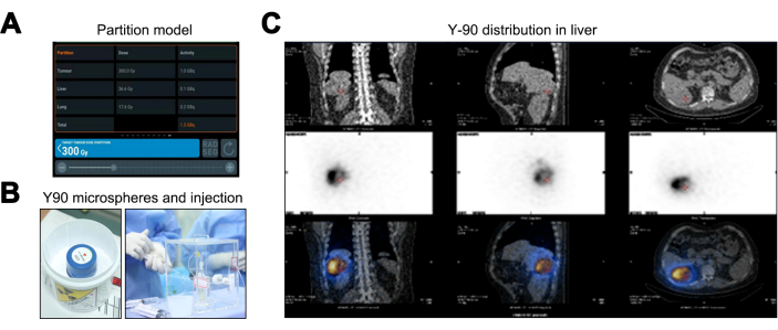
The partition model, in comparison to the Medical Internal Radiation Dose (MIRD) and Body Surface Area (BSA) methods, provides superior personalized radiation dose estimation by factoring in the TNR, enhancing individualized treatment planning. Results from the partition model indicate radiation doses of 36.6 Gy for the normal liver (below the 40 Gy threshold), 17.6 Gy for the lung tissue (within the 20 Gy limit), and a peak dose of 300 Gy for the tumor, necessitating a Y-90 microsphere activity of 1.36 GBq (Figure 3A). Post Y-90 TARE therapy (Figure 3B), a PET/CT scan was performed, indicating no off-target spread or coverage discrepancies (Figure 3C).
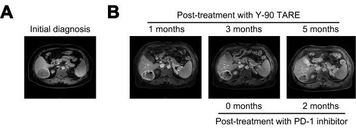
At 1 month after the Y-90 TARE treatment, the tumor was reduced to 6 cm, and the AFP level decreased to 21,155 ng/mL. At 3 months post-treatment, the tumor showed no significant changes. Given the persistent high risk of recurrence, treatment with a PD-1 inhibitor was initiated. At 5 months following Y-90 TARE therapy (2 months after starting PD-1 inhibitor treatment), the lesion had further reduced to 4 cm, and the AFP level had dramatically decreased to 1.84 ng/mL (Figure 4 and Table 1).
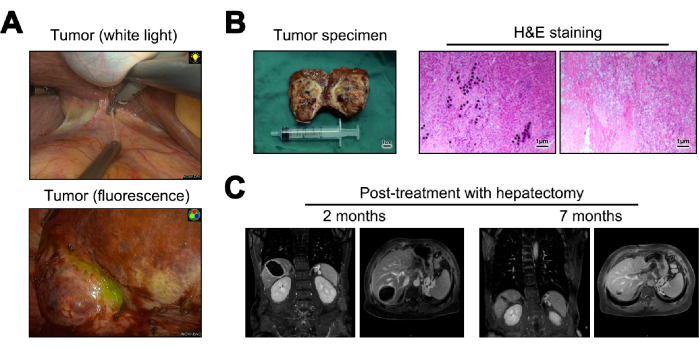
Images of the tumor during hepatectomy under both white light and fluorescence are presented (Figure 5A). The tumor specimens which were obtained from hepatectomy were transformed into frozen sections for gross pathology. When examined microscopically, they revealed no tumor cells, deposition of Y-90 microspheres, significant lymphocytic infiltration, and cirrhotic changes in the adjacent normal liver tissue23 (Figure 5B). At 12 months post-operation, follow-up, and assessment for recurrence were conducted, with MRI imaging indicating no evidence of recurrence or metastasis (Figure 5C).

Figure 1: Magnetic-Resonance Imaging (MRI) imaging at initial diagnosis. (A) Coronal section of MRI T1 Weighted Imaging (T1WI) signal, (B) transverse sections of MRI T1WI, T2 Weighted Imaging (T2WI), and Diffusion Weighted Imaging (DWI) signals. Please click here to view a larger version of this figure.

Figure 2: Injection and distribution of Technetium-99m Macroaggregated Albumin (Tc-99m MAA). (A) Illustration of the injection process of Technetium-99m macroaggregated albumin (Tc-99m MAA). (B) Presentation of the dose distribution of 99mTc MAA in the liver. Please click here to view a larger version of this figure.

Figure 3: Yttrium-90 Transarterial Radioembolization (Y-90 TARE) treatment process. (A) Depiction of the data calculated using the Partition model. (B) The packaging and injection of the Y-90 microspheres. (C) Single-photon Emission Computed Tomography/Computed Tomography (SPECT/CT) to validate the dosage distribution of the Y-90 microspheres. Please click here to view a larger version of this figure.

Figure 4: Tumor comparison. (A) Presentation of the MRI image at initial diagnosis, while (B) displays the MRI images at 1-, 3-, and 5-months post-treatment with Y-90 TARE. Programmed Cell Death-1(PD-1) inhibitor treatment was performed 3 months after Y-90 TARE. The patient underwent treatment with a PD-1 inhibitor 3 months after the Y-90 TARE procedure. Please click here to view a larger version of this figure.

Figure 5: Hepatectomy and subsequent follow-up. (A) Presentation of the tumor observed intraoperatively. (B) Illustration of the postoperative tumor specimen and Hematoxylin and Eosin (H&E) staining. (C) MRI images at 2- and 12-months post-surgery. Please click here to view a larger version of this figure.
| Post-treatment with Y-90 TARE | |||||
| Initial diagnosis | 1 months | 3 months | 5 months | ||
| AFP (ng/mL) | 66840 | 21155 | 19535 | 1.84 | |
Table 1: Post-treatment AFP level measurement.
Discussion
For HCC patients presenting with high-risk recurrence factors, an adverse prognosis persists even subsequent to curative hepatectomy, underscoring the importance of effective neoadjuvant therapy to enhance survival rates24,25. Relative to interventional treatments, Y-90 TARE boasts a superior local control rate26. While Y-90 TARE can activate the body's anti-tumor response22, the combined use of Y-90 with PD-1 inhibitors in neoadjuvant therapy for liver cancer has not yet been reported. This study retrospectively reviews a case of neoadjuvant Y-90 TARE followed by anti-PD-1 monoclonal antibody treatment in an HCC patient with high-risk recurrence factors who achieved complete remission. It presents a detailed treatment protocol for reference.
Several key points in the protocol of this study warrant attention. Firstly, given the potential for degradation and redistribution of MAA99, SPECT/CT imaging should be performed within 1-2 h post-MA99 Injection. Secondly, it is imperative to meticulously calculate the dose of Y-90 microspheres to prevent ectopic placement and excessive dosage, which could lead to hepatic and pulmonary damage. Finally, considering post-neoadjuvant surgery, a non-anatomical resection ensuring clear margins may be preferable to shorten the surgical duration and minimize surgery-related immunosuppression.
In the present study, the patient exhibited symptoms of sleep disturbances and constipation following neoadjuvant therapy. These were addressed using eszopiclone for sleep disorders and bisacodyl enteric-coated tablets for constipation. This suggests that adverse reactions related to Y-90 TARE and PD-1 inhibitor are minimal and can be pharmacologically managed. Furthermore, Y-90 TARE and PD-1 inhibitor did not induce liver tissue or lesion edema, severe adhesion, or increased fragility, the latter of which could precipitate significant bleeding or incomplete resection during subsequent surgical removal. Consequently, Y-90 TARE did not interfere with or impact subsequent surgical procedures.
Based on the levels of AFP and the changes in the lesion, we sequentially administered anti-PD-1 monoclonal antibody therapy following Y-90 TARE. After 5 months, the lesion achieved a pathological complete response (pCR), indicating that the timing and choice of treatment were appropriate. Adjusting the treatment strategy before the median response period in Y-90 TARE can effectively reduce the risk of disease progression27. However, although the degree of lesion resolution is conspicuously correlated with survival post-hepatic carcinoma resection28, whether subsequent surgical intervention is warranted for cases of pCR induced by Y-90 TARE remains a subject for further investigation. Besides, the optimal timing and dosage of Y-90 TARE and anti-PD-1 monoclonal antibody treatment, as well as the best timing and approach for subsequent surgery, remain to be further validated. Additionally, the high cost of the entire treatment process may impose a significant financial burden on patients.
The occurrence of a pCR following Y-90 TARE and PD-1 inhibitor treatment in our case is postulated to correlate with several factors in the current study. Initially, the intra-tumoral radiation dose is considered; we employed a conventional methodology based on Tc-99m MAA (partition model) for evaluating the Y-90 treatment dose24. Notably, owing to the patient's high TNR, the radiation dose permeating the lesion in this study was elevated, with Y-90 microspheres comprehensively covering the tumor, thereby achieving a curative effect. Secondly, significant immune cell infiltration within the tumor, indicating a pivotal role of the patient's anti-tumor immunity towards pCR, cannot be overlooked. Considering that this was the patient's initial diagnosis and the tumor was not in an advanced stage, intrinsic anti-tumor immunity persisted. Subsequent to tumor cell death induced by Y-90 TARE and PD-1 inhibitor, an inflammatory response may be triggered, enhancing tumor antigen exposure, activating host anti-tumor immunity, and culminating in immune cell infiltration and its consequential tumoricidal action29. Additionally, our prior research discerned a correlation between peritumoral hepatic inflammation and tumor resistance, which could potentially exacerbate hepatic immune tolerance30,31. Although hepatic cirrhotic alterations were present in the peritumoral tissue of the patient in this study, no significant inflammatory response was observed, and all hepatitis B-related examinations were unremarkable. This suggests that the patient's hepatic immune microenvironment belongs to a potentially modifiable subgroup, for which Y-90 TARE and PD-1 inhibitor represent a crucial strategy in enhancing the hepatic immune microenvironment.
The implementation of Y-90 TARE treatment necessitates stringent conditions and collaborative efforts across multiple departments, including nuclear medicine, interventional radiology, hepatobiliary surgery, imaging, and oncology. The potential for radiation-induced complications such as pneumonia, gastric ulcers, and acute pancreatitis underscores the need for meticulous dose calculations. Preoperative simulation evaluations and dose estimations conducted by the nuclear medicine department can extend the Y90 TARE treatment cycle, potentially leading to tumor progression. Y90 TARE may not be suitable for all liver cancers, particularly those with multiple intrahepatic metastases and diffuse or small diameter (<0.5 cm) liver cancers, due to challenges in differentiating tumor areas from normal liver tissue, which can lead to dose misestimation. Reports on the use of Y90 in conjunction with PD-1 inhibitors for neoadjuvant therapy in hepatocellular carcinoma are limited. Large-scale clinical studies are still required to substantiate its efficacy and therapeutic details.
In summary, the combination of Y-90 TARE and a PD-1 inhibitor presents a safe and effective approach for the neoadjuvant treatment of HCC patients. This strategy not only alleviates local tumor burden and minimizes micro-metastases to the greatest extent possible but also does so without increasing the risk of disease progression. Consequently, it holds the potential to extend the postoperative recurrence-free period for patients. We offer a replicable and feasible protocol for the neoadjuvant treatment of patients with high-risk HCC.
Disclosures
The authors declare that the research was conducted in the absence of any commercial or financial relationships that could be construed as a potential conflict of interest.
Acknowledgements
This study was funded by the National Natural Science Foundation of China (82303287), Guangdong Basic and Applied Basic Research Foundation (2021A1515110083), Science and Technology Projects in Guangzhou (202201010267), Fundamental Research Funds for the Central Universities (21621058).
Materials
| Name | Company | Catalog Number | Comments |
| ARTIS pheno Cutting-edge robotic imaging system | SIEMENS HEALTHINEERS | 10849000 | |
| DISPOSABLE LAPAROSCOPIC INSTRUMENT | Hangzhou Kangji Medical Instruments | 108Y series | |
| Endoscopic fluorescence camera system | Stryker | PC9000 | |
| ENSEAL G2 Curved Tissue Sealer | Ethicon from Johnson | NSLG2C14 | |
| Fluorescent laparoscopic lens | Stryker | SC9534 | |
| laparoscopic stapler | Ethicon from Johnson | PCEE45A | |
| Optima NM/CT 640 SPECT/CT | GE healthcare | 200439 | |
| SURGICEL FIBRILLAR Hemostat | Ethicon from Johnson | 1962 | |
| Tc-99m macroaggregated albumin [MAA](DAXIMAGE-MAA) | JUBILANT RADIOPHARMA | 877003-93-5 | |
| Tislelizumab Injection(PD-1 inhibitor) | BeiGene, Ltd | 10-848 | |
| ultrasound knife | Ethicon from Johnson | HARHD36 | |
| Xeleris Functional Imaging Workstation | GE healthcare | 5436592-22 | |
| Y-90 resin microspheres(SIR-Spheres) | Sirtex Medical Limited | SIR-Y001 |
References
- Su, H., et al. Molecular mechanism of CK19 involved in the regulation of postoperative recurrence of HBV-associated primary hepatocellular carcinoma in Guangxi. Ann Transl Med. 9 (24), 1780 (2021).
- Zheng, R., et al. Cancer incidence and mortality in China, 2016. J Natl Cancer Center. 2 (1), 1-9 (2022).
- Sung, H., et al. Global cancer statistics 2020: GLOBOCAN estimates of incidence and mortality worldwide for 36 cancers in 185 countries. CA. 71 (3), 209-249 (2021).
- Heimbach, J. K., et al. AASLD guidelines for the treatment of hepatocellular carcinoma. Hepatology (Baltimore, Md.). 67 (1), 358-380 (2018).
- Sato, T., et al. A prospective study of fully covered metal stents for different types of refractory benign biliary strictures. Endoscopy. 52 (5), 368-376 (2020).
- Zhou, H., Song, T. Conversion therapy and maintenance therapy for primary hepatocellular carcinoma. Biosci Trends. 15 (3), 155-160 (2021).
- Xu, X. F., et al. Risk factors, patterns, and outcomes of late recurrence after liver resection for hepatocellular carcinoma. JAMA Surg. 154 (3), 209-217 (2019).
- Song, T., Lang, M., Ren, S., Gan, L., Lu, W. The past, present, and future of conversion therapy for liver cancer. Am J Cancer Res. 11 (10), 4711-4724 (2021).
- Sangro, B., Salem, R., Kennedy, A., Coldwell, D., Wasan, H. Radioembolization for hepatocellular carcinoma: A review of the evidence and treatment recommendations. Am J Clin Oncol. 34 (4), 422 (2011).
- Chinese Medical Doctor Association, Clinical Guidelines Committee of Chinese College of Interventionalists, Chinese Research Hospital Association, Society for Hepatopancreatobiliary Surgery. Expert consensus on the standardized procedure of selective internal radiation therapy with Yttrium-90 microspheres for liver malignancies (2024 edition). Zhonghua Yi Xue Za Zhi. 104 (7), 486-498 (2024).
- Vilgrain, V., et al. Efficacy and safety of selective internal radiotherapy with yttrium-90 resin microspheres compared with sorafenib in locally advanced and inoperable hepatocellular carcinoma (SARAH): an open-label randomised controlled phase 3 trial. Lancet Oncol. 18 (12), 1624-1636 (2017).
- Villalobos, A., et al. Safety and efficacy of concurrent Atezolizumab/Bevacizumab or Nivolumab combination therapy with Yttrium-90 radioembolization of advanced unresectable hepatocellular carcinoma. Curr Oncol (Toronto, Ont.). 30 (12), 10100-10110 (2023).
- Kulik, L. M., et al. Safety and efficacy of 90Y radiotherapy for hepatocellular carcinoma with and without portal vein thrombosis. Hepatology (Baltimore, Md.). 47 (1), 71-81 (2008).
- Singh, P., Anil, G. Yttrium-90 radioembolization of liver tumors: what do the images tell us. Cancer Imaging. 13 (4), 645 (2013).
- Kim, Y. C., et al. Radiation safety issues in Y-90 microsphere selective hepatic radioembolization therapy: Possible radiation exposure from the patients. Nuc Med Mol Imag. 44 (4), 252-260 (2010).
- Kennedy, A., et al. Recommendations for radioembolization of hepatic malignancies using yttrium-90 microsphere brachytherapy: a consensus panel report from the radioembolization brachytherapy oncology consortium. Int J Radiation Oncol, Biol, Phys. 68 (1), 13-23 (2007).
- Molvar, C., Lewandowski, R. Yttrium-90 radioembolization of hepatocellular carcinoma-performance, technical advances, and future concepts. Semin Intervent Radiol. 32 (4), 388-397 (2015).
- Aliseda, D., et al. Liver resection and transplantation following Yttrium-90 radioembolization for primary malignant liver tumors: A 15-year single-center experience. Cancers. 15 (3), 733 (2023).
- Wehrenberg-Klee, E., Goyal, L., Dugan, M., Zhu, A. X., Ganguli, S. Y-90 radioembolization combined with a PD-1 inhibitor for advanced hepatocellular carcinoma. Cardiovas Intervent Radiol. 41 (11), 1799-1802 (2018).
- Maleux, G., et al. Predictive factors for adverse event outcomes after transarterial radioembolization with Yttrium-90 resin microspheres in Europe: Results from the prospective observational CIRT study. Cardiovas Intervent Radiol. 46 (7), 852-867 (2023).
- Lencioni, R., Llovet, J. M. Modified RECIST (mRECIST) assessment for hepatocellular carcinoma. Semin Liver Dis. 30 (1), 52-60 (2010).
- Yu, Q., Khanjyan, M., Fidelman, N., Pillai, A. Contemporary applications of Y90 for the treatment of hepatocellular carcinoma. Hepatol Comm. 7 (10), e0288 (2023).
- Zhou, J., et al. Guidelines for the diagnosis and treatment of primary liver cancer (2022 Edition). Liver Cancer. 12 (5), 405 (2023).
- Pasciak, A. S., Erwin, W. D. Effect of voxel size and computation method on Tc-99m MAA SPECT/CT-based dose estimation for Y-90 microsphere therapy. IEEE Trans Med Imag. 28 (11), 1754-1758 (2009).
- Sun, H. C., Zhu, X. D. Downstaging conversion therapy in patients with initially unresectable advanced hepatocellular carcinoma: An overview. Front Oncol. 11, 772195 (2021).
- Dhondt, E., et al. 90Y radioembolization versus drug-eluting bead chemoembolization for unresectable hepatocellular carcinoma: Results from the TRACE phase II randomized controlled trial. Radiology. 303 (3), 699-710 (2022).
- Taswell, C. S., et al. For hepatocellular carcinoma treated with Yttrium-90 microspheres, dose volumetrics on post-treatment Bremsstrahlung SPECT/CT predict clinical outcomes. Cancers. 15 (3), 645 (2023).
- Xu, L., Chen, L., Zhang, W. Neoadjuvant treatment strategies for hepatocellular carcinoma. World J Gastrointes Surg. 13 (12), 1550-1566 (2021).
- Chew, V., et al. Immune activation underlies a sustained clinical response to Yttrium-90 radioembolisation in hepatocellular carcinoma. Gut. 68 (2), 335-346 (2019).
- Jiang, Y., et al. Inflammatory microenvironment of fibrotic liver promotes hepatocellular carcinoma growth, metastasis and sorafenib resistance through STAT3 activation. J Cell Mol Med. 25 (3), 1568-1582 (2021).
- Jiang, Y., et al. TANK-binding kinase 1 (TBK1) serves as a potential target for hepatocellular carcinoma by enhancing tumor immune infiltration. Front Immunol. 12, 612139 (2021).
Reprints and Permissions
Request permission to reuse the text or figures of this JoVE article
Request PermissionExplore More Articles
This article has been published
Video Coming Soon
Copyright © 2025 MyJoVE Corporation. All rights reserved