Se requiere una suscripción a JoVE para ver este contenido. Inicie sesión o comience su prueba gratuita.
Obtaining Primary Ovarian Cancer Cells from Solid Specimens: A Method to Culture Epithelial Ovarian Cancer Cells from Ovarian Tumor Specimens
En este artículo
Overview
In this video, we describe a method for isolating and characterizing primary ovarian cancer cells from solid ovarian tumor tissue specimens. The epithelial cancer cells obtained using this protocol are suitable for understanding the processes that may lead to ovarian cancer initiation and development.
Protocolo
All procedures involving human participants have been performed in compliance with the institutional, national, and international guidelines for human welfare and have been reviewed by the local institutional review board.
1. Tissue Processing
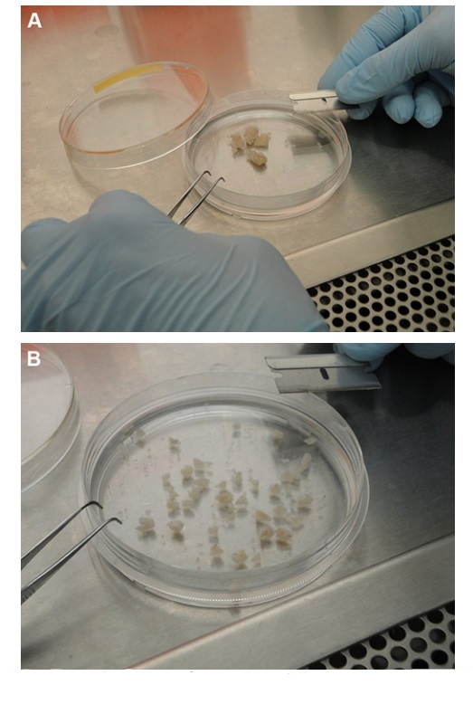
- Working under sterile conditions, transfer the samples onto a Petri dish (60 mm x 60 mm) containing 10 ml of fresh, ice-cold PBS and using a sterile razor blade, further cut into the smallest pieces possible (2 mm or less) (Figures 1A and 1B).
- Transfer the minced tissues into a 15 ml conical tube containing 10 ml of prewarmed (37 °C for 30 min) dispase II (2.4 U/ml) in DMEM and incubate at 5% CO2 and 37 °C for 30 min. To ensure optimal digestion of the specimens, manually agitate the cell slurry every 5 min.
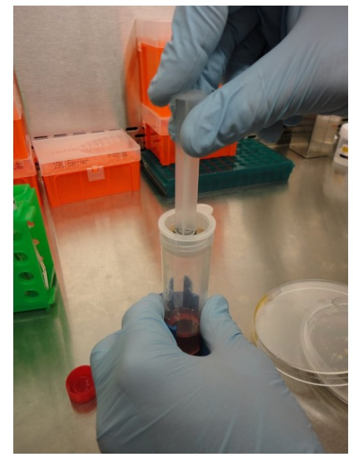
- After 30 min incubation, transfer (using a 10 ml serological pipette) the cell slurry onto a cell strainer (70 μm mesh) placed on top of a 50 ml conical tube and apply a gentle pressure against the mesh using a syringe plunger. Discard any undissociated tissue (remaining on the top of the mesh) and collect the obtained cell suspension in the 50 ml sterile conical tube. Centrifuge at 320 x g for 7 min at 4 °C (Figure 2).
- Discard the supernatant and resuspend the cell pellet in 10 ml of DMEM containing 10% FBS.
- Incubate the cell suspension in a Petri dish at 5% CO2 and 37 °C (Figure 3).
- Change the medium after 24 hr from the initial plating. This allows for removal of cellular debris and the majority of the erythrocytes present in the culture.
- Change the medium every three days for the following two weeks, after which the cultures of primary EOC are ready for downstream applications.
Access restricted. Please log in or start a trial to view this content.
Resultados

Figure 1. Processing of clinical specimens. A) Solid specimen transferred onto a Petri dish containing 10 ml of fresh, ice-cold 1x PBS. B) Clinical specimens further cut into pieces about 2 mm in size

Figure 2. S...
Access restricted. Please log in or start a trial to view this content.
Divulgaciones
No conflicts of interest declared.
Materiales
| Name | Company | Catalog Number | Comments |
| 1x PBS, sterile | Invitrogen | 14190-144 | |
| Screw lid, polypropylene specimen container, sterile | Thermo Scientific | ||
| DMEM medium, sterile | Invitrogen | 11995-065 | |
| Fetal bovine serum, sterile | Thermo Scientific Hyclone | SH30396.03 | |
| 100x Penicillin-streptomycin, liquid | Invitrogen | 15140-122 | |
| Dispase II, sterile | Roche | 04942 078 001 | |
| Small fine-tip forceps and scalpel blade, sterile | Fisher Scientific | forceps: 08-953E, blades: 08-927-5D | |
| Small dissecting scissors, sterile | Fisher Scientific | 08-945 | |
| 15 ml Conical capped tubes, sterile | BD Falcon | 352097 | |
| 50 ml Conical capped tubes, sterile | BD Falcon | 352027 | |
| 10 ml Serological pipette, sterile | Corning | 13-678-11E | |
| 6 ml Syringe plunger, sterile | Tyco Healthcare | 8881516911 | |
| Nylon filter (70 μm ) cell strainer, sterile | BD Falcon | 352350 | |
| 5 cm Petri dish, sterile | Corning | 430166 | |
| 10 cm Petri dish, sterile | Corning | 430167 |
Referencias
Access restricted. Please log in or start a trial to view this content.
This article has been published
Video Coming Soon
Source: Pribyl, L. J. et al. Method for Obtaining Primary Ovarian Cancer Cells From Solid Specimens. J. Vis. Exp. (2014)
ACERCA DE JoVE
Copyright © 2025 MyJoVE Corporation. Todos los derechos reservados