Se requiere una suscripción a JoVE para ver este contenido. Inicie sesión o comience su prueba gratuita.
An In Vitro Method for the Differentiation of Megakaryocytes and Platelet Formation
En este artículo
Overview
This video demonstrates a technique for the differentiation of megakaryocytes and the formation of platelets. Hematopoietic stem progenitor cells — isolated from human umbilical cord blood — differentiate into mature megakaryocytes in the presence of thrombopoietin. The mature megakaryocytes produce cytoplasmic extensions termed proplatelets, which give rise to individual platelets.
Protocolo
All procedures involving human participants have been performed in compliance with the institutional, national, and international guidelines for human welfare and have been reviewed by the local institutional review board.
1. Megakaryocyte Differentiation
- Seed 5 × 10⁵ cells/mL CD34+ cells in 2 mL of SFM supplemented with 50 ng/mL recombinant human thrombopoietin (rhTPO) per well in a 12-well plate. Incubate cells at 37 °C, 5% CO2 in a humidified atmosphere. If cells are confluent before they are required for analysis, harvest the cells and split them into multiple wells with fresh media and rhTPO.
NOTE: Two or three wells should be prepared specifically to monitor differentiation at different time points (e.g., days 7, 9, and 10). - Harvest cells from the wells set aside to monitor differentiation without disturbing the cells in the other wells.
- Stain cells with 20 µL anti-GPIIb/CD41-FITC antibody and 10 µL anti-GPIX/CD42a-Alexa Fluor 647 antibody in a final volume of 100 µL. Set up a control tube using the respective isotype control antibodies. Incubate for 15 - 30 min at 4 °C.
- Add 1 mL of SB to wash. Centrifuge at 400 × g for 10 min. Discard the supernatant and resuspend the pellet in 200 - 300 µL of SB. Analyze by flow cytometry.
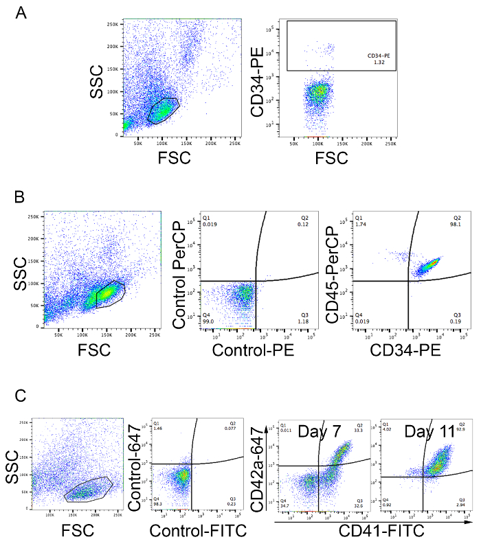
- For each fluorophore, analyze the isotype controls to set the gating for FITC and Alexa Flour 647 positive populations. Analyze the stained cell samples to determine the percent of CD41+/CD42a+ double positive cells, which represent mature MK (Figure 1C).
- For microscopic visualization of cell surface markers stain cells as described in 1.3.
- Add 1 mL of SB to wash. Centrifuge at 400 × g for 10 min. Resuspend the pellet in 100 µL of SB and spin onto a glass slide at 1,000 x g for 5 min. Fix cells on the slide by dipping them in methanol for 30 s. Air dry, add 20 µL of mounting media containing DAPI (see Table of Materials), cover with a coverslip, and visualize using a fluorescent microscope (Figure 2A).
- For visualization of intracellular antigens, resuspend cells in PBS and fix with paraformaldehyde (1% final concentration) for 15 min at room temperature. To permeabilize the cells, add triton-X 100 (0.1%) and incubate for 15 min. Wash cells with 2 mL PBS/0.1% triton-X 100. Resuspend in 100 µL of PBS/0.1% triton-X 100, add anti-vWf and anti-CD62p antibodies (1:200 dilution), and incubate for 30 min at room temperature.
- Wash with 2 mL PBS/0.1% triton-X 100 and centrifuge at 400 × g for 10 min, resuspend in 100 µL of the same buffer, and add anti-mouse IgG-Alexa 594 and anti-rabbit IgG-Alexa 488 (1:100). Incubate for 30 min at room temperature, wash with 2 mL PBS/0.1% triton-X 100, resuspend in 100 µL of the same buffer, and add 20 µL of anti-CD42b-APC. Then spin the cells onto glass slides as described in step 1.6.1 and prepare samples for microscopic visualization as described in step 1.6.1.
- For ploidy determination, harvest cells at days 12 or 13 of differentiation. Add 1 mL of SB to wash. Centrifuge at 400 × g for 10 min and stain with 20 µL anti-GPIIb/CD41-FITC antibody in a final volume of 100 µL. Incubate at 4 °C for 30 min.
- Wash once with 1 mL of SB and resuspend the pellet in 300 µL of hypotonic citrate buffer (1.25 mM sodium citrate, 2.5 mM sodium chloride, 3.5 mM dextrose) containing 20 µg/ml propidium iodide and 0.05% Triton-X 100. Incubate for 15 min at 4 °C protected from light.
- Add RNase to a final concentration of 20 µg/mL and incubate for 30 min at 4 °C protected from light. Determine the intensity of propidium iodide by flow cytometry by collecting 30,000 to 50,000 events of the CD41-FITC+ population (Figure 2C).
2. Proplatelet Counting, Platelet Enumeration, and Platelet Activation
- Harvest cells (from step 1.1) at days 8 or 9 of differentiation and seed at 1 × 10⁴ cells/well in 48-well plates in 200 µL of fresh SFM supplemented with 50 ng/mL rhTPO. Culture for 5 days at 37 °C, 5% CO2.
NOTE: For quantitation purposes, seed wells in triplicate. This low density is required for visualization and counting of proplatelet-bearing MK. Proplatelets usually start appearing after 2 days of culture. The peak is between days 4 and 5. - Count the number of proplatelet-bearing MK in the whole well on an inverted light microscope using 10X or 20X objectives.
NOTE: A heated (37 °C) microscope stage is preferable since keeping the cells at room temperature for extended periods causes shrinkage of the proplatelet extensions. Proplatelets are observed as long extensions from the MK body. Each MK may have several proplatelet protrusions. As proplatelets develop, the body of the MK decreases in size. - Harvest cells and centrifuge at 400 x g for 10 min at room temperature. Stain cells with 20 µL anti-human CD41-FITC antibody, as described in steps 1.3 and 1.4. Calculate the percentage of proplatelet-bearing MK (pbMK):
pbMK (%) = [(Proplatelet-bearing MKs/ well) / (Total CD41+ cells/ well)] x 100 - To count platelets released into the culture medium, gently mix the cells with a Pasteur pipette and collect 100 µL at days 14 or 15 of culture.
- Stain with 20 µL anti-human CD41-FITC antibody for 20 - 30 min at 4 °C. Set up a control tube using the respective isotype control antibody.
- Add 150 µL of SB and 50 µL of counting beads.
- For flow cytometric analysis, set the FSC and SSC to log scale. Use normal human blood platelets from platelet-rich plasma stained with CD41-FITC as described in section 2.4.1 to set the gating for platelets (Figure 3A).
- Analyze the stained platelets by counting beads by flow cytometry. Collect 1,000 events of counting beads using the FSC versus SSC scatter plot (Figure 3A). Calculate the number of platelets based on CD41-FITC positive events (Figure 3B) using the formula:
Platelets per µL=[(number of CD41-FITC positive events)/1,000 beads)] x [(number of beads in 50 µL)/sample volume)]
- To analyze platelet activation, gently mix the cells with a Pasteur pipette and collect 100 µL at days 14 or 15 of culture. Add 1 mL of Tyrode's buffer (137 mM NaCl, 2.7 mM KCl, 1 mM MgCl2, 1.8 mM CaCl2, 0.2 mM Na2HPO4, 12 mM NaHCO3, 5.5 mM D-glucose, pH 6.5) and centrifuge at 200 x g for 5 min to pellet cells.
- Collect the supernatant and centrifuge at 800 x g for 10 min to pellet platelet-sized particles.
- Discard the supernatant and resuspend in 100 µL of Tyrode's buffer. Add 20 µL of PAC1-FITC antibody and adenosine diphosphate (ADP) to a final concentration of 20 µM. Incubate at room temperature for 20 min. Analyze by flow cytometry to determine the percentage of FITC-positive events. Use fresh human platelets treated in the same manner as a positive control (Figure 3C).
Resultados

Figure 1: Flow cytometry plots of CD34+ cell isolation and MK differentiation in culture. (A) Mononuclear cells (gated as shown in the left panel) purified from human cord blood were stained with anti-CD34-PE antibody to determine the percentage of the CD34+ cells in the sample (1.3%, right panel). (B) After separation, the positive fraction was staine...
Divulgaciones
Materiales
| Name | Company | Catalog Number | Comments |
| Cell Culture Reagents | |||
| Recombinant Human TPO | Miltenyi Biotec | 130-094-013 | |
| StemSpan SFEM II | Stem Cell Technologies | 9605 | Serum-free media for CD34+ cells |
| Flow Cytometry and Cell Staining Reagents | |||
| PE Mouse anti-Human CD34 | BD Biosciences | 340669 | Clone 8G12. This can be used for CD34 purity check. Final antibody concentration 1:10 dilution. |
| PerCP mouse anti-human CD45 | BD Biosciences | 347464 | 1:10 dilution |
| PerCP isotype control | BD Biosciences | 349044 | 1:10 dilution |
| FITC Mouse anti-Human CD41a | BD Biosciences | 340929 | Final antibody concentration 1:5 dilution. |
| APC Mouse anti-Human CD42b | BD Biosciences | 551061 | This antibody can also be used to detect mature MK (the percentage of positive cells in usually lower than with anti CD42a). Final antibody concentration 1:10 dilution. |
| Alexa Fluor 647 Mouse anti-Human CD42a | AbD Serotec | MCA1227A647T | Currently distributed by Bio-Rad. Final antibody concentration 1:10 dilution. |
| Alexa Fluor 647 Mouse Negative Control | AbD Serotec | MCA928A647 | Currently distributed by Bio-Rad. Isotype control antibody |
| Anti von Willebrand factor rabbit polyclonal | Abcam | AB6994 | 1:200 dilution |
| V450 mouse anti-humna CD41a | BD Biosciences | 58425 | 1: 20 dilution |
| V450 isotype control | BD Biosciences | 580373 | 1:20 dilution |
| PAC1-FITC antibody | BD Biosciences | 340507 | 1:10 dilution |
| Anti CD62p mouse monoclonal | Abcam | AB6632 | 1:200 dilution |
| Alexa Fluor 488 goat anti rabbit IgG | Invitrogen | A11008 | 1:100 dilution |
| Alexa Fluor 594 goat anti mouse IgG | Invitrogen | A11020 | 1:100 dilution |
| Ig Isotype Control cocktail-C | BD Biosciences | 558659 | Isotype control antibody |
| Propidium iodide | Sigma Aldrich | P4864 | |
| CountBright Absolute Counting Beads | Molecular Probes, Invitrogen | C36950 | Counting beads |
| Materials | |||
| Falcon 5mL round bottom polypropylene FACS tubes, with Snap Cap, Sterile | In Vitro technologies | 352063 | |
| Glass slides | Menzel-Glaser | J3800AMNZ | |
| Mounting media with DAPI | Vector Laboratories | H-1200 | Antifade mounting medium with DAPI |
| Equipment | |||
| Inverted microscope | Leica | DMIRB inverted microscope | |
| Fluorescent microscope | Zeiss | Vert.A1 | |
| Cell analyser | BD Biosciences | FACS Canto II | |
| Cytospin centrifuge | ThermoScientific | Cytospin 4 | |
| Software | |||
| Cell analyser software | BD Biosciences | FACS Diva Software | |
| Single cell analysis software | Tree Star | FlowJo | |
| Fluorescent microscope software | Zeiss | Zen 2 blue edition |
This article has been published
Video Coming Soon
Source: Perdomo, J., et al. Megakaryocyte Differentiation and Platelet Formation from Human Cord Blood-derived CD34+ Cells. J. Vis. Exp. (2017)
ACERCA DE JoVE
Copyright © 2025 MyJoVE Corporation. Todos los derechos reservados