Évaluations dynamiques de la réserve de débit coronaire après une reperfusion d’ischémie myocardique chez la souris
Dans cet article
Résumé
Le présent protocole décrit une vue parasternale longitudinale modifiée pour une localisation rapide et précise de l’artère descendante antérieure gauche. Cette approche se veut plus simple et plus conviviale tout en facilitant l’examen des changements dynamiques de la réserve de débit coronaire suite à une ischémie-reperfusion myocardique chez la souris.
Résumé
Après une ischémie cardiaque, la perfusion myocardique est souvent insuffisante, même si le flux a été rétabli avec succès et complètement dans une artère en amont. Ce phénomène, connu sous le nom de « phénomène de non-refusion », est attribué à un dysfonctionnement microvasculaire coronarien et a été associé à de mauvais résultats cliniques. Dans la pratique clinique, une réduction de la réserve de débit coronaire (CFR) est fréquemment utilisée comme indicateur de maladie coronarienne. Le CFR est défini comme le rapport entre la vitesse d’écoulement de pointe induite par des facteurs pharmacologiques ou métaboliques et la vitesse d’écoulement au repos.
Ce protocole s’est concentré sur l’évaluation des changements dynamiques de la CFR avant et après l’ischémie-reperfusion (IR) à l’aide de mesures Doppler à ondes de pouls. Dans cette étude, des souris normales ont montré la capacité d’augmenter la vitesse de pointe du flux sanguin coronaire jusqu’à deux fois plus élevée que les valeurs au repos sous stimulation à l’isoflurane. Cependant, après ischémie-reperfusion, le taux de létalité à 1 h a significativement diminué par rapport à la valeur de référence avant l’opération. Au fil du temps, le taux de létalité s’est redressé progressivement, mais il est demeuré inférieur au niveau normal. Malgré la préservation de la fonction systolique, la détection précoce du dysfonctionnement microvasculaire est cruciale, et l’établissement d’un guide pratique pourrait aider les médecins dans cette tâche, tout en facilitant l’étude de la progression des maladies cardiovasculaires au fil du temps.
Introduction
Les maladies coronariennes sont l’une des principales causes de mortalité dans le monde1. Même lorsque l’artère coronaire coupable est rouverte par une intervention coronarienne percutanée primaire (ICP) après une ischémie cardiaque, la perfusion microvasculaire coronaire reste souvent diminuée. De plus, il n’y a aucune garantie de reperfusion dans les capillaires en aval alimentant le myocarde1. Ce phénomène, connu sous le nom de « phénomène de non-refusion », est lié à une progression clinique et à un mauvais pronostic. Par conséquent, l’obtention d’une refusion microvasculaire adéquate après une thérapie de reperfusion réussie devient essentielle pour la récupération du myocarde. Par conséquent, l’évaluation précoce de la fonction microvasculaire après la revascularisation est cruciale pour les pratiques cliniques.
Diverses techniques, telles que le fil-guide invasif intracoronaire de température/pression à l’indice de résistance microcirculatoire (IMR) et de résistance microvasculaire hyperémique (HMR), la résonance magnétique cardiovasculaire non invasive (CMR), la tomodensitométrie par émission monophotonique (SPECT) et la tomographie par émission de positons (TEP), peuvent être utilisées pour évaluer la fonction microvasculaire2. Cependant, ces méthodes sont invasives ou semi-invasives, coûteuses et souvent difficiles à trouver, ce qui limite leur utilité clinique. D’autre part, l’évaluation de la réserve de débit coronaire (CFR) par échocardiographie Doppler transthoracique offre une approche non invasive, relativement simple et rentable sans exposer les patients aux rayonnements ionisants, comme on le voit dans d’autres méthodes3.
Bien que des études antérieures aient utilisé l’échocardiographie Doppler transthoracique pour mesurer la faucologie chez les souris et les rats, il reste des défis pour les opérateurs à localiser les angles complexes entre la plate-forme, les souris et la sonde. Ce protocole résout ce problème en fournissant une méthode plus facile pour localiser l’artère coronaire descendante antérieure gauche (DAL) et mesurer rapidement le taux de létalité à l’aide de la vue parasternale longitudinale modifiée (PLAX).
De plus, le taux de létalité obtenu dans l’artère liée à l’infarctus infarctus (IRA) distale à la lésion coupable a montré une forte corrélation avec l’état de perfusion évalué par échocardiographie de contraste myocardique (ECM)4. Il a également été identifié comme un marqueur prédictif de la viabilité et de la récupération de la fonction ventriculaire gauche (VG) après un infarctus aigu du myocarde (IAM)5. De plus, le taux de létalité a été établi comme un marqueur fiable de la mortalité toutes causes confondues et des résultats cardiovasculaires indésirables 6,7. Des rapports antérieurs ont décrit l’utilisation de l’échocardiographie pour évaluer la létalité dans des modèles d’infarctus du myocarde chez le rat8. Néanmoins, le taux de létalité au stade précoce de l’ischémie-reperfusion n’a pas été étudié de manière approfondie. Par conséquent, cette étude fournit une valeur de référence pour le diagnostic de la dysfonction microvasculaire et l’évaluation de l’effet thérapeutique de l’ischémie-reperfusion par des tests dynamiques chez des souris IR au stade précoce de la reperfusion.
Protocole
Toutes les expériences ont été approuvées par le Comité de protection et d’utilisation des animaux de l’Université de Pékin. Des souris C57 mâles âgées de 8 à 12 semaines ont été utilisées pour la présente étude. Les animaux proviennent d’une source commerciale (voir la Table des matières).
1. Préparation des animaux
- Retirez les poils de la zone précordiale à l’aide d’une crème dépilatoire (voir tableau des matériaux). Ensuite, procédez à l’acquisition des images échographiques en suivant l’étape 2.
2. Imagerie par ultrasons avant la chirurgie IR
- Anesthésier la souris en la plaçant dans la chambre d’induction de l’isoflurane (voir le tableau des matériaux) et initier l’administration d’isoflurane à une concentration de 1,5 % avec un débit de 1,5 à 2,0 L/min d’O2.
- Positionnez la souris anesthésiée en position couchée sur le dos sur la plateforme de surveillance physiologique (plateforme d’imagerie du système d’échographie, voir Tableau des matériaux). Appliquez une petite quantité de gel conducteur sur la feuille de cuivre de la table de surveillance physiologique et fixez les pattes de la souris avec du ruban adhésif pour obtenir des informations physiologiques sur l’ECG et la respiration. Maintenez la température corporelle à 37-38 °C à l’aide de la plate-forme chauffante intégrée.
REMARQUE : Basculez l’interrupteur de commande de l’appareil d’anesthésie gazeuse de la chambre au masque et maintenez la dose d’anesthésique constante à 1,5 % pendant ce temps. La fréquence cardiaque des souris à ce stade devrait être d’environ 350-450 battements/min. - Appliquez une pommade vétérinaire ophtalmique sur les yeux avant l’imagerie pour prévenir la sécheresse.
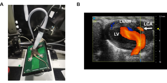
- Effectuer une échocardiographie transthoracique à l’aide d’une sonde linéaire9 à 40 MHz (p. ex., sonde MS550 pour les souris) (figure 1A).
REMARQUE : L’appareil à ultrasons est équipé de la sonde MS550. - Appliquez généreusement du gel de transmission d’ultrasons (voir tableau des matériaux) pour assurer une qualité d’image optimale.
- Ajustez la position de la sonde et de la plate-forme pour formerune vue parasternale longitudinale modifiée (PLAX) 9 (Figure 1A) afin d’examiner l’artère descendante antérieure gauche (DAL) en mode B. Brièvement, imagez le cœur à l’aide d’une vue parasternale grand axe, la plate-forme étant alignée latéralement et positionnée à un petit angle entre la sonde et la plate-forme.
- Localisez la section cardiaque appropriée en mode B et activez le Doppler couleur sur l’écran tactile (Figure 1B). Le DAL (flèche blanche) peut être identifié dans la paroi ventriculaire gauche (Figure 1B). La couleur rouge en temps réel indique la direction du flux sanguin (c’est-à-dire que le flux sanguin est vers la sonde).
REMARQUE : En ajustant l’axe des x, le Doppler couleur permet de visualiser l’ensemble du DAL (du sinus aortique au site de la branche distale) dans cette fenêtre d’image. - Déplacez légèrement la sonde pour trouver la bonne position et réduire l’influence du flux sanguin dans les veines pulmonaires.
REMARQUE : Pour réduire l’influence du flux sanguin de la veine pulmonaire, ajustez légèrement la sonde et différenciez le DAL de la veine pulmonaire. Le DAL se dirige dans la paroi ventriculaire gauche, tandis que la veine pulmonaire se draine dans l’oreillette gauche. Localisez le DAL à l’aide du Doppler couleur et utilisez le mode B pour sélectionner la section appropriée. - Après avoir visualisé le LAD en mode Doppler couleur, appuyez sur le bouton Pulse Wave (PW) et passez en mode PW. Positionnez la ligne indicatrice jaune sur l’artère coronaire, en vous assurant qu’elle est parallèle à la direction de l’écoulement. Appuyez ensuite sur le bouton Enregistrer le clip pour enregistrer les données de référence.
- Augmentez la concentration d’isoflurane à 3 % et attendez 30 s. Assurez-vous que la vitesse d’écoulement augmente progressivement avec le temps. Appuyez fréquemment sur le bouton Enregistrer le clip pour capturer la vitesse de flux sanguin la plus élevée.
REMARQUE : Le stimulus isoflurane fait travailler le cœur plus fort, ce qui peut entraîner un mouvement du cœur et du DAL. Une fois l’image en mode PW capturée ou enregistrée dans le cinéma, cliquez sur le mode PW pour vous assurer que la ligne indicatrice jaune se trouve sur le DAL. En cas de déplacement, ajustez légèrement la ligne indicatrice jaune au point de flux sanguin et poursuivez l’enregistrement. Ce processus nécessite une maîtrise et un changement rapides. - Après avoir recueilli des images du flux sanguin coronaire, éteignez l’anesthésique et ajustez la sonde à la vue PLAX normale. Attendez que la fréquence cardiaque de la souris augmente lentement jusqu’à environ 500 bpm, puis mesurez la fonction cardiaque de la souris en basculant la sonde sur la section parasternale à axe court (PSAX).
- Une fois l’imagerie échocardiographique terminée, retirez l’animal de la plate-forme et laissez-le se rétablir dans sa cage d’origine. Retirez le gel et laissez l’animal sécher pour éviter l’hypothermie.
- Utilisez les outils de fonction Peak Vel et LV pour obtenir les vitesses diastoliques maximales et la fonction systolique cardiaque à partir des images, respectivement.
- Calculez le CFR comme le rapport entre la vitesse de débit coronaire de pointe diastolique (CFV) au débit maximal et la CFV de pointe diastolique au départ.
3. Procédure d’ischémie-reperfusion myocardique
REMARQUE : La mesure initiale était de référence, puis une intervention chirurgicale a été effectuée sur le même animal.
- Anesthésier les souris avec du pentobarbital sodique (60 mg/kg) et administrer l’analgésique carprofène (5 mg/kg, injection sous-cutanée). Utilisez un coussin chauffant préchauffé (37 °C) tout au long de l’intervention chirurgicale. Assurez-vous que la profondeur de l’anesthésie est appropriée en l’absence de réflexe de retrait, de pincement des orteils et de clignement des yeux. Placez la souris en décubitus dorsal sur le coussin chauffant et appliquez une pommade ophtalmique sur les yeux pour éviter qu’ils ne se dessèchent. Effectuer une intubation endotrachéale pour la ventilation mécanique : désinfecter la zone avec 3 gommages alternés de bétadine ou de chlorhexidine et d’alcool à 70% avant et après l’épilation des poils du cou. Localisez la trachée et insérez doucement le cathéter. Ensuite, raccordez les souris au ventilateur (volume courant inspiratoire de 250 μL à 120 respirations/min) (voir tableau des matériaux).
- Désinfectez la peau de la zone précordiale avec 3 gommages alternés de bétadine ou de chlorhexidine et d’alcool à 70%. Exposez le cœur en effectuant une thoracotomie gauche10 au quatrième espace intercostal.
- Lister l’artère descendante antérieure gauche (DAL) avec un nœud coulant à l’aide d’une suture en soie 5-0 (voir le tableau des matériaux), en insérant 1 à 2 mm sous la racine de l’appendice auriculaire gauche. Assurez-vous que la direction de la couture est parallèle au bord inférieur de l’appendice auriculaire gauche. Confirmer visuellement l’ischémie chez toutes les souris par assombrissement local de la couleur du myocarde ischémique.
- Occlure l’artère DAL pendant 30 min11. Après 30 min, relâchez la ligature et vérifiez la reperfusion en observant la rougeur de la zone précédemment décolorée du muscle cardiaque pendant 1 à 2 min.
- Après l’occlusion/reperfusion coronaire, fermez la poitrine en couches et laissez les souris récupérer pendant environ une demi-heure11.
4. Imagerie par ultrasons après une chirurgie IR
- Mesurez à nouveau le taux de létalité à 1 h, 3 h, 5 h, 8 h, 24 h et 48 h après la reperfusion, respectivement, en suivant les étapes décrites à la section 2. Comparez ces mesures aux valeurs de létalité obtenues avant la procédure d’ischémie-reperfusion (IR).
5. Analyse statistique
- Effectuer des analyses statistiques à l’aide d’un logiciel d’analyse statistique approprié (voir la Table des matériaux).
REMARQUE : Pour comparer des variables continues entre des échantillons appariés, utilisez des tests t appariés si les données sont normalement distribuées. Pour les données non distribuées normalement ou lorsque les hypothèses de normalité ne sont pas satisfaites, utilisez l’ANOVA de Friedman. Considérons p < 0,05 comme statistiquement significatif.
Résultats Représentatifs
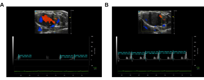
Cette étude a utilisé des souris C57 mâles (poids corporel ~18-20 g) pour caractériser le changement dynamique du taux de létalité. L’image PLAX modifiée a été utilisée pour évaluer les caractéristiques de l’écoulement artériel coronaire de la DAL (figures 1A, B). Le CFR a été calculé comme le rapport entre la vitesse d’écoulement maximale pendant la vasodilatation maximale induite par l’isoflurane à 3 % et la vitesse d’écoulement maximale à une concentration initiale de 1,5 % d’isoflurane12,13. Toutes les mesures et tous les calculs ont été répétés sur trois cycles cardiaques consécutifs et la moyenne a été calculée, avec des résultats représentatifs illustrés à la figure 2.
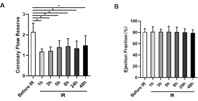
Avant la chirurgie IR, le taux de létalité de base a été mesuré et les souris avaient une valeur normale de létau près de 2,14 ± 0,43. Cependant, après la chirurgie IR, le taux de létalité a significativement diminué à 1 h de reperfusion par rapport à avant la chirurgie IR (1,18 ± 0,14 vs 2,14 ± 0,43) (Figure 3A). Cette réduction indique que la microcirculation n’a pas été immédiatement rétablie, même après l’ouverture du récipient coupable. Comme le temps de reperfusion était prolongé, les valeurs de létalité sont restées à un niveau constamment bas, avec des valeurs de 1,21 ± 0,20 à 3 h, 1,39 ± 0,33 à 5 h, 1,44 ± 0,38 à 8 h, 1,34 ± 0,36 à 24 h et 1,48 ± 0,47 à 48 h après la reperfusion, ce qui suggère que l’hypoperfusion pourrait persister pendant au moins deux jours (figure 3A). De plus, il n’y avait pas de signification statistique entre les valeurs du CFR à 1 h, 3 h, 5 h, 8 h, 24 h et 48 h.
La fonction cardiaque des souris a également été surveillée, et il a été observé qu’il n’y avait pas de changements significatifs dans la fonction cardiaque ventriculaire gauche lorsque le taux de létalité était significativement réduit chez les souris (figure 3B).

Figure 1 : Vue parasternale longitudinale modifiée. (A) illustre le positionnement de la sonde et de la plate-forme lors de l’obtention de la vitesse de l’artère coronaire de la DAL. (B) montre l’emplacement du capteur de vitesse de l’onde de pouls sur l’artère coronaire LAD. La couleur bleue indique un mouvement d’éloignement de la sonde à ultrasons, tandis que la couleur orange indique un mouvement vers la sonde à ultrasons. Veuillez cliquer ici pour voir une version agrandie de cette figure.

Figure 2 : Visualisation et enregistrement de l’imagerie de la vitesse des ondes de pouls de la DAL. (A) Image de l’onde de pouls pendant la condition de repos de l’artère coronaire de la DAL. (B) Image de l’onde de pouls coronaire hyperémique maximale de l’artère coronaire LAD. La couleur bleue illustre le mouvement d’éloignement de la sonde d’échographie, tandis que la couleur orange illustre le mouvement vers la sonde d’échographie. Veuillez cliquer ici pour voir une version agrandie de cette figure.

Figure 3 : Mesure de la réserve de débit coronaire et de la fraction d’éjection. (A) Analyse statistique de la létalité chez les animaux avant l’ischémie et à 1 h, 3 h, 5 h, 8 h, 24 h et 48 h après la reperfusion, respectivement (n = 9). (B) Évaluation de la fraction d’éjection de chaque groupe à chaque point temporel (n = 9). *p < 0,05 ; données présentées sous forme de ± moyenne ET, analysées à l’aide de tests t appariés et de l’ANOVA de Friedman. Veuillez cliquer ici pour voir une version agrandie de cette figure.
Discussion
Cette étude présente un protocole qui utilise une vue parasternale longitudinale modifiée pour évaluer dynamiquement la flétrissure après ischémie-reperfusion. Les principaux résultats indiquent une diminution significative de la létalité chez les souris IR, la réduction la plus prononcée étant observée 1 h après la reperfusion. Cependant, la fonction cardiaque n’a pas été affectée dans les 48 heures.
Le létologie sert d’indicateur de l’approvisionnement en sang myocardique, offrant une approche non invasive pour évaluer à la fois la sténose de l’artère coronaire et la circulation microvasculaire coronaire. Des études cliniques ont démontré que des valeurs de létalité plus faibles sont associées à de moins bons pronostics14,15,16, et une valeur seuil de létalité de 1,75 a été établie comme optimale pour la stratification du risque14. Une méta-analyse récente a en outre montré que le risque de décès augmente de 16 % pour chaque diminution de 0,1 unité de létalité, indiquant que l’hypercholestérolémie représente un continuum de risque, avec des niveaux plus faibles prédisposant les patients à de moins bons résultats cliniques17. Dans cette étude, le taux de létalité a montré une tendance à augmenter avec l’allongement du temps de reperfusion, mais il est resté inférieur à ce qu’il était avant l’intervention, ce qui souligne l’importance de surveiller les patients non seulement immédiatement après l’ouverture du vaisseau coupable par ICP, mais aussi à 48 h. De plus, le létalité sert de mesure de la dysfonction microvasculaire coronaire, intégrant les effets hémodynamiques des maladies focales, diffuses et des petits vaisseaux sur la perfusion des tissus myocardiques18. Par conséquent, le létau de létalité est une technique non invasive cruciale pour diagnostiquer les maladies coronariennes microvasculaires. Comme le taux de létalité sur le DAL est un indicateur fort et indépendant de la mortalité 6,7, cette étude vise à fournir des valeurs de référence pour les décisions cliniques. De plus, l’utilisation d’appareils à ultrasons peut potentiellement réduire le besoin d’angiographie coronarienne dans un environnement de limitation des coûts des soins de santé. Grâce à une formation appropriée et à une mise à niveau de la technologie, la stratification des risques peut être adaptée aux besoins individuels des patients.
La vue PLAX modifiée offre plus de commodité et de temps aux chercheurs scientifiques. L’amélioration continue de cette technologie facilitera son application plus large dans d’autres maladies microvasculaires coronariennes. Les étapes clés de ce protocole comprennent la visualisation de l’artère coronaire et l’obtention d’images de vitesse PW de haute qualité. La vitesse du flux sanguin augmente progressivement avec l’augmentation de la concentration de l’anesthésique, il est donc recommandé de capturer en continu pour éviter de manquer la vitesse maximale du flux sanguin. Comme l’augmentation de la concentration anesthésique peut modifier la fréquence cardiaque, il est conseillé de revenir brièvement en mode Doppler couleur pendant la mesure afin d’assurer un positionnement cohérent avant et après la mesure.
Il est essentiel de reconnaître les limites, y compris les limites inhérentes à la mesure par ultrasons de la CFR. En raison de la courbure de l’artère coronaire, il n’est pas possible d’afficher l’ensemble de l’artère dans son intégralité, ce qui conduit à la mesure dans un seul segment. Les opérateurs doivent viser à mesurer le début de l’artère coronaire afin d’identifier le point de vitesse maximale du flux sanguin coronaire aussi précisément que possible. De plus, le taux de létalité devrait idéalement être déterminé en fonction des changements dans le volume du flux sanguin coronaire, mais cette étude utilise la vitesse du flux sanguin au lieu du volume du flux sanguin, négligeant l’effet du diamètre des vaisseaux. Cependant, des études antérieures ont démontré une forte corrélation entre le CFR et le CFVR (réserve de vitesse de flux coronaire)19. Des recherches plus approfondies sur la fonction microvasculaire coronaire pourraient aider à comprendre les altérations complexes de l’ischémie et améliorer notre compréhension de la dysfonction microvasculaire coronaire.
Déclarations de divulgation
Les auteurs déclarent qu’ils n’ont aucune relation commerciale ou financière concurrente connue qui aurait pu sembler influencer les travaux rapportés dans cet article.
Remerciements
Ce travail a été soutenu par la Fondation nationale des sciences naturelles de Chine (subvention n° 82270352), le projet de recherche clinique sur la construction d’un service de recherche à Pékin (2022-YJXBF-04-03), le financement national de la recherche clinique hospitalière de haut niveau (2022-NHLHCRF-YSPY-01), Founds for Health Improvement and Research de Capital (n° 2022-1-4062), le programme national de construction de disciplines de spécialité clinique clés (subvention n° 2020-QTL-009) et la Fondation de la Société chinoise de cardiologie (n° 2020-QTL-009). CSCF2021B02).
matériels
| Name | Company | Catalog Number | Comments |
| 5-0 silk suture | Ningbo MEDICAL Needle Co., Ltd. | 210322 | |
| C57 mice | Peking University Health Science Center Department of Laboratory Animal Science | ||
| Depilating agent | Nair | NAR-255-1 | |
| Electrode gel | Cofoe | ||
| High Frequency Ultrasound | FUJIFILM VisualSonics, Inc. | Vevo3100 | |
| Isoflurane | REWARD | R510-22-10 | |
| Linear array high frequency transducer | FUJIFILM VisualSonics, Inc. | MS550 | |
| Rodent Ventilator | Shanghai Alcott Biotech | ALC-V9 | |
| Small Animal Anesthesia Machine | REWARD | R530 | |
| SPSS | IBM Corp, Armonk, NY, USA | version 23.0 | statistical analysis software |
| Ultrasound Gel | Cofoe | ||
| Vevo Lab Software | FUJIFILM VisualSonics, Inc. | Verison 5.7.0 |
Références
- O'Farrell, F. M., Mastitskaya, S., Hammond-Haley, M., Freitas, F., Wah, W. R., Attwell, D. Capillary pericytes mediate coronary no-reflow after myocardial ischaemia. Elife. 6, e29280 (2017).
- Dimitrow, P. P. Transthoracic Doppler echocardiography-noninvasive diagnostic window for coronary flow reserve assessment. Cardiovascular ultrasound. 1, 4 (2003).
- Picano, E. Stress echocardiography: a historical perspective. The American Journal of Medicine. 114 (2), 126-130 (2003).
- Lim, D. S., Kim, Y. H., Lee, H. S. Coronary flow reserve is reflective of myocardial perfusion status in acute anterior myocardial infarction. Catheterization and Cardiovascular Interventions: Official Journal of The Society For Cardiac Angiography & Interventions. 51 (3), 281-286 (2000).
- Feldman, L. J., Himbert, D., Juliard, J. M. Reperfusion syndrome: relationship of coronary blood flow reserve to left ventricular function and infarct size. Journal of the American College of Cardiology. 35 (5), 1162-1169 (2000).
- Cortigiani, L., et al. Coronary flow reserve during dipyridamole stress echocardiography predicts mortality. JACC Cardiovascular imaging. 5 (11), 1079-1085 (2012).
- Han, B., Wei, M. Proximal coronary hemodynamic changes evaluated by intracardiac echocardiography during myocardial ischemia and reperfusion in a canine model. Echocardiography (Mount Kisco, NY). 25 (3), 312-320 (2008).
- Kelm, N. Q., Beare, J. E., LeBlanc, A. J. Evaluation of coronary flow reserve after myocardial ischemia reperfusion in rats. Journal of Visualized Experiments. 148, e59406 (2019).
- Batra, A., Warren, C. M., Ke, Y. Deletion of P21-activated kinase-1 induces age-dependent increased visceral adiposity and cardiac dysfunction in female mice. Molecular and Cellular Biochemistry. 476 (3), 1337-1349 (2021).
- Lv, B., Zhou, J., He, S. Induction of myocardial infarction and myocardial ischemia-reperfusion injury in mice. Journal of Visualized Experiments. 179, e63257 (2022).
- Huang, G., Lu, X., Duan, Z. PCSK9 knockdown can improve myocardial ischemia/reperfusion injury by inhibiting autophagy. Cardiovascular Toxicology. 22 (12), 951-961 (2022).
- Lenzarini, F., Di Lascio, N., Stea, F., Kusmic, C., Faita, F. Time course of isoflurane-induced vasodilation: a Doppler ultrasound study of the left coronary artery in mice. Ultrasound in Medicine & Biology. 42 (4), 999-1009 (2016).
- Chowdhury, S. A. K., Rosas, P. C. Echocardiographic characterization of left ventricular structure, function, and coronary flow in neonate mice. Journal of Visualized Experiments. 182, e63539 (2022).
- Sadauskiene, E., Zakarkaite, D., Ryliskyte, L. Non-invasive evaluation of myocardial reperfusion by transthoracic Doppler echocardiography and single-photon emission computed tomography in patients with anterior acute myocardial infarction . Cardiovascular Ultrasound. 9, 16 (2011).
- Bax, M., de Winter, R. J., Schotborgh, C. E. Short- and long-term recovery of left ventricular function predicted at the time of primary percutaneous coronary intervention in anterior myocardial infarction. Journal of the American College of Cardiology. 43 (4), 534-541 (2004).
- Lee, S., Otsuji, Y., Minagoe, S. Noninvasive evaluation of coronary reperfusion by transthoracic Doppler echocardiography in patients with anterior acute myocardial infarction before coronary intervention. Circulation. 108 (22), 2763-2768 (2003).
- Kelshiker, M. A., Seligman, H., Howard, J. P. Coronary flow reserve and cardiovascular outcomes: a systematic review and meta-analysis. European Heart Journal. 43 (16), 1582-1593 (2022).
- Taqueti, V. R., Di Carli, M. F. Coronary microvascular disease pathogenic mechanisms and therapeutic options: JACC state-of-the-art review. Journal of the American College of Cardiology. 72 (21), 2625-2641 (2018).
- Wikström, J., Grönros, J., Gan, L. M. Adenosine induces dilation of epicardial coronary arteries in mice: relationship between coronary flow velocity reserve and coronary flow reserve in vivo using transthoracic echocardiography. Ultrasound in Medicine & Biology. 34 (7), 1053-1062 (2008).
Réimpressions et Autorisations
Demande d’autorisation pour utiliser le texte ou les figures de cet article JoVE
Demande d’autorisationThis article has been published
Video Coming Soon