A subscription to JoVE is required to view this content. Sign in or start your free trial.
Generation of Mouse Proximal Colon Loop Model: A Surgical Procedure to Generate Proximal Colon Loop and Measure Intestinal Permeability by Detection of Fluorescent Markers in Serum Following Intraluminal Injection
In This Article
Overview
In this video, we describe a surgical procedure to generate mouse proximal colon loop or pcLoop segment followed by intraluminal injection of fluorescent markers into the segment to study the intestinal epithelial permeability.
Protocol
All procedures involving animal models have been reviewed by the local institutional animal care committee and the JoVE veterinary review board.
1. Generation of the proximal colon loop (pcLoop)
- Skin preparation: Scrub fur of the abdominal midline with alcohol swabs or gauze sponge soaked with 70% Ethanol. Do not wet a wide area of fur with alcohol to prevent hypothermia.
- Using scissors, perform a midline laparotomy. Make a vertical incision in the middle of the abdomen (about 2 cm in length) and expose the peritoneum. Be careful to not injure intra-abdominal organs.
- Place pre-cut wet cotton gauze over the exposed intra-abdominal cavity.
- Use wet cotton swabs to mobilize and exteriorize the caecum. Carefully place the caecum on the wet cotton gauze.
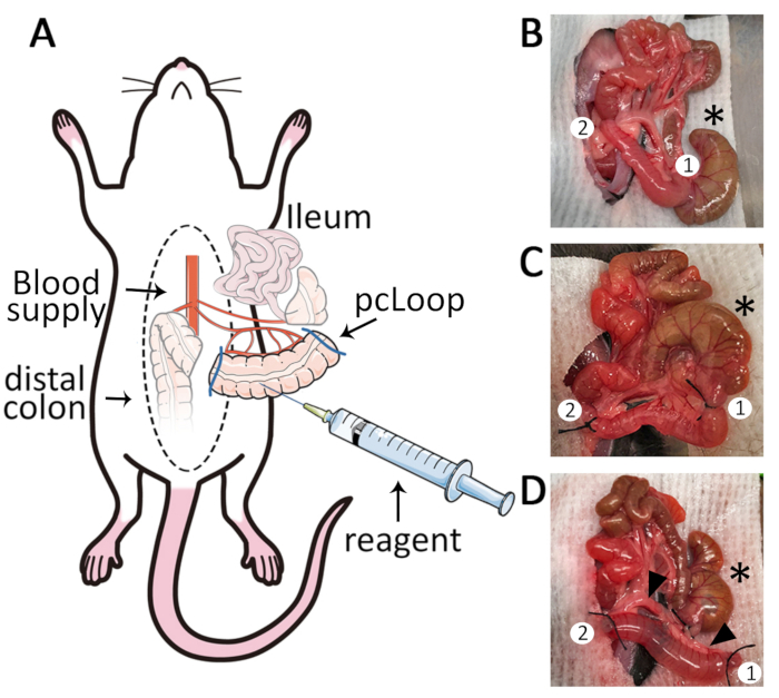
NOTE: Caecum is localized in the left caudal quadrant of the abdominal cavity in a majority of mice independent of the sex of the animal. - Using wet cotton swabs, exteriorize the entire ileum and place it on the top of a wet cotton gauze. Identify the proximal colon and the blood supply located in the mesocolon. Mobilize the proximal colon and by using fine tip forceps create the first ligature in an area free of vessels in the mesocolon at about 0.5 cm distal from the caecum (Figure 1B).
- Measure 2 cm from the first ligature and create a second ligature at an area free of blood supply in the mesocolon (Figure 1C).
- Using fine scissors carefully cut next to each ligation to isolate a 2 cm long pcLoop.
NOTE: With fine scissors carefully cut next to each ligation to isolate the 4 cm ileal loop, keeping intact blood supply and mesenteric membrane., it is important to cut off both ends to isolate a pcLoop that is gently cleaned of luminal contents. Carefully cut through the colonic tissue and mesocolon to prevent small vessels from bleeding into the intestinal lumen. If necessary, use thermal cautery to limit bleeding at the incision site. - Gently flush the pcLoop with warm HBSS to remove feces using a flexible yellow feeding tube attached to 10 mL syringe (Prepare 10 mL syringe filled with warm HBSS and attach to a yellow feeding tube. This syringe will be used for moisturizing exposed tissues during surgery).
- Ligate the two cut ends of the flushed pcLoop using silk suture.
- Use a 1 mL syringe with 30G needle to slowly inject 200 μL of reagent such as FITC-dextrans (1 mg / mL in HBSS). Keep the unused FITC-dextran solution protected from light to prepare the standard curve after serum collection). The pcLoop will inflate causing a moderate distension of the mucosa (Figure 1D).
NOTE: Inject the reagent into the pcLoop lumen on the opposite side of the mesenteric artery. Ensure consistency between animals and create a 2 cm long pcLoop to ensure equal distension of the mucosa. - Utilize wet cotton swabs to gently place back the ligated pcLoop, ileum and caecum into the abdominal cavity.
- Use a needle holder, anatomical forceps and 3.0 non-absorbable silk sutures with reverse cutting needle to close the abdominal wall.
- Place the animal in a temperature-regulated anesthesia chamber for the incubation period.
תוצאות

Figure 1: The proximal colon loop model. (A) Schematic overview of the pcLoop model. Median laparotomy is performed on mice under anesthesia placed on a temperature-controlled surgery board. (B) Exteriorization of the caecum (*), proximal colon, mesocolon and ileum. Two adequate sites for ligation are identified (1,2). (C) The first ligature (1) ...
Disclosures
Materials
| Name | Company | Catalog Number | Comments |
| Equipment and Material | |||
| BD Alcohol Swabs | BD | 326895 | |
| BD 1ml Tuberculin Syringe Without Needle | BD | 309659 | |
| BD PrecisionGlide Needle, 25G X 5/8" | BD | 305122 | |
| BD PrecisionGlide Needle, 30G X 1/2" | BD | 305106 | |
| Cotton Tip Applicator (cotton swab), 6", sterile | FisherScientific | 25806 2WC | |
| Dynarex Cotton Filled Gauze Sponges, Non-Sterile, 2" x 2" | Medex | 3249-1 | |
| EZ-7000 anesthesia vaporizer (Classic System, including heating units) | E-Z Systems | EZ-7000 | |
| Kimwipes, small (tissue wipe) | FisherScientific | 06-666 | |
| Moria Fine Scissors | FST | 14370-22 | |
| Puralube Vet Ointment, Sterile Ocular Lubricant | Dechra | 12920060 | |
| Ring Forceps (blunt tissue forceps) | FST | 11103-09 | |
| Roboz Surgical 4-0 Silk Black Braided, 100 YD | FisherScientific | NC9452680 | |
| Semken Forceps (anatomical forceps) | FST | 1108-13 | |
| Sofsilk Nonabsorbable Coated Black Suture Braided Silk Size 3-0, 18", Needle 19mm length 3/8 circle reverse cutting | HenrySchein | SS694 | |
| Student Fine Forceps, Angled | FST | 91110-10 | |
| 10ml Syringe PP/PE without needle | Millipore Sigma | Z248029 | |
| Reagents | |||
| Isoflurane | Halocarbon | 12164-002-25 | |
| Fluorescein Isothiocyanate-Dextran, average molecular weight 4.000 | Sigma | 60842-46-8 |
This article has been published
Video Coming Soon
Source: Boerner, K. et al. Functional Assessment of Intestinal Permeability and Neutrophil Transepithelial Migration in Mice using a Standardized Intestinal Loop Model. J. Vis. Exp. (2021).
Copyright © 2025 MyJoVE Corporation. All rights reserved