Войдите в систему

Peripheral Vascular Exam

Обзор

Source: Joseph Donroe, MD, Internal Medicine and Pediatrics, Yale School of Medicine, New Haven, CT

The prevalence of peripheral vascular disease (PVD) increases with age and is a significant cause of morbidity in older patients, and peripheral artery disease (PAD) is associated with cardiovascular and cerebrovascular complications. Diabetes, hyperlipidemia, hypertension, and tobacco use are important disease risk factors. When patients become symptomatic, they frequently complain of limb claudication, defined as a cramp-like muscle pain that worsens with activity and improves with rest. Patients with chronic venous insufficiency (CVI) often present with lower extremity swelling, pain, skin changes, and ulceration.

While the benefits of screening asymptomatic patients for PVD are unclear, physicians should know the proper exam technique when the diagnosis of PVD is being considered. This video reviews the vascular examination of the upper and lower extremities and abdomen. As always, the examiner should use a systematic method of examination, though in practice, the extent of the exam a physician performs depends on their suspicion of underlying PVD. In a patient who has or is suspected to have risk factors for vascular disease, the vascular exam should be thorough, beginning with inspection, followed by palpation, and then auscultation, and it should include special maneuvers, such as determining the ankle brachial index. Maneuvers that make use of a handheld Doppler are demonstrated in a companion video.

Figure 1
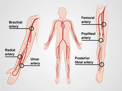
Figure 1. The major arm and leg arteries.

Процедура

1. Preparation

  1. Wash your hands prior to examining the patient.
  2. Have the patient put on a gown. This examination should never occur through clothing.
  3. Check the blood pressure in both arms.

2. The Upper Extremities

  1. Have the patient lie supine on the exam table, with the head raised to a comfortable position.
  2. Begin with inspection by exposing the entirety of both arms. Note symmetry, color, hair pattern, size, skin changes, nail changes, varicosities, muscle wasting, and trauma (Table 1).
  3. Palpate by using the back of the fingers to assess skin temperature. Examine from distal to proximal, comparing one side to the other.
  4. Assess capillary refill by applying firm pressure over the distal 1st or 2nd digit for 5 sec. Release pressure and count how many seconds it takes for the normal skin color to return. A normal capillary refill time (CRT) is less than 2 sec, and values greater than 5 sec increase the likelihood of vascular disease. Additionally, CRT may be prolonged in hypovolemia and cooler ambient temperatures.
  5. Palpate for edema over the dorsum of the hands using firm pressure for at least 2 sec. If present, palpate proximally, noting the extent and distribution of the edema, and whether or not it is pitting. Grade the edema as trace, mild, moderate, or severe.
  6. Palpate the major arteries and note the symmetry, the intensity, and the regularity of the pulse. Useful terminology to describe the pulse intensity includes absent, diminished, normal, or bounding. If unsure, compare the patient's pulse to your own pulse. Use anatomical landmarks to find the pulse. If no pulse is felt, vary the pressure, then adjust your position, as there is variability in the path of each artery.
    1. Palpate the radial arteries, which lie lateral to the flexor carpi radialis tendon.
    2. Palpate the ulnar arteries, which are just lateral to the flexor carpi ulnaris tendon.
    3. Palpate the brachial arteries in the antecubital fossa, medial to the biceps tendon. The artery can be followed proximally in the medial groove between the biceps and triceps muscles.

3. The Abdomen

  1. Lower the head of the table so the patient is lying flat.
  2. Inspect the abdomen for dilated veins. Dilated veins around the umbilicus may be due to portal hypertension or obstruction of the inferior vena cava (IVC).
    1. For dilated superficial veins, determine the direction of filling by using a finger to compress the vein proximally.
    2. Use a second finger to strip the blood distally from the vein and then leave the finger in place, thus compressing two points along the flattened vein.
    3. Remove the proximal finger and note the speed at which the vein refills.
    4. Repeat the process; however, remove the distal finger and compare the filling speed. Note the direction of the fast filling, which is away from the source of venous hypertension.
  3. Palpate for the abdominal aorta, just above the umbilicus and slightly left of the midline. Use 3 to 4 finger pads of both hands to apply slow and steady downward pressure. The hands should point cephalad and slightly toward each other.
  4. Once the pulse is encountered, gradually bring the fingertips closer together until the lateral walls of the aorta are felt. Measure the distance between the fingers.
  5. Next,auscultatefor bruits using the diaphragm of the stethoscope, applying moderate pressure. A bruit with a systolic and diastolic component is more likely to be pathologic than a systolic bruit alone.
    1. Auscultate the renal arteries above the umbilicus and 1" to 2" lateral to the midline.
    2. Auscultate the abdominal aorta above the umbilicus and to the left of the midline.
    3. Auscultate the iliac arteries below the umbilicus and 1" to 2" lateral to the midline.

4. The Lower Extremities.

  1. Begin with inspection by exposing the entirety of both legs, but leave the genitalia covered. Look for changes as described in Step 2.2 and Table 1.
  2. Palpate for temperature, CRT, edema, and arteries, as described in Step 2.3.
  3. Palpate the dorsalis pedis (DP) arteries, just lateral to the extensor hallucis longus tendon. One or both DP arteries may be congenitally absent in a small percentage of patients.
  4. Palpate the posterior tibialis (PT) arteries at the posterior-inferior aspect of the medial malleolus.
  5. Palpate the popliteal arteries, beginning with the leg slightly flexed at the knee. Place both thumbs on the patellar ligament and wrap your fingers around the knee, such that the fingertips land in the middle of the popliteal fossa. If there is difficulty identifying the pulse, gradually flex the knee in 15° intervals while continuing to palpate. If unable to encounter the pulse in this position, have the patient turn to the prone position, flex the knee, and support the lower extremity. Place your hands on either side of the knee and use the thumbs to palpate the popliteal artery.
  6. Palpate the femoral arteries, just inferior to the inguinal ligament, approximately midway between the anterior superior iliac spine and the symphysis pubis.
  7. Auscultate the femoral arteries using the bell or diaphragm of the stethoscope, using light pressure, so as not to artificially induce a bruit.
Finding Peripheral Arterial Disease Chronic Venous Insufficiency
Edema Absent or mild Present, unilateral, or bilateral
Ulcers Well demarcated, often distal leg, dorsum of foot, toes (trauma sites) Irregular margins, often over anterior shin and medial malleolus
Hair Distribution Decreased No change
Color Pallor (acute), dependent hyperemia (chronic), distal gangrene (severe) Brown-red hyperpigmentation
Nails Decreased growth, thickened Thickened, darkened, onychomycosis
Varicose Veins Absent Present
Muscle Atrophy May be present Difficult to detect due to significant edema
Skin Appearance Thin, shiny, atrophic Thickened, scaly
Temperature Cool No change

Table 1. Skin changes associated with peripheral vascular disease.

5. Special Maneuvers

  1. Use the Allen test prior to cannulating the radial artery to ensure adequate collateral flow through the palmar arch from the ulnar artery.
    1. Begin by palpating the ulnar and radial arteries on the side.
    2. Ask the patient to make a tight fist.
    3. Apply sufficient pressure over the ulnar and radial arteries to occlude them.
    4. Ask the patient to open the fist, and note the pallor of the palm.
    5. Release the ulnar artery. If sufficient collateral flow is present, the palm should become pink again within 3 to 5 sec.
  2. Use Buerger's test to assess for PAD of the lower extremities, and it may also be useful for predicting the severity of the disease. With the patient supine, elevate the legs to 60° for 2 min or until the pallor of the distal extremity is noted.
    1. Lower the legs and allow them to dangle below the table's edge. Observe for 2 min or until a hyperemia is observed over the dorsum of the foot, indicating arterial insufficiency.
  3. Perform the following maneuvers in patients with varicose veins to localize the site of incompetent valves.
    1. Perform the Brodie-Trendelenburg test with the patient in the supine position.
      1. Elevate the leg of interest and strip the blood proximally out of the great saphenous vein (GSV).
      2. Compress the GSV just below the sapheno-femoral junction (SFJ), and ask the patient to stand.
      3. Observe the filling of the GSV, which under normal circumstances, fills distal to proximal and takes 20 to 30 sec. Rapid filling with the GSV occluded suggests insufficiency of the perforating veins.
      4. Release the pressure over the GSV. Accelerated filling suggests venous insufficiency at the level of the SFJ.
    2. Perform the cough test to detect reflux at the SFJ. With the patient standing, palpate over the SFJ with light pressure.
      1. Instruct the patient to cough. A palpable thrill suggests retrograde flow and venous insufficiency.
    3. To perform the Perthes test, place a tourniquet around the leg, just below the knee.
      1. Instruct the patient to perform 10 heel raises. Emptying of the varicose veins suggests incompetence above the level of the tourniquet (SFJ, sapheno-popliteal junction, or thigh perforating veins). If the veins remain distended, the site of insufficiency is the calf perforating veins.
Заявка и Краткое содержание

Peripheral vascular disease is an important cause of morbidity, particularly in older patients. The detection and subsequent treatment of PVD can improve quality of life and potentially mitigate cardiovascular and cerebrovascular complications. General screening for peripheral vascular disease of the extremities is not a current recommendation by the US Preventive Service Task Force (USPSTF). However, the USPSTF does recommend ultrasound screening for abdominal aortic aneurysms in males who have smoked and are aged 65 to 75. Additionally, the American Heart Association/American College of Cardiology recommends a comprehensive vascular exam in anyone at risk of PVD.

The most important findings that make PAD more likely in a patient include characteristic ulcers, asymmetric temperature difference in the foot, absent pulses, and limb bruits. The most important finding that argues against significant PAD is the presence of at least one pedal pulse on a given leg. A positive Buerger's test increases the likelihood of more extensive disease. Of the physical exam maneuvers to localize the site of reflux in patients with varicose veins, Perthes and Brodie-Trendelenburg tests are the most helpful for ruling out a particular location as the site of reflux. The overall accuracy of these venous reflux maneuvers is limited, however, and detection of the site of reflux is improved through use of a handheld Doppler.

This video reviewed a systematic method and proper technique of vascular examination of the extremities and abdomen, and included a review of special diagnostic maneuvers that should be performed if PVD is suspected. Like all aspects of the physical exam, practice is critical for improving accuracy, and an understanding of the relevant anatomy is important to a successful examination and interpretation of the exam findings.

Теги
Peripheral Vascular ExamPeripheral Vascular DiseasePVDPeripheral Artery DiseasePADChronic Venous InsufficiencyCVILimb ClaudicationLower Extremity SwellingPainSkin ChangesUlcerationVascular ExaminationUpper ExtremitiesAbdomenLower Extremities

Перейти к...

0:00

Overview

1:40

Peripheral Vascular Exam Procedure

9:09

Summary

Видео из этой коллекции:

article

Now Playing

Peripheral Vascular Exam

Physical Examinations I

67.5K Просмотры

article

General Approach to the Physical Exam

Physical Examinations I

115.0K Просмотры

article

Observation and Inspection

Physical Examinations I

92.8K Просмотры

article

Palpation

Physical Examinations I

82.3K Просмотры

article

Percussion

Physical Examinations I

99.1K Просмотры

article

Auscultation

Physical Examinations I

60.0K Просмотры

article

Proper Adjustment of Patient Attire during the Physical Exam

Physical Examinations I

82.8K Просмотры

article

Blood Pressure Measurement

Physical Examinations I

106.6K Просмотры

article

Measuring Vital Signs

Physical Examinations I

113.5K Просмотры

article

Respiratory Exam I: Inspection and Palpation

Physical Examinations I

155.2K Просмотры

article

Respiratory Exam II: Percussion and Auscultation

Physical Examinations I

210.9K Просмотры

article

Cardiac Exam I: Inspection and Palpation

Physical Examinations I

174.8K Просмотры

article

Cardiac Exam II: Auscultation

Physical Examinations I

138.8K Просмотры

article

Cardiac Exam III: Abnormal Heart Sounds

Physical Examinations I

91.0K Просмотры

article

Peripheral Vascular Exam Using a Continuous Wave Doppler

Physical Examinations I

38.3K Просмотры

JoVE Logo

Исследования

Образование

О JoVE

Авторские права © 2025 MyJoVE Corporation. Все права защищены