A subscription to JoVE is required to view this content. Sign in or start your free trial.
In Vivo Biomechanical Testing of Nerve: A Procedure for Biomechanical Analysis of Brachial Plexus Nerve Injury in a Neonatal Pig Model
In This Article
Overview
In this video, we demonstrate the biomechanical testing of the brachial plexus nerve in a pig model to measure the threshold tensile strength of the nerve when subjected to stretching. This technique helps in determining the structural and mechanical properties of the nerve.
Protocol
All procedures involving animal models have been reviewed by the local institutional animal care committee and the JoVE veterinary review board.
1. Biomechanical Testing
- Set-up of the biomechanical testing device
NOTE: A custom-built mechanical testing device was designed and fabricated to perform in vivo stretch of the brachial plexus (BP) nerve (Figure 1).- Attach the base of the set-up to a cart.
- Attach the electromechanical actuator onto the base using large C-clamps. The actuator can provide 150 lb of force, a 10" stroke, and a speed of 15 mm/s. The speed can be reduced to 0.2 mm/s and still function as desired.
- Attach the 200 N load cell to the actuator.
- Attach (screw-in) a clamp to the load cell that consists of padded plexiglass, which prevents the stress concentration at the clamping site.
- Attach a camera to a tripod. Ensure that the camera has the capability of recording up to 120 f/s at a resolution of 658 x 4926 pixels.
- Attach USB cables from the camera, actuator, and load cell to the computer to integrate and synchronize all components of the set-up.
- Plug the computer, actuator, and load cell into a power source.
- Calibrate the load cell prior to recording the applied loads. To do so, perform the steps below:
- Set the actuator at a 90° angle using the adjustable handle and check the angle with a protractor.
- Open the software that works with the load cell (Materials Table). Press the Start button to show a live readout of voltage.
- Hang weights from the clamp ranging from 0–1,000 g in increments of 100 g from the setup and record the measured voltages.
- Calculate the linear equation of the voltages and weights by finding the slope (m) and intercept (b). This is done using a spreadsheet program and the included slope function to calculate b from Equation 1 below. Insert Equation 2 shown below into the mechanical setup code.
Equation 1: b = y – mx
Where: y is the weight, x is the voltage, m is the slope, and b is the intercept (constant).
Equation 2: y = mx + b
Where: y is the weight, x is the voltage, m is the slope, and b is the constant.
- Testing: the BP nerve is cut and anchored to the testing set-up by a custom-built clamp.
- Place an anesthetized neonatal pig in supine position with exposed BP nerve.
- Cut the BP nerve using fine scissors.
- Clamp the cut side of the BP nerve in the custom-built clamp (Figure 1).
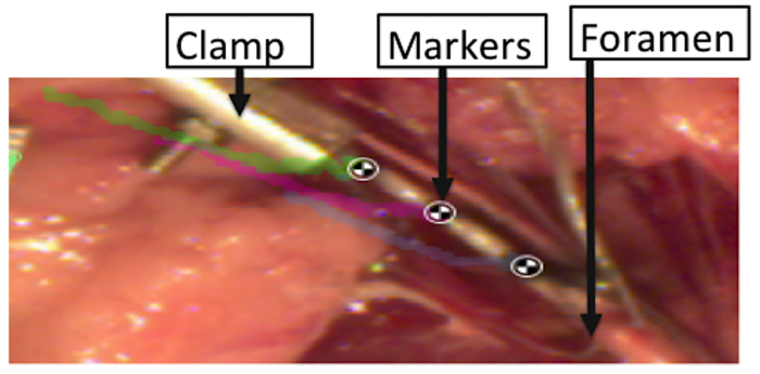
- Manually place black acrylic paint or India ink on the clamped BP segment (Figure 2).
- Place a calibration grid, which is a 1 cm ruler, flat within the animal to set the scale for data analysis.
- Use the camera's software to view the camera's placement directly over the tested segments, thereby allowing the monitoring of the motion/displacement of the markers and determining the actual tissue strain at any time point.
- Record initial measurements such as the height at which the nerve inserts into the body from the table and the height of the clamp from the table, the angle of the actuator, and the full length of the tissue.
- Open the programming software (a table containing the graphical user interface [GUI] (Figure 3).
- Run the GUI by pressing the Run button.
- Initialize the system by pressing the Initialize button.
- Tare the system by pressing the Tare button.
- Stretch the BP segment by pressing the start test button. This pulls the tissue at an assigned rate of 500 mm/min until complete failure occurs in any segment of the BP. The program also saves a video file, the applied tensile load, displacement of the tissue, and duration of the test.
- Record the failure site, which is the point at which the tissue ruptures.
Results

Figure 1: Details of in vivo mechanical testing device including the actuator, load cell, and clamp.

Figure 2: Markers placed over the BP segments to record strains sustained by the tissue during the stretch.
Disclosures
Materials
| Name | Company | Catalog Number | Comments |
| Omega Subminature Tension & Compression Load Cell | Omega | LCM201-200N | 200N load cel |
| Basler acA640-120uc camera | Basler | acA640-120uc | - |
| Feedback Linear Actuator | Progressive Automations | PA-14P | 10" stroke, 150lb force, 15mm/s speed |
| Motion Tracking Software | Kinovea | N/A | Open Source |
| Proramming Software - MATLAB | Mathworks | N/A | version 2018A |
| Scissors | Fine Science Tools Inc | 14094-11 or 14060-09 | - |
This article has been published
Video Coming Soon
Source: Singh, A., et al. Methods for In Vivo Biomechanical Testing on Brachial Plexus in Neonatal Piglets. J. Vis. Exp. (2019).
Copyright © 2025 MyJoVE Corporation. All rights reserved