A subscription to JoVE is required to view this content. Sign in or start your free trial.
hFTAA Staining of Amyloid Deposits: A Technique to Visualize Amyloid Fibrils Using Oligothiophene-Based Dye for Fluorescence Imaging in Tissue Sections
Overview
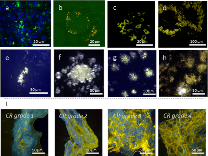
This video describes a technique to stain amyloids in tissue sections using a luminescent-conjugated oligothiophene dye - heptamer-formyl thiophene acetic acid, or hFTAA - for fluorescence imaging. Amyloids are β-sheet-rich abnormal protein aggregates. Their detection in tissue samples is helpful in the diagnosis and treatment of amyloid-associated diseases or proteinopathies.
Protocol
1. LCO Staining Solution
NOTE: If hFTAA is purchased from a commercial vendor, please follow the vendor's instructions instead of section 1.
- Resuspend the lyophilized hFTAA in 2 mM NaOH to prepare a stock solution of 1 mg/mL. Keep the stock in a glass vial at 4 °C. The stock can be stored for one year.
- On the day of staining, prepare a working solution by diluting the stock 1:10,000 in Phosphate-buffered saline (PBS).
2. Preparation of Tissue Samples
NOTE: Many tissue types can be imaged using hFTAA as an am....
Representative Results

Figure 1: Various tissue types and proteins aggregates showing h-FTAA staining of: (a) Mallory-Denk bodies consisting of keratin aggregates in a liver (counterstained with DAPI), (b) p62-positive r inclusions in sporadic inclusion-body myositis (s-IBM) muscle tissue, (c) Amyloid of islet amyloid polypeptide in human pancreas, (d) Amyloid of Immunoglobulin light .......
References
Reprints and Permissions
Request permission to reuse the text or figures of this JoVE article
Request PermissionThis article has been published
Video Coming Soon
Source: Nyström, S. et al., Imaging Amyloid Tissues Stained with Luminescent Conjugated Oligothiophenes by Hyperspectral Confocal Microscopy and Fluorescence Lifetime Imaging. J. Vis. Exp. (2017).
ABOUT JoVE
Copyright © 2025 MyJoVE Corporation. All rights reserved