A subscription to JoVE is required to view this content. Sign in or start your free trial.
Protein Phase Separation Assay: An Optogenetic Method for Mutant RNA-Binding Protein Phase Separation in Spinal Motor Neurons of Zebrafish Larvae
Overview
In this video, we describe a phase separation assay, wherein intracellular proteins comprising intrinsically disordered regions fused to a photosensitive oligomerization domain are induced via blue-light exposure to self-associate into membrane-less liquid-like condensates.
Protocol
All procedures involving animal models have been reviewed by the local institutional animal care committee and the JoVE veterinary review board
1. Preparation of LED for blue light illumination
- Turn on an LED panel by using the associated application installed on a tablet/phone. Put the probe of a spectrometer into an empty well of a 6-well dish and adjust the LED light to the wavelength peaking at ~456 nm through the application. Place the optical sensor of an optical power meter in the empty well and adjust the power of the LED light (~0.61 mW/cm2). The LED light setting can be saved and is retrieva....
Representative Results

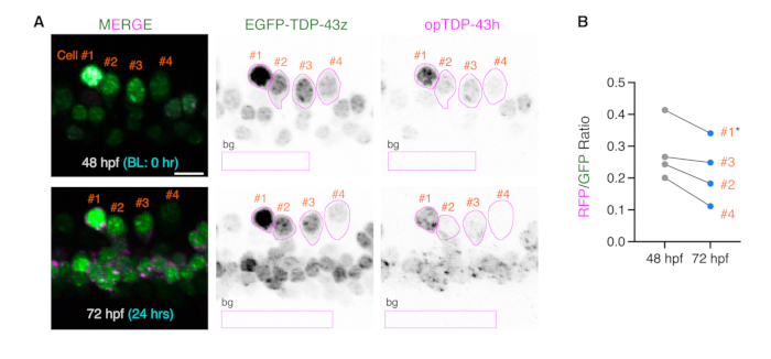
Figure 1: Ratiometric comparisons of opTDP-43h and EGFP-TDP-43z before and after light stimulation. (A) ROIs covering the somas of four single mnr2b-positive cells at 48 and 72 hpf were drawn based on the EGFP-TDP-43z signal and are shown in magenta. The rectangular ROIs (bg) were used to subtract the background signal (background ROI). Figures are adapted from Asakawa et al<.......
Reprints and Permissions
Request permission to reuse the text or figures of this JoVE article
Request PermissionThis article has been published
Video Coming Soon
Source: Asakawa, K. et al. Optogenetic Phase Transition of TDP-43 in Spinal Motor Neurons of Zebrafish Larvae. J. Vis. Exp. (2022)
ABOUT JoVE
Copyright © 2025 MyJoVE Corporation. All rights reserved