A subscription to JoVE is required to view this content. Sign in or start your free trial.
Comet Assay for DNA Damage Detection in Cells
Overview
The video demonstrates the comet assay for detecting DNA damage following exposure to harmful agents. Under alkaline conditions, the DNA unwinds and denatures, and upon electrophoresis, the undamaged and damaged DNA migrate differently in the agarose gel, forming a comet-shaped structure. The slowly migrating undamaged DNA forms the circular comet head, while the rapidly migrating damaged DNA fragments form the elongated comet tail.
Protocol
1. Preparation of materials for the comet assay
- Preparation of the microscope slides
- Pour 1% (w/v) normal melting point agarose [dissolved in double-distilled water (ddH2O)] in a 50 mL tube and microwave to dissolve the agarose in the ddH2O. Store at 37 °C to prevent solidification prior to coating slides. Should solidification occur, discard and prepare fresh.
- Pre-coat microscope slides by dipping the slides into the 50 mL tube containing 1% (w/v) normal melting point agarose.
- Wipe the back of the slides quickly after dipping the slides.
NOTE: Failure to wipe the back of....
Representative Results

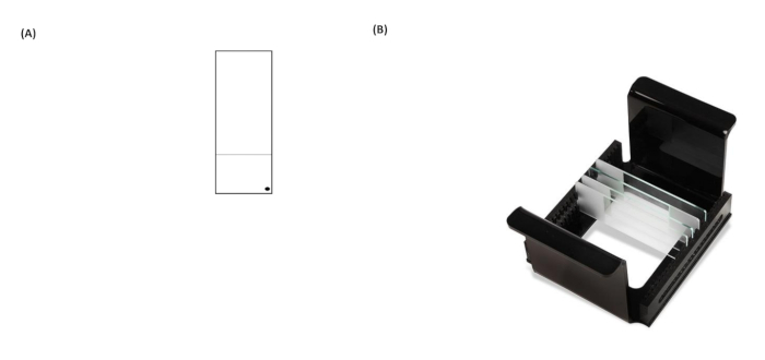
Figure 1: Representative images of a comet assay slide and HTP rack (microscope slide carrier). (A) For correct orientation, the pre-coated face of the microscope slide is recognized by a black dot in the right-hand corner of a microscope slide. (B) The image of the HTP rack illustrates how the slides are kept in a tight vertical orientation, with tabs on the carrier to fix its orientation within.......
Reprints and Permissions
Request permission to reuse the text or figures of this JoVE article
Request PermissionThis article has been published
Video Coming Soon
Source: Ji, Y., et al. A High-Throughput Comet Assay Approach for Assessing Cellular DNA Damage. J. Vis. Exp. (2022).
ABOUT JoVE
Copyright © 2025 MyJoVE Corporation. All rights reserved