A subscription to JoVE is required to view this content. Sign in or start your free trial.
Method Article
Intracranial Injection of Adeno-associated Viral Vectors
In This Article
Summary
Here we present the intracranial injection of AAV vectors for fluorescent labeling of neurons and glia in the visual cortex.
Abstract
Intracranial injection of viral vectors engineered to express a fluorescent protein is a versatile labeling technique for visualization of specific subsets of cells in different brain regions both in vivo and in brain sections. Unlike the injection of fluorescent dyes, viral labeling offers targeting of individual cell types and is less expensive and time consuming than establishing transgenic mouse lines. In this technique, an adeno-associated viral (AAV) vector is injected intracranially using stereotaxic coordinates, a micropipette and an automated pump for precise delivery of AAV to the desired area with minimal damage to the surrounding tissue. Injection parameters can be tailored to individual experiments by adjusting the animal age at injection, injection location, volume of injection, rate of injection, AAV serotype and the promoter driving gene expression. Depending on the conditions chosen, virally-induced transgene expression can allow visualization of groups of cells, individual cells or fine cellular processes, down to the level of dendritic spines. The experiment shown here depicts an injection of double-stranded AAV expressing green fluorescent protein for the labeling of neurons and glia in the mouse primary visual cortex.
Protocol
1. Virus Handling and Storage
- Proper protection and handling techniques should be chosen based on the biosafety level of the virus to be used. These practices can be found in Biosafety in Microbiological and Biomedical Laboratories 5th edition, available on the CDC website
(http://www.cdc.gov/od/OHS/biosfty/bmbl5/bmbl5toc.htm). The use of AAV vectors is approved for Biosafety Level 1 (BSL-1). For the experiment shown here, a lab coat and gloves will be worn when handling the virus, in accordance with procedures for handling BSL-1 agents. - To preserve the activity of the virus it is best to divide it into small aliquots to avoid repeated freezing and thawing.
- Prepare a Biosafety Cabinet (BSC) Class II for aliquoting virus by clearing the BSC of any unnecessary objects and sterilizing the surface with 70% EtOH. Place a beaker containing 10% bleach solution in the hood for collecting AAV-contaminated waste. Also placed in the BSC are sterile 0.5mL tubes and a container of dry ice.
- Thaw viral stock on ice outside the BSC.
- Vortex virus and open the tube inside the BSC.
- Pipette the desired aliquot volume (e.g. 5 μL) into a 0.5mL tube, close the tube and place it in the dry ice to flash-freeze the virus.
- When all virus has been aliquoted, dispose of the virus tube and pipette tips in the waste container containing 10% bleach.
- Remove the waste container from the hood and add additional 10% bleach, then pour the bleach down the sink. Dispose of plastic waste in a biohazard container as instructed by your institute's biosafety officer.
- Clean any equipment or surfaces that came in contact with the virus with 10% bleach. Discard gloves.
- Store aliquots in -80°C freezer.
2. Surgery
- Cover the surgical area with absorbent lab bench paper. Surgical tools should be sterilized and surgery done under aseptic conditions in accordance with your institution's biosafety and animal use guidelines.
- Choose an area adjacent to the surgical area that will be dedicated to loading the micropipettes with virus, and cover it with absorbent lab bench paper. Place an aliquot of virus in a container of ice in this area, and allow the virus to thaw on ice as the surgery is being performed.
- Setup a waste container of 10% bleach in the dedicated virus handling area for disposing of pipette tips, etc. that come in contact with the virus.
- Pull a glass Wiretrol micropipette to a tip diameter of approximately 20 microns. Place a small drop of mineral oil on the blunt end of the micropipette and insert the wire plunger provided with the micropipettes.
- Secure the micropipette in the clamp of the micropump arm.
- Inject a mouse subcutaneously with buprenorphine at a dose of 0.5 mg/kg. Anesthetize the mouse with Avertin via intraperitoneal injection with a dose of 200 mg/kg and remove the hair from the top of the head with surgical scissors, making sure to leave margins large enough to prevent hair from entering the incision.
- Attach a heating blanket to the base of a stereotax to maintain a body temperature of 37°C throughout the surgery and secure the mouse in the stereotax.
- Bathe the head with three alternating scrubs of ethanol and betadine to sterilize the area.
- Place a drop of Tobradex eye ointment on each eye to keep the eyes moist during surgery.
- Make an incision down the midline of the head and pull the skin back to expose the skull.
- Carefully remove the fascia from the skull using fine-tipped forceps.
- Locate the area to be injected using stereotaxic coordinates and mark the skull with a surgical pen.
- Using a dental drill with a 1.4mm burr, thin an area of the skull approximately 2mm in diameter until the skull cracks, dividing the thinned area into several segments.
- Throughout this procedure, keep the skull moist with applications of sterile saline.
- Perform a craniotomy by gently removing the thinned skull segments using extra-fine tipped forceps.
3. Injection Preparation
- With a KimWipe placed over the lid, open the tube of virus and pipette 1.5uL (for a 1uL injection) of virus onto a small piece of parafilm.
- Place the tip of the micropipette into the viral stock and manually withdraw the plunger. If difficulty is experienced drawing viral stock into the micropipette, the tip can be enlarged slightly be piercing a KimWipe with the micropipette to break a small portion of the glass.
- Lower micropump arm onto plunger until a small amount of virus is dispelled from the tip of the micropipette. Remove this small drop with a cotton tip applicator and discard applicator in waste container.
- Apply a drop of mineral oil to the tip of the micropipette to prevent clogging as the micropipette is lowered into the brain.
4. Virus Injection
- Using X and Y stereotaxic coordinates, position the micropipette over the area to be injected. In this experiment, the stereotaxic coordinates used to locate primary visual cortex are 2.7mm posterior to bregma and 2.5mm lateral to the midline. Very slowly lower the micropipette (at an approximate rate of 1mm/1minute) to the proper Z position.
- Enter the desired injection parameters into the SYS Micro4 micropump controller box and initiate the injection. For this experiment, 1 microliter over 10 minutes will be used as a rate of injection.
- When the injection is finished, leave the pipette to rest for one-two minutes to prevent efflux of virus during removal. After this period, very slowly remove the micropipette from the brain (same rate as above).
- Suture the scalp and seal it with tissue glue. Inject the animal subcutaneously with buprenorphine at a dose of 0.1 mg/kg every 8-12 hours over the next 72 hours, or as long as the animal is exhibiting signs of pain. Allow the animal to recover under a heat lamp until it is ambulating and ready to be returned to its cage. Imaging experiments can be initiated after the desired incubation time (days to weeks after viral injection).
5. Cleanup
- Rinse the micropipette with 10% bleach and discard it in a sharps container.
- Dispose of waste container in the same manner as Section 1.
- Discard lab bench paper into biohazard bin and wipe down all surfaces and instruments that may have come in contact with the virus with 10% bleach.
- Unused virus may be frozen again, keeping in mind that repeated freeze/thaw cycles cause degradation of the virus.
6. Representative Results

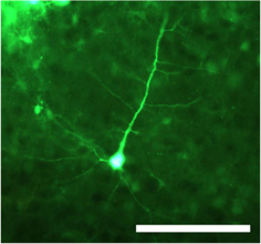
Figure 1. Transduced neuron after injection of double-stranded adeno-associated virus serotype 1 (dsAAV S1) carrying green fluorescent protein (GFP) under control of the CMV promotor. The cell body as well as proximal and distal dendrites are clearly visible in fixed sections imaged using an epifluorescent microscope. Scale bar= 100 μm.

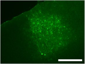
Figure 2. Labeling typical of an intracranial virus injection in primary visual cortex using dsAAV S1 showing the extent of viral spread as well as labeled neurons, glia and processes. Scale bar= 250 μm.

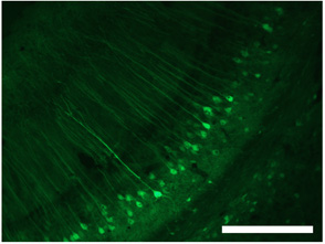
Figure 3. Labeling of cells in hippocampus using dsAAV S1. Scale bar= 250 μm. These figures are adapted from Lowery et al. 20091
Discussion
Virally-mediated gene delivery holds great potential for the study of neurological processes and treatment of brain disorders1,2,3. The great versatility of this technique can also be exploited to fluorescently label cells for imaging both in vitro and in vivo4. Here we demonstrate a detailed procedure for the transduction of neurons and glia in mouse visual cortex using a double-stranded adeno-association virus expressing enhanced green fluorescent protein.
Disclosures
The animal and experimental protocols were approved by the University of Rochester University Committee on Animal Resources (UCAR) in accordance with the PHS Policy on Humane Care and Use of Laboratory Animals.
Acknowledgements
This work was made possible by grants from the NIH (EY012977), a Career Award in the Biomedical Sciences from the Burroughs Wellcome Fund, the Whitehall Foundation, and the Sloan Foundation (A.K.M.).
Materials
| Name | Company | Catalog Number | Comments |
| St–lting Mouse and Neonatal Rat Adaptor | Stoelting Co. | 51625 | Regular stereotax for securing animals for surgery may be substituted |
| Extra Fine Bonn Scissors, 8.5cm, straight tip, cutting edge 13mm | Fine Science Tools | 14084-08 | |
| Eye Dressing Forceps, 10cm, tip width 0.5mm, curved | Fine Science Tools | 11152-10 | |
| Dumont #5/45 Forceps- Dumoxel Standard Tip, 11cm, angled | Fine Science Tools | 11251-35 | Extra-fine tipped forceps for performing craniotomy |
| Standard Pattern Forceps, straight, 2.5mmx1.35mmtip, 12cm | Fine Science Tools | 11000-12 | |
| Microtorque Control Box and Tech2000 handpiece | Ram Products, Inc. | TECH2000ON/OFF | Dental drill |
| Micro Drill Stainless Steel Burrs 1.4mm tip diameter | Fine Science Tools | 19008-14 | |
| Wiretrol micropipettes, to deliver 1-5 Ul | VWR international | 5-000-1001 or 53480-287 | |
| Mineral oil | VWR international | 29447-338 | |
| Manual Micromanipulator and Tilting Base (right-handed) | World Precision Instruments, Inc. | M3301-M3-R | Used for determining stereotaxic co-ordinates |
| UltraMicroPump (UMP3) (one) with SYS-Micro4 Controller | World Precision Instruments, Inc. | UMP3-1 | |
| Sutures | VWR international | 95056-952 | |
| P-97 Flaming/Brown Micropipette Puller | Sutter Instrument Co. | P-97 | |
| Tobradex | Available from your institution’s veterinary services |
References
- Kaplitt, M. G., Leone, P., Samulski, R. J., Xiao, X., Pfaff, D. W., O'Malley, K. L., During, M. J. Long-term gene expression and phenotypic correction using adeno-associated virus vectors in the mammalian brain. Nat Genet. 8, 148-154 (1994).
- Harding, T. C., Dickinson, P. J., Roberts, B. N., Yendluri, S., Gonzalez-Edick, M., Lecouteur, R. A., Jooss, K. U. Enhanced gene transfer efficiency in the murine striatum and an orthotopic glioblastoma tumor model, using AAV-7- and AAV-8-pseudotyped vectors. Hum Gene Ther. 17, 807-820 (2006).
- Lo, W. D., Qu, G., Sferra, T. J., Clark, R., Chen, R., Johnson, P. R. Adeno-associated virus-mediated gene transfer to the brain: duration and modulation of expression. Hum Gene Ther. 10, 201-213 (1999).
- Lowery, R. L., Zhang, Y., Kelly, E. A., Lamantia, C. E., Harvey, B. K., Majewska, A. K. Rapid long-term labeling of cells in the developing and adult rodent visual cortex using double-stranded adeno-associated viral vectors. Dev Neurobiol. 69, 674-688 (2009).
Reprints and Permissions
Request permission to reuse the text or figures of this JoVE article
Request PermissionExplore More Articles
This article has been published
Video Coming Soon
Copyright © 2025 MyJoVE Corporation. All rights reserved