A subscription to JoVE is required to view this content. Sign in or start your free trial.
Immunoprecipitation Assay to Study Enzyme Receptor Interactions in Transfected Cells
In This Article
Overview
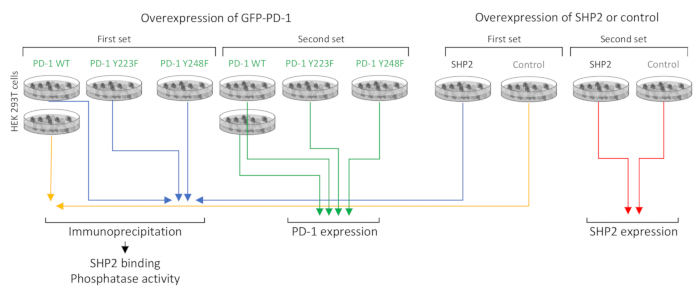
In this video, we demonstrate a technique to perform the co-immunoprecipitation of GFP-tagged wild-type and mutated PD-1 proteins using beads coated with anti-GFP antibodies. The bead-bound PD-1-GFP proteins are incubated with SHP2 proteins that interact with PD-1, enabling the identification of specific structural motifs in PD-1 that play a critical role in SHP2 interaction.
Protocol
1. Immunoprecipitation
NOTE: The following steps should be performed on ice or at 4 °C.
- Supplement the lysis buffer (50 mM Tris-HCl at pH 7.2, 250 mM NaCl, 0.1% NP-40, 2 mM EDTA, 10% glycerol) with protease inhibitors (dissolve 1 tablet in 10 mL of lysis buffer) and with 1 mM sodium orthovanadate. Add 500 µL of ice-cold lysis buffer to the cells and remove and collect the cells from the plates immediately using a cell scraper.
NOTE: It is important to add the sodium orthovanadate only to the lysis buffer that will be used for the PD-1-GFP-transfected plates since the SHP2-transf....
Representative Results

Figure 1: Experimental conditions and strategy. GFP-PD-1 WT (wild type), GFP-PD-1 Y223F (ITIM mutant), or GFP-PD-1 Y248F (ITSM mutant) were expressed in HEK 293T cells that were subsequently treated with pervanadate. Phosphorylated GFP-PD-1 proteins collected by GFP immunoprecipitation were mixed with lysates from cells overexpressing SHP2, and the levels and activity of SHP2 bound to each vers.......
This article has been published
Video Coming Soon
Source: Strazza, M., et al. Co-immunoprecipitation Assay for Studying Functional Interactions Between Receptors and Enzymes. J. Vis. Exp. (2018).
ABOUT JoVE
Copyright © 2025 MyJoVE Corporation. All rights reserved