Absolute Quantum Yield Measurement of Powder Samples
Summary
In this video we will demonstrate measuring and calculating absolute quantum yield and chromaticity coordinates directly in powder samples using the Hitachi F-7000 Quantum Yield Measuring System.
Abstract
Measurement of fluorescence quantum yield has become an important tool in the search for new solutions in the development, evaluation, quality control and research of illumination, AV equipment, organic EL material, films, filters and fluorescent probes for bio-industry.
Quantum yield is calculated as the ratio of the number of photons absorbed, to the number of photons emitted by a material. The higher the quantum yield, the better the efficiency of the fluorescent material.
For the measurements featured in this video, we will use the Hitachi F-7000 fluorescence spectrophotometer equipped with the Quantum Yield measuring accessory and Report Generator program. All the information provided applies to this system.
Measurement of quantum yield in powder samples is performed following these steps:
- Generation of instrument correction factors for the excitation and emission monochromators. This is an important requirement for the correct measurement of quantum yield. It has been performed in advance for the full measurement range of the instrument and will not be shown in this video due to time limitations.
- Measurement of integrating sphere correction factors. The purpose of this step is to take into consideration reflectivity characteristics of the integrating sphere used for the measurements.
- Reference and Sample measurement using direct excitation and indirect excitation.
- Quantum Yield calculation using Direct and Indirect excitation. Direct excitation is when the sample is facing directly the excitation beam, which would be the normal measurement setup. However, because we use an integrating sphere, a portion of the emitted photons resulting from the sample fluorescence are reflected by the integrating sphere and will re-excite the sample, so we need to take into consideration indirect excitation. This is accomplished by measuring the sample placed in the port facing the emission monochromator, calculating indirect quantum yield and correcting the direct quantum yield calculation.
- Corrected quantum yield calculation.
- Chromaticity coordinates calculation using Report Generator program.
The Hitachi F-7000 Quantum Yield Measurement System offer advantages for this application, as follows:
- High sensitivity (S/N ratio 800 or better RMS). Signal is the Raman band of water measured under the following conditions: Ex wavelength 350 nm, band pass Ex and Em 5 nm, response 2 sec), noise is measured at the maximum of the Raman peak. High sensitivity allows measurement of samples even with low quantum yield. Using this system we have measured quantum yields as low as 0.1 for a sample of salicylic acid and as high as 0.8 for a sample of magnesium tungstate.
- Highly accurate measurement with a dynamic range of 6 orders of magnitude allows for measurements of both sharp scattering peaks with high intensity, as well as broad fluorescence peaks of low intensity under the same conditions.
- High measuring throughput and reduced light exposure to the sample, due to a high scanning speed of up to 60,000 nm/minute and automatic shutter function.
- Measurement of quantum yield over a wide wavelength range from 240 to 800 nm.
- Accurate quantum yield measurements are the result of collecting instrument spectral response and integrating sphere correction factors before measuring the sample.
- Large selection of calculated parameters provided by dedicated and easy to use software.
During this video we will measure sodium salicylate in powder form which is known to have a quantum yield value of 0.4 to 0.5.
Protocol
1. System Configuration
- F-7000 Fluorescence Spectrophotometer equipped with High sensitivity extended range photomultiplier R-928F detector.
- F-7000 Accessories: Rhodamine B, light diffuser, red filter and sub-standard light source were used to generate the spectral correction factors for the instrument.
- Quantum Yield measuring accessory, which includes: 60 mm Integrating sphere, Aluminum Oxide white tiles, Spectralon white standard, Powder cells (2ea), Aluminum Oxide powder and Quantum Yield software.
- Report Generator program and the appropriate template will be used for the calculation of chromaticity coordinates.
2. System Setup
- Turn on the Hitachi F-7000 fluorescence spectrophotometer and allow the xenon lamp to warm up for one hour. Start using the standard sample compartment (cuvette holder) installed in the instrument.
3. Acquisition of Integrating Sphere Correction Factors
When measuring the Integrating Sphere correction factors, the software automatically selects the measuring test parameters listed in Table 1.
| ANALYTICAL CONDITIONS | |
| Measurement | Wavelength scan |
| Scan mode | Synchronous |
| Data mode | Fluorescence |
| EM WL | 200 nm |
| EX Start WL | 200 nm |
| EX End WL | 900 nm |
| Scan speed | 240 nm/min |
| Delay | 5.0 s |
| EX Slit | 5.0 nm |
| EM Slit | 20 nm |
| PMT Voltage | 250 V |
| Corrected spectra | ON |
| Response | Auto |
Table 1.
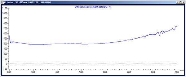
3.1. Acquisition of Diffuser data
- Place diffuser in the standard sample compartment and close the sample compartment.
- Click on the window Quantum Yield Correction Factor Measurement and then, on Diffuser Measurement.
- Enter the file name: "IS_factor_F70_diffuser" for the diffuser data and click OK (Video 1).
- After measurement, the file will be saved in the "Correct" folder of FL Solutions. Figure 1 is an example of the Diffuser Measurement data.
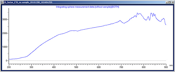
3.2. Acquisition of correction factor for no sample (reference)
- Remove the standard sample compartment from the instrument, store diffuser, then install the integrating sphere.
- Fill the powder cell with Aluminum Oxide powder up to a height of at least 25 mm to make sure the powder fully covers the port of the integrating sphere. Tap the bottom of the cell carefully to compact the powder.
- Place the aluminum oxide white tile in the reference port (P2) of the integrating sphere (the one facing the emission monochromator) and the powder cell with Aluminum Oxide in the sample port (P1) of the Integrating sphere (the one facing the excitation monochromator).
- Click on the Quantum Yield Correction Factor Measurement window, then on Integrating Sphere Measurement (without sample) (Video 2).
- The software will remind you to set the samples. Enter the name "IS with no sample" and click OK.
- After measurement, the file will be saved in the folder "Correct" of FL Solutions. Figure 2 is an example of the Integrating Sphere with no sample measurement data.
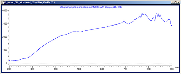
3.3. Acquisition of correction factor in the presence of a sample:
- Remove the cell with aluminum oxide powder and replace it with the Spectralon white standard. (The Spectralon standard should be facing the excitation monochromator (P1)).
- Click on the Quantum Yield Correction Factor Measurement window and then on Integrating Sphere Measurement (with sample). The software will remind you to set the Standard Reflectance Material for measurement.
- Enter the name: "IS with sample" for the Integrating Sphere with sample data file and click OK (Video 3).
- After measurement, the file will be saved in the "Correct" folder of FL Solutions. Figure 3 is an example of the Integrating Sphere with sample measurement data.
NOTE: In case you need to use cutoff filters to block second order scattering light on the emission side which interfere with the fluorescence peak, you will need to measure a baseline using the diffuser only and also the diffuser and the appropriate filter. These data files will be used by the software when calculating quantum yield. In practice we recommend measuring all the cutoff filters as part of the initial set up of the system.
4. Sample Measurement (Sodium Salicylate Powder)
Quantum yield measurement involves the acquisition of an emission spectrum for both no-sample (reference) and in the presence of a sample. Select the analytical measurement parameters as follows:
- Click on the "Method" button and in the General tab select wavelength scan as the measurement mode and enter the appropriate information on the operator and accessories (Video 4).
- Click on the "Instrument" tab and enter the measuring parameters for the instrument as shown in Table 2 (Video 5).
| ANALYTICAL CONDITIONS | |
| Measurement | Wavelength scan |
| Scan mode | Emission |
| Data mode | Fluorescence |
| EX WL | 350 nm |
| EM Start WL | 330 nm |
| EM End WL | 600 nm |
| Scan speed | 1200 nm/min |
| Delay | 0 s |
| EX Slit | 5.0 nm |
| EM Slit | 5.0 nm |
| PMT Voltage | 350 V |
| Corrected spectra | ON |
| Response | Auto |
| Corrected spectra | ON |
Table 2.
- No additional settings are required at this time since the Monitor, Processing and Report tab settings can be made after the data has been measured. We are just going to review them and then click the OK button, in order to set the selected measuring parameters in the instrument (Video 6).
- As an option, settings selected could be saved for future use. We will proceed now measuring the Aluminum Oxide reference standard using direct excitation.
- Place the powder cell with Al2O3 powder in the sample measuring port (P1) (in front of the excitation beam).
- Click on the "Sample" button and type the sample name: "P1_Baseline_Al2O3", then click on the box next to "Auto file". Select the folder and file name for the data: "P1_Baseline_Al2O3", then click "Save" and "OK" (Video 7).
- Click on the "Measure" button for measuring the Al2O3 sample. (Video 8), after the data processing window opens, click on the "Auto Scale Axis" button adjusting the scale, to visualize the scattering peak with direct excitation (Figure 4).
- Now we will proceed measuring the sample of Sodium Salicylate using direct excitation. Click on the "Sample" icon and enter "P1_Sodium Salicylate" for the sample and file name, then click the OK button (Video 9).
- Place the Sodium Salicylate sample in the powder cell and in the P1 port of the integrating sphere (the port facing the excitation light beam) and click on the "Measure" button. (Video 10) When the data processing window opens, click on the "Auto Scale Axis" button to adjust the scale and visualize the scattering and fluorescence peaks.
- At this time we will repeat measurements for Aluminum Oxide and Sodium Salicylate with the samples placed in the P2 port of the integrating sphere, in order to read them using indirect excitation.
- First click on the "Sample" button and type the sample and file name: "P2_Baseline_Al2O3" for both of them (Video 11).
- Move the Aluminum Oxide white tile from P2 to P1 of the integrating sphere and place a cell filled with Aluminum Oxide powder in P2.
- Click on the "Measure" button to read the sample (Video 12).
- To complete sample measurement we need to measure the sodium salicylate sample using indirect irradiation. First we type the sample and file names as in previous steps. The name will be P2_Sodium salicylate (Video 13).
- Place the sodium salicylate sample in the P2 port of the integrating sphere and click the Measure button (Video 14).
5. Quantum Yield Calculation
First we will proceed loading the integrating sphere correction factors.
- Click on the Quantum Yield Calculation button to open the quantum yield calculation program (Video 15).
- Click on the Quantum Yield Correction Factor Setting button (Video 16).
- Click on the Integrating Sphere Correction tab and click the box in front of "Integrating sphere correction", then click on the Filter Correction tab and make sure the "Filter correction" box is unselected, click again on the Integrating Sphere Correction tab (Video 17).
- Click on the Load button of the Diffuse Measurement Data section, then select file "IS_factor_F70 no sample" (Video 18).
- Select the "IS_factor_F70_diffuser" file and then click on the load button (Video 19).
- Click on the Load button of the Integrating sphere measurement data (without sample) section (Video 20).
- Select the "IS_factor_F70 no sample" file and then click on the load button (Video 21).
- Click on the Load button of the Integrating sphere measurement data (with sample) section (Video 22).
- Select the "IS_factor_F70 with sample" file and then on the load button (Video 23).
- Normalized wavelength can be left at 600nm or adjusted to the wavelength value where the integrating sphere correction is equal to 1. To do this, make sure the box in front of "Display quantum yield calculation window" is selected and click the OK button of the "Quantum Yield Factor Setting" window, which will close this window (Video 24).
- Now click on the "Integrating Sphere Correction" tab of the "Quantum Yield Calculation" window and adjust the cursor until the Integrating sphere correction reading is "1", making note of the wavelength (Video 25).
- Click on the "Quantum Yield Correction Factor Setting" and if required change the Normalized wavelength to the reading obtained in the previous step and click the OK button (Video 26).
The next step is to load the Baseline and Sample data files
- Click on the "Quantum Yield Calculation" tab (Video 27).
- Load "Data without sample" (file P1_Baseline_ Al2O3) for direct irradiation by clicking on the load button and load "Data with sample" (file P1_Sodium salicylate) for direct irradiation (Video 28).
- Next we will proceed to select the "scattering" and "fluorescence" regions (Video 29).
- Now we will calculate the quantum yield for direct irradiation of the sample. Click on the "Calculation" button and read the results (Video 30). We will need this data for the final calculation of the quantum yield for the sample taking into consideration indirect excitation.
- Click on Text file and save the data under the file name "QY Direct Irradiation" (Video 31).
- Using data files P2_Baseline_Al2O3 and P2_Sodium salicylate, we will calculate quantum yield for indirect excitation (Video 32) Now we will save this data for the final Quantum Yield calculation.
- Click on the Text file and save the data text file under the name "QY Indirect Irradiation" (Video 33) Now we will open the two text files in Excel with the Quantum Yield data for direct and indirect excitation. Finally we will calculate the Quantum Yield for the sample including the effect of Indirect Excitation, using the following formula:
Φ=Φd-(1-Ad)ΦI
Where:
Φ is the corrected quantum yield taking into consideration indirect excitation
Φd is the Internal Quantum Yield using Direct Excitation. (Internal quantum yield = Amount of Fluorescence / Amount of absorbed excitation light.)
Ad is the absorptance for direct excitation. (This is the ratio of the amount of excitation beam absorbed by the sample). (Absorbance = (ArEx - CsEx)/ ArEx, where ArEx is the amount of excitation light and CsEx is the amount of reflected light)
ΦI is the Internal Quantum Yield using Indirect Excitation
Φ=0.536 - (1 - 0.848)0.420 Calculated
Φ=0.47216
6. Calculation of Chromaticity
- We will use the optional software Report Generator along with a template prepared for the calculation of chromaticity.
- Open the data file P1_Sodium salicylate (Video 34).
- Click the "Property" button, then the "Report tab". In "Output" select "Use print generator sheet" from the dropdown menu. In "Print items" select template "FL70Std01_Color-chart.xls" and then click the "Open" button. There is no need to select the wavelength range and interval, since this is done automatically by Report Generator (Video 35).
- The next step is to create the report. Click on the "Report" tab and the macro for creating the report will be executed and saved in the "Reports" folder in Excel format under the sample name (Video 36).
- At this time we can open the report to see the color data (Video 37).
7. Secrets to Success
- Use fresh samples.
- Be aware that material from different manufacturers could give different results.
- Tap the bottom of the powder cell to compact the sample and present a uniform surface for measurement.
- Protect samples from light. They deteriorate with light exposure.
- Try using a faster scanning speed to minimize sample exposure to light.
8. Representative Results
8.1. Sodium salicylate is known to have a quantum yield of 0.4 to 0.5

Figure 1. Click here to view larger figure.

Figure 2. Click here to view larger figure.

Figure 3. Click here to view larger figure.
Please click here to see Video 1.
Please click here to see Video 2.
Please click here to see Video 3.
Please click here to see Video 4.
Please click here to see Video 5.
Please click here to see Video 6.
Please click here to see Video 7.
Please click here to see Video 8.
Please click here to see Video 9.
Please click here to see Video 10.
Please click here to see Video 11.
Please click here to see Video 12.
Please click here to see Video 13.
Please click here to see Video 14.
Please click here to see Video 15.
Please click here to see Video 16.
Please click here to see Video 17.
Please click here to see Video 18.
Please click here to see Video 19.
Please click here to see Video 20.
Please click here to see Video 21.
Please click here to see Video 22.
Please click here to see Video 23.
Please click here to see Video 24.
Please click here to see Video 25.
Please click here to see Video 26.
Please click here to see Video 27.
Please click here to see Video 28.
Please click here to see Video 29.
Please click here to see Video 30.
Please click here to see Video 31.
Please click here to see Video 32.
Please click here to see Video 33.
Please click here to see Video 34.
Please click here to see Video 35.
Please click here to see Video 36.
Please click here to see Video 37.
Disclosures
Luis A. Moreno is employed by Hitachi High Technologies America that produces the instrument used in this article.
Materials
| Name | Company | Catalog Number | Comments |
| Name of the reagent | Company | Catalogue number | Comments |
| Sodium salicylate powder | Wako | 191-03142 | Mol. weight 160.10 |
References
- . . Quantum Yield Measurement of Sodium Salicylate. FL080002, 1 (2008).
- Lakowicz, J. R. Principles of Fluorescence Spectroscopy. Science and Business Media, LLC. 60, (2006).
- Horigome, J., Wakui, T., Shirasaki, T. A Simple Correction Method for Determination of Absolute Fluorescence Quantum Yields of Solid Samples with a conventional Fluorescence Spectrophotometer. Bunseki Kagako. 58 (6), 553-559 (2009).
Reprints and Permissions
Request permission to reuse the text or figures of this JoVE article
Request PermissionExplore More Articles
This article has been published
Video Coming Soon
ABOUT JoVE
Copyright © 2025 MyJoVE Corporation. All rights reserved