الفئران هند الاطراف طويل العظام تشريح ونخاع العظم عزل
In This Article
Summary
Here we present a protocol for the dissection of hind limb long bones (femurs and tibiae) from the laboratory mouse. We further describe a rapid technique for bone marrow isolation from these bones that utilizes centrifugation for removal of bone marrow from the bone marrow space.
Abstract
Investigation of the bone and the bone marrow is critical in many research fields including basic bone biology, immunology, hematology, cancer metastasis, biomechanics, and stem cell biology. Despite the importance of the bone in healthy and pathologic states, however, it is a largely under-researched organ due to lack of specialized knowledge of bone dissection and bone marrow isolation. Mice are a common model organism to study effects on bone and bone marrow, necessitating a standardized and efficient method for long bone dissection and bone marrow isolation for processing of large experimental cohorts. We describe a straightforward dissection procedure for the removal of the femur and tibia that is suitable for downstream applications, including but not limited to histomorphologic analysis and strength testing. In addition, we outline a rapid procedure for isolation of bone marrow from the long bones via centrifugation with limited handling time, ideal for cell sorting, primary cell culture, or DNA, RNA, and protein extraction. The protocol is streamlined for rapid processing of samples to limit experimental error, and is standardized to minimize user-to-user variability.
Introduction
The study of long bones and the cells of the bone marrow is central to a myriad of research disciplines, including, but not limited to, bone biology, cancer biology, immunology, hematology, and biomechanics. The bone is a highly dynamic organ that together with the cartilage forms the skeleton to provide mechanical support against loading and protection of the internal organs. In addition, the mineral components of bone are a storage sink for the critical signaling molecules calcium and phosphorus, as well as other factors1. Finally, bones house the bone marrow and, together with metabolically active bone forming osteoblasts and bone resorbing osteoclasts, provide the stem cell niche necessary for the maintenance of hematopoietic and lymphoid cell populations.
Bone and bone marrow are affected in many disorders, often leading to bone marrow dysfunction, severe bone pain, and pathologic fracture. Bone is a common site of metastasis in many solid tumors, most notably breast cancer and prostate cancer, where tumor cells directly engage the bone marrow niche to initiate the vicious cycle of bone metastasis and displace hematopoietic stem cells2,3. Hematopoietic malignancies including myeloma and leukemia are characterized by bone marrow dysfunction as well as deregulation of healthy bone remodeling1. Other non-malignant skeletal disorders are also active areas of research, such as osteoarthritis, osteoporosis, scoliosis, and rickets. Even in an otherwise healthy individual, biomechanical failure in a bone leads to a painful fracture. All of these disorders represent active areas of research with the goal of identifying new preventative measures and treatment regimens to reduce morbidity and mortality.
To research the plethora of roles of the bone and the bone marrow, both under physiologic and pathologic conditions, it is critical for researchers to have a simple and efficient standardized method for dissection of the mouse long bones for rapid processing of large in vivo experiments. The dissection protocol outlined here is suitable for all long bone analyses including ex vivo imaging, histology, histomorphometry, and strength testing, among others. Similarly, a standardized bone marrow isolation method with high bone marrow cell recovery and low inter-user variability is important for experimental analysis such as fluorescence-activated cell sorting (FACS) or quantitative PCR (qPCR) as well as downstream applications such as primary cell culture of bone marrow cells.
Protocol
وقد وافق كل عمل الحيوانية رعاية الحيوان المؤسسية واستخدام اللجنة وفقا للتوصيات الواردة في دليل لرعاية واستخدام الحيوانات المختبرية من المعاهد الوطنية للصحة.
تشريح 1. هند الاطراف طويل العظام
- الموت ببطء الماوس وفقا للمبادئ التوجيهية المؤسسية.
- ضع الماوس في موقف ضعيف ويضعوا التي تعلق كل أربع أرجل من خلال منصات الماوس مخلب أدناه مفصل الكاحل.
- رذاذ الفأر مع 70٪ من الإيثانول، يسكب دقيق الساقين.
- إجراء شق صغير على يمين خط الوسط في أسفل البطن، وذلك فوق الورك.
- تمديد شق أسفل الساق والماضي مفصل الكاحل.
- سحب الجلد وقطع عضلة الفخذ ترتكز على نهاية القريبة من عظم الفخذ لفضح الجانب الأمامي من الفخذ ويعلقون الخروج من المحطة، ووضع دبوس في زاوية 45 درجة من المجلس.
- مع هؤلاء الاشخاص(ه) من مقص ضد الجانب الخلفي من الفخذ، وقطع أوتار الركبة بعيدا عن مفصل الركبة.
- سحب الجلد والعضلات في اوتار الركبة ترتكز على نهاية القريبة من عظم الفخذ لفضح الجانب الخلفي من الفخذ ويعلقون الخروج من المحطة، ووضع دبوس في زاوية 45 درجة من المجلس.
- مع ملقط، عقد نهاية البعيدة للعظم الفخذ، وذلك فوق مفصل الركبة. توجيه شفرات المقص على جانبي جسم الفخذ نحو مفصل الورك، والحرص على عدم قطع في عظمة الفخذ نفسها.
- بعد الوصول إلى رأس الفخذ، التي أشار إليها مقص فتح قليلا، تحريف مقص مع شفرة العليا من مقص تتحرك مباشرة على رأس الفخذ إلى خلع عظم الفخذ، والحرص على عدم التقاط العظم تحت رأس الفخذ.
- فهم الجزء العلوي من جسم الفخذ مع ملقط، وقطع الأنسجة اللينة بعيدا عن رأس الفخذ لتحريرها من الحق.
- سحب عظم الساق بأكملها، بما في ذلكعظم الفخذ والركبة، والساق، وحتى بعيدا عن الجسم، قطع بعناية بعيدا النسيج الضام والعضلات التي تربط المحطة على الجلد.
- تجهد مفصل الكاحل ومرة أخرى استخدام مقص في حركة التواء لخلع الساق.
- استيعاب النهاية البعيدة من الساق، مع الحرص على عدم قطع الأوتار، وسحب الساق وحتى بعيدا عن الجسم ومجلس دبوس.
- قطع أي نوع من الأنسجة الضامة المتبقية ربط العظام الطويلة على الماوس عند الركبة.
- إزالة أي عضلة إضافية أو النسيج الضام تعلق على عظم الفخذ والساق.
- للحصول على أي التطبيقات التي تتطلب العظام لتبقى سليمة (علم الأنسجة، histomorphometry واختبار النشاط الحيوي، الخ.)، والمضي قدما مع البروتوكولات في المنزل القياسية (كما هو الحال في 4-7). لعزل نخاع العظم، انتقل إلى القسم 2.
2. إعداد العظام الطويلة لنخاع العظم عزل
- باستخدام ملقط، فهم عظم الفخذ مع الرضفة تواجه بعيداالثانية نهاية الداني (رئيس الفخذ) إلى أسفل.
- تجهد مفصل الركبة واستخدام المقص في حركة التواء لخلع الساق والفخذ.
- قطع أي النسيج الضام عقد عظم الفخذ والساق معا.
- باستخدام ملقط، فهم عظم الفخذ مع الجانب الأمامي تواجه بعيدا والنهاية القريبة (الفخذ نهاية الرأس) أسفل.
- توجيه مقص فوق رمح الفخذ إلى اللقم.
- تناوب بلطف المقص ذهابا وإيابا لإزالة اللقم، الرضفة، والكردوس لفضح الكردوس.
- إزالة أي عضلة إضافية أو النسيج الضام تعلق على عظم الفخذ باستخدام ملقط، مقص، وKimwipes.
- باستخدام ملقط، فهم الساق مع الجانب الأمامي تواجه بعيدا والنهاية البعيدة (نهاية الكاحل) إلى أسفل.
- إذا الكردوس قصبي غير سليمة، توجيه مقص فوق رمح الساق إلى اللقم.
- تناوب بلطف المقص ذهابا وإيابا لإزالة اللقم والكردوس لفضح metaphيسيس.
- إزالة أي عضلة إضافية أو النسيج الضام تعلق على الساق باستخدام ملقط، مقص، وKimwipes.
3. نخاع العظم عزل
- دفع على إبرة G 18 من خلال الجزء السفلي من أنبوب 0.5 مل microcentrifuge ل.
- وضع العظام الطويلة (بحد أقصى 2 عظام الفخذ والظنبوب 2) في أنبوب، وصولا في نهاية الركبة نهاية أسفل وإغلاق الغطاء.
- عش أنبوب 0.5 مل microcentrifuge لفي أنبوب microcentrifuge 1.5 مل.
- أنابيب الطرد المركزي المتداخلة في ≥10،000 x ج في microcentrifuge لمدة 15 ثانية.
- تحقق من أن نخاع العظام قد خرجت العظام عن طريق التفتيش البصري. يجب أن تظهر عظام بيضاء، وينبغي أن يكون هناك بيليه البصرية كبير في أنبوب أكبر.
- تجاهل أنبوب 0.5 مل microcentrifuge لمع العظام.
- تعليق نخاع العظام في حل مناسب (على سبيل المثال، في برنامج تلفزيوني، وسائل الإعلام والثقافة، وFACS العازلة)، والمضي قدما في البروتوكول التجريبي (DNA، RNA، أو العزلة البروتين،تحليل نظام مراقبة الأصول الميدانية، أو زراعة الخلايا الأولية).
Representative Results
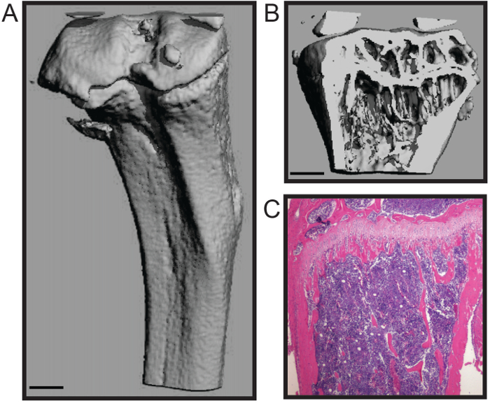
هو الأمثل لبروتوكول الموصوفة هنا لتشريح السريع لعظم الفخذ والساق الماوس مع حد أدنى من الأضرار التي لحقت الأنسجة والعظام. هذا الأسلوب هو مناسبة لعدد من التحليلات التحويلية، بما في ذلك دراسات الميكانيكا الحيوية، histomorphometry (الشكل 1A - B)، والأنسجة (الشكل 1C) 4،7. وhistomophometric ممثل micoCT 3D إعادة الإعمار (الشكل 1A - B) يدل على أن كلا من العظام إسفنجي وقذيفة القشرية يتم الاحتفاظ تسمح الكميات دقيقة من المعلمات الهيكلية موحدة لhistomorphometry العظام، بما في ذلك عدد التربيقي، سمك، والمباعدة بين الولادات. حجم العظام. وسماكة القشرية، ضمن تدابير أخرى 8. يظهر مقطع تمثيلي نسيجية والثابتة الفورمالين وdecalcified الساق H & E الملون (الشكل 1C). توضح الصورة وسلامة كل من ب متكلسةواحد ونخاع العظام الخلوي للتحليل النسيجي.
الإجراء نخاع العظام العزلة يحافظ على عقم مساحة النخاع العظمي، له معالجة منخفضة للحد من التلوث، ولا يتطلب قطع من العظام الطويلة، وبالتالي تقليل فقدان العائد نخاع العظام. هذا نخاع العظم هو مناسبة للعديد من التطبيقات المصب، بما في ذلك التدفق الخلوي 5 و PCR تحليل. وبالإضافة إلى ذلك، فإن هذا الإجراء يمكن أن تستخدم لعزل نخاع العظام للثقافة الخلية الأولية من خلايا نخاع العظام، بما في ذلك الخلايا الآكلة وبانيات (الشكل 2A - B) 4،6.

الشكل 1. Histomorphological والتحليلات النسيجية من الماوس لونغ العظام. إعادة الإعمار microCT ثلاثي الأبعاد من الساق الماوس تظهر (أ) قذيفة القشرية الخارجية و (ب ترونج>) العظام تربيقية (مقياس بار = 0.5 ملم). (C) نسيجية H & E وصمة عار على الساق decalcified ومقطوع (4X). صور من باب المجاملة كاثرين Weilbaecher، جامعة واشنطن مدرسة الطب، الولايات المتحدة الأمريكية. الرجاء انقر هنا لعرض نسخة أكبر من هذا الرقم.

الشكل 2. الابتدائية نخاع العظم الثقافة الخليوي لتمايز الخلايا الآكلة وبانيات. تلطيخ (A) TRAP لالآكلة متعددة النوى بعد 7 أيام في وسائل الإعلام osteoclastogenic (4X). (ب) الفوسفاتيز القلوية (اللون الأرجواني) لبانيات والأحمر الأليزارين (اللون الأحمر) وصمة عار لتمعدن بعد 21 يوما في وسائل الإعلام عظمي المنشأ. صور من باب المجاملة كاثرين Weilbaecher، جامعة واشنطن مدرسة الطب، الولايات المتحدة الأمريكية.ديزيل / ftp_upload / 53936 / 53936fig2large.jpg "الهدف =" _ فارغة "> الرجاء انقر هنا لعرض نسخة أكبر من هذا الرقم.
Discussion
We present a simple and efficient method for removal of mouse hind long bones and subsequent bone marrow isolation. This method maintains the high structural and cellular integrity of the bones and bone marrow and has low handling time, minimizing the likelihood of user-induced fracture or bone scoring that may influence downstream analyses. In addition, the centrifugation method for isolating bone marrow does not require cutting the bone to expose the bone marrow space or fluid to flush the bone marrow, reducing potential points of contamination. Moreover, the centrifuge technique is relatively high-throughput with lower hands-on time than other methods, thus reducing processing time.
High variation is inherent to in vivo mouse studies due to high mouse-to-mouse phenotypic variation. In order to maximize the research impact of expensive and labor-intensive mouse studies, it is critical to minimize technical experimental error9,10. Time from animal sacrifice to downstream analysis or tissue fixation introduces experimental variation that may overcome subtle changes and reduce large differences between groups. Therefore, rapid processing of samples is essential for accurate data analysis. The long bone dissection and bone marrow isolation techniques described here are optimized for rapid processing of animals and samples to reduce technical variation.
This protocol can be widely applied to many research fields, including investigation of the bone tissue itself or interrogation of the cells of the bone marrow. In addition, this straightforward approach to long bone dissection will enable researchers in related fields to directly interrogate bone contributions in order to expand our knowledge of bone marrow dysfunction in otherwise understudied pathologies.
Acknowledgements
This work was supported by NCI grant nos. U54CA143803, CA163124, CA093900, and CA143055 to K.J.P. The authors thank the current and past members of the Weilbaecher lab, especially Katherine Weilbaecher, Michelle Hurchla, and Hongju Deng, and members of the Brady Urological Institute, especially members of the Pienta laboratory for critical reading of the manuscript.
Materials
| Name | Company | Catalog Number | Comments |
| pinboard | |||
| pins | |||
| 70% ethanol | |||
| dissection sissors | |||
| dissection forceps | |||
| Kimwipes | Kimberly-Clark | 34120 | |
| 16 guage needle | |||
| 1.5 ml microcentrifuge tube | |||
| 0.5 ml microcentrifuge tube | |||
| microcentrifuge |
References
- McHayleh, W. M., Ellerman, J., Roodman, G. D. Ch. 80. Primer on the Metabolic Bone Diseases and Disorders of Mineral Metabolism. , 379-381 (2008).
- Weilbaecher, K. N., Guise, T. A., McCauley, L. K. Cancer to bone: a fatal attraction. Nat Rev Cancer. 11 (6), 411-425 (2011).
- Pedersen, E. A., Shiozawa, Y., Pienta, K. J., Taichman, R. S. The prostate cancer bone marrow niche: more than just 'fertile soil. Asian J Androl. 14 (3), 423-427 (2012).
- Amend, S. R., et al. Thrombospondin-1 regulates bone homeostasis through effects on bone matrix integrity and nitric oxide signaling in osteoclasts. J Bone Miner Res. 30 (1), 106-115 (2015).
- Hurchla, M. A., et al. The epoxyketone-based proteasome inhibitors carfilzomib and orally bioavailable oprozomib have anti-resorptive and bone-anabolic activity in addition to anti-myeloma effects. Leukemia. 27 (2), 430-440 (2013).
- Rauch, D. A., et al. The ARF tumor suppressor regulates bone remodeling and osteosarcoma development in mice. PLoS One. 5 (12), e15755 (2010).
- Su, X., et al. The ADP receptor P2RY12 regulates osteoclast function and pathologic bone remodeling. J Clin Invest. 122 (10), 3579-3592 (2012).
- Dempster, D. W., et al. Standardized nomenclature, symbols, and units for bone histomorphometry: a 2012 update of the report of the ASBMR Histomorphometry Nomenclature Committee. J Bone Miner Res. 28 (1), 2-17 (2013).
- Begley, C. G., Ellis, L. M. Drug development: Raise standards for preclinical cancer research. Nature. 483 (7391), 531-533 (2012).
- Festing, M. F., Altman, D. G. Guidelines for the design and statistical analysis of experiments using laboratory animals. ILAR J. 43 (4), 244-258 (2002).
Explore More Articles
This article has been published
Video Coming Soon
ABOUT JoVE
Copyright © 2025 MyJoVE Corporation. All rights reserved