Murino Hind Limb largo Bone La disección y el aislamiento de médula ósea
In This Article
Summary
Here we present a protocol for the dissection of hind limb long bones (femurs and tibiae) from the laboratory mouse. We further describe a rapid technique for bone marrow isolation from these bones that utilizes centrifugation for removal of bone marrow from the bone marrow space.
Abstract
Investigation of the bone and the bone marrow is critical in many research fields including basic bone biology, immunology, hematology, cancer metastasis, biomechanics, and stem cell biology. Despite the importance of the bone in healthy and pathologic states, however, it is a largely under-researched organ due to lack of specialized knowledge of bone dissection and bone marrow isolation. Mice are a common model organism to study effects on bone and bone marrow, necessitating a standardized and efficient method for long bone dissection and bone marrow isolation for processing of large experimental cohorts. We describe a straightforward dissection procedure for the removal of the femur and tibia that is suitable for downstream applications, including but not limited to histomorphologic analysis and strength testing. In addition, we outline a rapid procedure for isolation of bone marrow from the long bones via centrifugation with limited handling time, ideal for cell sorting, primary cell culture, or DNA, RNA, and protein extraction. The protocol is streamlined for rapid processing of samples to limit experimental error, and is standardized to minimize user-to-user variability.
Introduction
The study of long bones and the cells of the bone marrow is central to a myriad of research disciplines, including, but not limited to, bone biology, cancer biology, immunology, hematology, and biomechanics. The bone is a highly dynamic organ that together with the cartilage forms the skeleton to provide mechanical support against loading and protection of the internal organs. In addition, the mineral components of bone are a storage sink for the critical signaling molecules calcium and phosphorus, as well as other factors1. Finally, bones house the bone marrow and, together with metabolically active bone forming osteoblasts and bone resorbing osteoclasts, provide the stem cell niche necessary for the maintenance of hematopoietic and lymphoid cell populations.
Bone and bone marrow are affected in many disorders, often leading to bone marrow dysfunction, severe bone pain, and pathologic fracture. Bone is a common site of metastasis in many solid tumors, most notably breast cancer and prostate cancer, where tumor cells directly engage the bone marrow niche to initiate the vicious cycle of bone metastasis and displace hematopoietic stem cells2,3. Hematopoietic malignancies including myeloma and leukemia are characterized by bone marrow dysfunction as well as deregulation of healthy bone remodeling1. Other non-malignant skeletal disorders are also active areas of research, such as osteoarthritis, osteoporosis, scoliosis, and rickets. Even in an otherwise healthy individual, biomechanical failure in a bone leads to a painful fracture. All of these disorders represent active areas of research with the goal of identifying new preventative measures and treatment regimens to reduce morbidity and mortality.
To research the plethora of roles of the bone and the bone marrow, both under physiologic and pathologic conditions, it is critical for researchers to have a simple and efficient standardized method for dissection of the mouse long bones for rapid processing of large in vivo experiments. The dissection protocol outlined here is suitable for all long bone analyses including ex vivo imaging, histology, histomorphometry, and strength testing, among others. Similarly, a standardized bone marrow isolation method with high bone marrow cell recovery and low inter-user variability is important for experimental analysis such as fluorescence-activated cell sorting (FACS) or quantitative PCR (qPCR) as well as downstream applications such as primary cell culture of bone marrow cells.
Protocol
Todos los animales de trabajo fue aprobado por el Cuidado de Animales institucional y el empleo de acuerdo con las recomendaciones descritas en la Guía para el Cuidado y Uso de Animales de Laboratorio de los Institutos Nacionales de Salud.
La disección 1. extremidad posterior del hueso largo
- La eutanasia el ratón de acuerdo con las directrices institucionales.
- Coloque el ratón en una posición supina y colocar fijando las cuatro patas a través de las almohadillas de las patas del ratón debajo de la articulación del tobillo.
- Pulverizar el ratón con un 70% de etanol, rociar a fondo las piernas.
- Hacer una pequeña incisión a la derecha de la línea media en el abdomen inferior, justo por encima de la cadera.
- Extender la incisión en la pierna y más allá de la articulación del tobillo.
- Tire hacia atrás la piel y cortar el músculo cuádriceps anclado al extremo proximal del fémur para exponer la cara anterior del fémur y el pasador hacia fuera de la pierna, colocando el pasador en un ángulo de 45 grados con respecto a la placa.
- Con el blade de la tijera contra el lado posterior del fémur, cortar los tendones de la corva lejos de la articulación de la rodilla.
- Tirar de la piel y los músculos isquiotibiales anclados al extremo proximal del fémur para exponer el lado posterior del fémur y el pasador hacia fuera de la pierna, colocando el pasador en un ángulo de 45 grados con respecto a la placa.
- Con las pinzas, mantenga el extremo distal del fémur, justo por encima de la articulación de la rodilla. Guía de las cuchillas de las tijeras a cada lado del eje femoral hacia la articulación de la cadera, teniendo cuidado de no cortar en la propia fémur.
- Después de alcanzar la cabeza del fémur, indicado por las tijeras se abren ligeramente, torcer la tijera con la hoja superior de la tijera se mueven directamente sobre la cabeza del fémur para dislocar el fémur, con cuidado de no romper el hueso debajo de la cabeza del fémur.
- Agarre la parte superior del eje femoral con el fórceps, cortar el tejido blando lejos de la cabeza femoral para liberarla de acetábulo.
- Tire todo el hueso de la pierna, incluyendofémur, la rodilla, y la tibia, arriba y lejos del cuerpo, cuidadosamente cortar el tejido conectivo y muscular que conecta la pierna a la piel.
- Extender demasiado la articulación del tobillo y otra vez utilizar las tijeras en un movimiento giratorio para dislocar la tibia.
- Agarrando el extremo distal de la tibia, teniendo cuidado de no cortar los tendones, tire de la tibia hacia arriba y lejos del cuerpo y el tablón de anuncios.
- Cortar cualquier tejido conectivo restante fijar el hueso largo para el ratón en la rodilla.
- Eliminar cualquier músculo adicional o tejido conectivo unido al fémur y la tibia.
- Para las aplicaciones que requieren que el hueso se mantienen intactos (histología, la histología, las pruebas biomecánicas, etc.), Proceder con protocolos estándar en la casa (como en el 4-7). Para aislar la médula ósea, pase a la sección 2.
2. Preparación de hueso largo de médula ósea Aislamiento
- Usando las pinzas, agarre el fémur con la rótula de espaldas a unand el extremo proximal (cabeza femoral) hacia abajo.
- Extender demasiado la articulación de la rodilla y el uso de las tijeras en un movimiento giratorio para dislocar la tibia y el fémur.
- Cortar cualquier tejido conectivo que sostiene el fémur y la tibia juntos.
- El uso de las pinzas, sujete el fémur con el lado anterior de espaldas y el extremo proximal (extremo de la cabeza femoral) hacia abajo.
- Guiar la tijera hasta la diáfisis femoral a los cóndilos.
- gire suavemente las tijeras de ida y vuelta para eliminar los cóndilos, la rótula y la epífisis para exponer la metáfisis.
- Retire cualquier músculo adicional o tejido conectivo unido al fémur usando fórceps, tijeras y Kimwipes.
- El uso de las pinzas, sujete la tibia con el lado anterior de espaldas y el extremo distal (extremo de tobillo) hacia abajo.
- Si la epífisis tibial está intacto, guiar a las tijeras de la diáfisis de la tibia de los cóndilos.
- gire suavemente las tijeras de ida y vuelta para eliminar los cóndilos y la epífisis para exponer el metaphanalysis.
- Retire cualquier músculo adicional o tejido conectivo unido a la tibia utilizando fórceps, tijeras y Kimwipes.
Aislamiento 3. médula ósea
- Empuje una aguja de 18 G a través de la parte inferior de un tubo de 0,5 ml de microcentrífuga.
- Coloque los huesos largos (máximo de 2 fémures y tibias 2) en el tubo, hasta la rodilla extremo hacia abajo termina hacia abajo y cierre la tapa.
- Nido del tubo de microcentrífuga de 0,5 ml en un tubo de microcentrífuga de 1,5 ml.
- Centrifugar los tubos anidados en ≥10,000 xg en una microcentrífuga durante 15 segundos.
- Compruebe que la médula ósea se ha hecho girar hacia fuera de los huesos mediante inspección visual. Los huesos deben aparecen de color blanco y no debe haber una gran pastilla visual en el tubo más grande.
- Desechar el tubo de microcentrífuga de 0,5 ml con los huesos.
- Suspender la médula ósea en solución apropiada (por ejemplo, PBS, medios de cultivo, tampón FACS) y proceder con el protocolo experimental (DNA, RNA, o el aislamiento de proteínas,análisis FACS, o cultivo de células primarias).
Representative Results
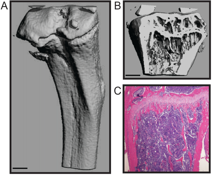
El protocolo descrito aquí está optimizado para una rápida disección del fémur y la tibia de ratón con un mínimo de daños en el tejido óseo. Esta técnica es adecuado para un número de análisis aguas abajo, incluyendo estudios de biomecánica, histomorfometría (Figura 1A - B), y la histología (Figura 1C) de 4,7. El representante histomophometric reconstrucción micoCT 3D (Figura 1A - B) demuestra que tanto el hueso esponjoso y cortical shell se mantienen que permite la cuantificación exacta de los parámetros estructurales normalizados para la histomorfometría de hueso, incluyendo el número trabecular, el grosor, y el espaciamiento; volumen de hueso; y el grosor cortical, entre otras medidas 8. El corte histológico muestra un representante fijado en formalina y descalcificadas tibia H & E manchadas (Figura 1C). La imagen demuestra la integridad tanto de la b calcificadouno y la médula ósea celular para el análisis histológico.
El procedimiento de aislamiento de la médula ósea conserva la esterilidad del espacio de la médula ósea, tiene baja manejo para reducir la contaminación, y no requiere el corte del hueso largo, reduciendo así la pérdida de rendimiento de la médula ósea. Esta médula ósea es conveniente para muchas aplicaciones posteriores, incluyendo citometría de flujo 5 y los análisis de PCR. Además, este procedimiento se puede utilizar para aislar de la médula ósea para el cultivo celular primario de células de médula ósea, incluyendo los osteoclastos y los osteoblastos (Figura 2A - B) 4,6.

Figura 1. histomorfológico y análisis histológicos de ratón hueso largo. MicroCT la reconstrucción tridimensional de una tibia de ratón que muestra (A) la concha cortical externa y (B trong>) hueso trabecular (barra de escala = 0,5 mm). (C) histológico tinción con HE de una tibia descalcificada y seccionada (4x). Imágenes cortesía de Katherine Weilbaecher, Facultad de Medicina, Universidad de Washington, EE.UU.. Haga clic aquí para ver una versión más grande de esta figura.

Figura 2. Primaria de Médula Ósea cultivo celular para la diferenciación de los osteoclastos y osteoblastos. Tinción (A) para los osteoclastos multinucleadas TRAP después de 7 días en los medios de comunicación osteoclastogénicos (4x). (B) La fosfatasa alcalina (color púrpura) de los osteoblastos y rojo de alizarina (color rojo) para la mineralización de manchas después de 21 días en los medios de comunicación osteogénico. Imágenes cortesía de Katherine Weilbaecher, Facultad de Medicina, Universidad de Washington, EE.UU..iles / ftp_upload / 53936 / 53936fig2large.jpg "target =" _ blank "> Haga clic aquí para ver una versión más grande de esta figura.
Discussion
We present a simple and efficient method for removal of mouse hind long bones and subsequent bone marrow isolation. This method maintains the high structural and cellular integrity of the bones and bone marrow and has low handling time, minimizing the likelihood of user-induced fracture or bone scoring that may influence downstream analyses. In addition, the centrifugation method for isolating bone marrow does not require cutting the bone to expose the bone marrow space or fluid to flush the bone marrow, reducing potential points of contamination. Moreover, the centrifuge technique is relatively high-throughput with lower hands-on time than other methods, thus reducing processing time.
High variation is inherent to in vivo mouse studies due to high mouse-to-mouse phenotypic variation. In order to maximize the research impact of expensive and labor-intensive mouse studies, it is critical to minimize technical experimental error9,10. Time from animal sacrifice to downstream analysis or tissue fixation introduces experimental variation that may overcome subtle changes and reduce large differences between groups. Therefore, rapid processing of samples is essential for accurate data analysis. The long bone dissection and bone marrow isolation techniques described here are optimized for rapid processing of animals and samples to reduce technical variation.
This protocol can be widely applied to many research fields, including investigation of the bone tissue itself or interrogation of the cells of the bone marrow. In addition, this straightforward approach to long bone dissection will enable researchers in related fields to directly interrogate bone contributions in order to expand our knowledge of bone marrow dysfunction in otherwise understudied pathologies.
Acknowledgements
This work was supported by NCI grant nos. U54CA143803, CA163124, CA093900, and CA143055 to K.J.P. The authors thank the current and past members of the Weilbaecher lab, especially Katherine Weilbaecher, Michelle Hurchla, and Hongju Deng, and members of the Brady Urological Institute, especially members of the Pienta laboratory for critical reading of the manuscript.
Materials
| Name | Company | Catalog Number | Comments |
| pinboard | |||
| pins | |||
| 70% ethanol | |||
| dissection sissors | |||
| dissection forceps | |||
| Kimwipes | Kimberly-Clark | 34120 | |
| 16 guage needle | |||
| 1.5 ml microcentrifuge tube | |||
| 0.5 ml microcentrifuge tube | |||
| microcentrifuge |
References
- McHayleh, W. M., Ellerman, J., Roodman, G. D. Ch. 80. Primer on the Metabolic Bone Diseases and Disorders of Mineral Metabolism. , 379-381 (2008).
- Weilbaecher, K. N., Guise, T. A., McCauley, L. K. Cancer to bone: a fatal attraction. Nat Rev Cancer. 11 (6), 411-425 (2011).
- Pedersen, E. A., Shiozawa, Y., Pienta, K. J., Taichman, R. S. The prostate cancer bone marrow niche: more than just 'fertile soil. Asian J Androl. 14 (3), 423-427 (2012).
- Amend, S. R., et al. Thrombospondin-1 regulates bone homeostasis through effects on bone matrix integrity and nitric oxide signaling in osteoclasts. J Bone Miner Res. 30 (1), 106-115 (2015).
- Hurchla, M. A., et al. The epoxyketone-based proteasome inhibitors carfilzomib and orally bioavailable oprozomib have anti-resorptive and bone-anabolic activity in addition to anti-myeloma effects. Leukemia. 27 (2), 430-440 (2013).
- Rauch, D. A., et al. The ARF tumor suppressor regulates bone remodeling and osteosarcoma development in mice. PLoS One. 5 (12), e15755 (2010).
- Su, X., et al. The ADP receptor P2RY12 regulates osteoclast function and pathologic bone remodeling. J Clin Invest. 122 (10), 3579-3592 (2012).
- Dempster, D. W., et al. Standardized nomenclature, symbols, and units for bone histomorphometry: a 2012 update of the report of the ASBMR Histomorphometry Nomenclature Committee. J Bone Miner Res. 28 (1), 2-17 (2013).
- Begley, C. G., Ellis, L. M. Drug development: Raise standards for preclinical cancer research. Nature. 483 (7391), 531-533 (2012).
- Festing, M. F., Altman, D. G. Guidelines for the design and statistical analysis of experiments using laboratory animals. ILAR J. 43 (4), 244-258 (2002).
This article has been published
Video Coming Soon
ABOUT JoVE
Copyright © 2025 MyJoVE Corporation. All rights reserved