Sign In

A subscription to JoVE is required to view this content. Sign in or start your free trial.

In This Article

  • Summary
  • Abstract
  • Introduction
  • Protocol
  • Results
  • Discussion
  • Disclosures
  • Acknowledgements
  • Materials
  • References
  • Reprints and Permissions

Summary

We present a method for the acquisition of fluorescence reporter time courses from single cells using micropatterned arrays. The protocol describes the preparation of single-cell arrays, the setup and operation of live-cell scanning time-lapse microscopy and an open-source image analysis tool for automated preselection, visual control and tracking of cell-integrated fluorescence time courses per adhesion site.

Abstract

Live-cell Imaging of Single-Cell Arrays (LISCA) is a versatile method to collect time courses of fluorescence signals from individual cells in high throughput. In general, the acquisition of single-cell time courses from cultured cells is hampered by cell motility and diversity of cell shapes. Adhesive micro-arrays standardize single-cell conditions and facilitate image analysis. LISCA combines single-cell microarrays with scanning time-lapse microscopy and automated image processing. Here, we describe the experimental steps of taking single-cell fluorescence time courses in a LISCA format. We transfect cells adherent to a micropatterned array using mRNA encoding for enhanced green fluorescent protein (eGFP) and monitor the eGFP expression kinetics of hundreds of cells in parallel via scanning time-lapse microscopy. The image data stacks are automatically processed by newly developed software that integrates fluorescence intensity over selected cell contours to generate single-cell fluorescence time courses. We demonstrate that eGFP expression time courses after mRNA transfection are well described by a simple kinetic translation model that reveals expression and degradation rates of mRNA. Further applications of LISCA for event time correlations of multiple markers in the context of signaling apoptosis are discussed.

Introduction

In recent years, the importance of single-cell experiments has become apparent. Data from single cells allow the investigation of cell-to-cell variability, the resolution of intracellular parameter correlations and the detection of cellular kinetics that remain hidden in ensemble measurements1,2,3. In order to investigate cellular kinetics of thousands of single cells in parallel, new approaches are needed that enable monitoring the cells under standardized conditions over a time period of several hours up to several days followed by a quantitative data analysis 4. Here, we present Live-cell Imaging of Single-Cell Arrays (LISCA), which combines the use of microstructured arrays with time-lapse microscopy and automated image analysis.

Several methods for generating microstructured single-cell arrays have been established and published in literature5,6. Here, we briefly describe Microscale Plasma-Initiated Protein Patterning (µPIPP). A detailed protocol of the single-cell array fabrication using µPIPP is also found in reference7. The use of single-cell arrays enables alignment of thousands of cells on standardized adhesion spots presenting defined microenvironments for each cell and thus reduces a source of experimental variability (Figure 1A). Single-cell arrays are used to monitor the time courses of fluorescent markers purposed to indicate a variety of cellular processes. Long-term microscopy in scanning time-lapse mode allows for monitoring a large area of the single-cell arrays and hence sampling single-cell data in high-throughput over an observation time of several hours or even days. This generates time-line stacks of images from each position of the array (Figure 1B). In order to reduce the large amount of image data and to extract the desired single-cell fluorescence time courses in an efficient way, automated image processing is required that takes advantage of the positioning of cells (Figure 1C).

The challenge of LISCA is to adapt the experimental protocols and computational tools to form a high-throughput assay that generates quantitative and reproducible data of cellular kinetics. In this article we provide a step-by-step description of the individual methods and how they are combined in a LISCA assay. As an example, we discuss the time course of enhanced green fluorescent protein (eGFP) expression after artificial mRNA delivery. eGFP expression following mRNA delivery is described by reaction rate equations modeling translation and degradation of mRNA. Fitting the model function for the time course of eGFP concentration to the LISCA readout of the fluorescence intensity for each individual cell over time yields best estimates of model parameters such as the mRNA degradation rate. As a representative result we discuss the mRNA delivery efficiency of two different lipid-based transfection agents and how their parameter distributions differ.

figure-introduction-3315
Figure 1: Representation of the LISCA workflow combining (A) micro-patterned single-cell arrays (B) scanning time-lapse microscopy and (C) automated image analysis of recorded image series. The single-cell arrays consist of a two-dimensional pattern of cell-adhesive squares with a cell-repellent interspace leading to an arrangement of the cells on the micropattern, as can be seen in the phase-contrast image as well as the fluorescence image of eGFP expressing cells (A). The entire microstructured area is imaged in a scanning time-lapse mode repeatedly taking images at a sequence of positions (B). Recorded image series are processed to read out the fluorescence intensity per cell over time (C). Scale bars: 500 µm (A), 200 µm (C). Please click here to view a larger version of this figure.

Protocol

figure-protocol-25
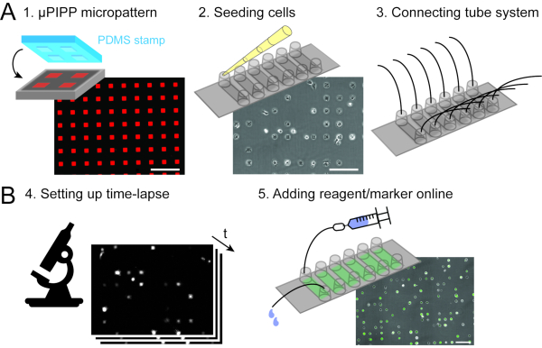
Figure 2: Data acquisition combining single-cell microarrays (A) with scanning time-lapse microscopy (B). As preparation of the time-lapse experiment, a single-cell array with a 2D micropattern of adhesion squares is prepared (1), followed by cell seeding and the alignment of the cells on the micropattern (2) as well as the connection of a perfusion system to the six-channel slide, which enables liquid handling during the time-lapse measurement (3). A scanning time-lapse experiment is set up (4) and the cells are transfected on the microscope by injecting an mRNA lipoplex solution through the perfusion system during the time-lapse experiment (5). Scale bars: 200 µm. Please click here to view a larger version of this figure.

1. Microstructured single-cell array fabrication (Figure 2A)

  1. Prepare the materials needed for µPIPP array fabrication.
    1. Prepare sterile phosphate-buffered saline (PBS) at pH 7.4.
    2. Prepare sterile ultrapure water with a resistivity of at least 18 MΩcm at 25 °C.
    3. Prepare PLL(20 kDa)-g[3.5] PEG(2 kDa) (PLL-PEG) working solution with a 2 mg/mL concentration of PLL-PEG in ultrapure water containing 150 mM NaCl and 10 mM 4-(2-hydroxyethyl)-1-piperazineethanesulfonic acid (HEPES).
    4. Prepare an extracellular matrix protein solution for surface coating: 1 mg/mL fibronectin (FN) in PBS.
    5. Prepare a silicon wafer with a micropattern fabricated by photolithography8 that functions as a reusable master. The micropattern consists of squares with an edge length of 30 µm, a depth of 12 µm and an inter-square distance of 60 µm, arranged in six stripes each having a width of 6 mm and a height of 18 mm.
  2. Mix a polydimethylsiloxane (PDMS) monomer with 9% crosslinker (mass %) using a silicone elastomer kit and degas it for about 30 min until it is bubble free using a desiccator. Cast the silicon wafer with a roughly 3-5 mm thick PDMS layer and degas it again for about 30 min until it is bubble-free.
    1. Put the silicon wafer with the PDMS in a baking oven at 50 °C to cure the PDMS for at least 4 h.
  3. Cut the PDMS stamps.
    1. Use a scalpel and cut out of the PDMS layer one PDMS masterpiece that contains the six micropattern stripes.
    2. Place the PDMS masterpiece on a bench with the micropattern facing upward.
    3. Cut each of the six micropattern stripes of the PDMS masterpiece with a razorblade into a PDMS stamp. Take care that the edges of the PDMS stamps are open by cutting off some of the patterned area.
  4. Place the PDMS stamps on a coverslip of a six-channel slide (Figure 3-1).
    1. Use an uncoated coverslip and mark the channel positions of the six-channel slide by carefully scratching the protection foil of the coverslip. Then place the coverslip on the bench with the protection foil facing downward.
    2. Place the PDMS stamps with the micropattern facing downward on the coverslip at the marked channel positions using tweezers.
    3. Check the attachment of the PDMS stamps under a microscope. If a PDMS stamp is fully attached to the coverslip, the squares in contact appear darker than the interspace. The attachment of the PDMS stamp to the coverslip is crucial for the micropattern quality.
  5. Place the coverslip with the six PDMS stamps on it into a plasma cleaner and treat it with oxygen plasma (pressure 0.2 mbar, ~40 W for 3 min) to make the surfaces between the PDMS stamps and the coverslip hydrophilic (Figure 3-2).
  6. Carry out all further steps of the micropattern fabrication in a biosafety cabinet. Use 15 µL of the PLL-PEG solution and pipette one drop of it next to each PDMS stamp so that the PLL-PEG solution is absorbed into the hydrophilic pattern of the PDMS stamp (Figure 3-3). Let the PLL-PEG incubate for 20 min at room temperature.
  7. Rinse 1 mL of ultrapure water over the coverslip with the PDMS stamps on it and remove the PDMS stamps using tweezers (Figure 3-4). Then rinse the coverslip a second time with 1 mL of ultrapure water and let it dry.
  8. When the coverslip has dried completely, stick a six-channel sticky slide to the coverslip (Figure 3-4). Take care that the micropatterned areas align with the bottom of the channels.
  9. Functionalize the adhesion squares with FN.
    1. Fill 40 µL of PBS into each channel.
    2. Prepare a 100 µg/mL FN solution in PBS.
    3. Add 40 µL of the FN solution to each channel (Figure 3-5). Mix the FN solution with the PBS in the channel thoroughly by removing 40 µL from one reservoir and adding it to the opposite reservoir of the same channel for 3 times to generate a homogeneous solution. Incubate the FN solution for 45 min at room temperature.
    4. Wash each channel three times with 120 µL of PBS (Figure 3-6).
  10. In order to check for the pattern quality, use a fluorescently labeled FN in step 9.2. (Figure 4A).
    ​NOTE: We recommend preparing the µPIPP array not more than one day before cell seeding as PLL-PEG and FN are not covalently bound to the substrate and the quality of the pattern may decrease over time. Store the prepared µPIPP array in the fridge.

figure-protocol-6140
Figure 3: Single-cell microarray fabrication by µPIPP. (1) PDMS stamps with a three-dimensional micropattern structure on the surface are arranged on a coverslip of a six-channel slide. (2) The coverslip with the PDMS stamps on it is treated with oxygen plasma to make the surfaces hydrophilic. (3) PLL-PEG is added. It is absorbed into the microstructure by capillary forces and makes the surfaces not covered by the PDMS stamp cell-repellent. (4) The coverslip is rinsed with water to remove the remaining PLL-PEG. Then, the PDMS stamps are removed and a six-channel sticky slide is stuck unto the coverslip. (5) Fibronectin, a protein of the extracellular matrix, is added to make the areas without PLL-PEG cell-adhesive. (6) The six-channel slide is washed with phosphate-buffered saline. Please click here to view a larger version of this figure.

2. Cell seeding (Figure 2A)

NOTE: For the following washing steps, add the respective liquid to one reservoir and then remove an equal volume of liquid from the opposite reservoir of a channel.

  1. Wash each channel with 120 µL of 37 °C fully supplemented cell growth medium. Before adding the cell suspension, ensure that only the channels are filled with medium but not the reservoirs.
  2. Detach HuH7 cells from a cell culture flask following your standard protocol for cell passaging and adjust the cell suspension concentration to 4 x 10⁵ cells/mL.
  3. Add 40 µL of cell suspension and mix the cell growth medium with the cell suspension by removing 40 µL from one reservoir and adding it to the opposite reservoir of the same channel for 3 times to reach a homogeneous cell distribution (Figure 4B).
  4. Remove 40 µL of suspension from the channel so that only the channel is filled with cell suspension.
  5. Put the slide in an incubator and check cell adhesion 1 h after seeding using a phase-contrast microscope.
  6. Add 120 µL of 37 °C warm cell growth medium.
  7. But the slide back in the incubator for further 3 h to enable cellular self-organization on the micropattern (Figure 4C).

figure-protocol-8739
Figure 4: Cellular self-organization and quality control of µPIPP array. (A) The microstructured surface consists of squared FN-coated adhesion spots shown in red surrounded by a cell-repellent polymer. (B) After cell seeding, the HuH7 cells are randomly distributed and (C) adhere mainly on the adhesion spots over a time period of 4 h. Reprinted with permission 7. Scale bars: 200 µm. Please click here to view a larger version of this figure.

3. Perfusion system (Figure 2A)

NOTE: The use of a perfusion system is only required if reagents or fluorescent markers need to be added during the course of the time-lapse measurement. Depending on your needs, you can connect each channel to a separate perfusion system or connect several channels in series to the same perfusion system. The number of perfusion systems corresponds to the number of independent experimental conditions. Connect the tubes under sterile conditions in a biosafety cabinet and avoid the inclusion of air bubbles in the perfusion system. If no perfusion system is used, add the reagents/markers in a biosafety cabinet before the time-lapse measurement. The perfusion system is in-house fabricated, the used material is listed in the Table of Materials. The assembly of the perfusion system has been described previously9.

  1. Use a 1 mL syringe (with replacement sporn) and fill the syringe with 1 mL of 37 °C cell growth medium.
  2. Connect the syringe to the inlet tube using the valve and fill the tube with medium.
  3. Connect the inlet tube to a reservoir of a channel and make sure that no air bubbles are trapped.
  4. To connect another channel in series to this perfusion system, connect a serial connector to the reservoir opposite to the inlet tube of the current channel. Proceed to the next channel and connect the free end of the serial connector to one of its reservoirs.
  5. Repeat the previous steps until the required number of channels are connected in series.
  6. Connect the outlet tube directly to the free reservoir of the current channel. Fill the connected tube with medium in order to check that the perfusion system does not leak.
  7. Repeat the previous steps until all six channels of the slide are connected to a perfusion system.
  8. Place the slide with the connected perfusion system(s) back in the incubator until further use or place it directly in the heating chamber of the microscope pre-warmed to 37 °C for time-lapse measurement.

4. Time-lapse microscopy (Figure 2B)

NOTE: For long-term measurements, maintain a stable temperature of 37 °C and a stable CO2 level. As an alternative to CO2-dependent cell growth medium, use L15 medium for which no gas incubation system is required.

NOTE: For quantitative imaging, use cell growth medium without phenol red during the time-lapse measurement to reduce background fluorescence and use the same settings of the time-lapse protocol as well as the same microscope for technical replicates.

  1. Set up a time-lapse protocol for recording a phase-contrast image and a fluorescence image with exposure times of 750 ms (depending on the camera), 10 min time interval between consecutive loops through the position list, and an observation time of 30 h, using a 10x objective and appropriate fluorescence filters.
  2. Put the six-channel slide with the cells on the single-cell arrays in the sample holder of the 37 °C warm heating chamber. If perfusion systems are connected to the six-channel slide, fix the tubes to the stage using some tape to ensure that the six-channel slide is not moved during liquid exchange. Insert the free ends of the outlet tubes through a hole of a 15 mL reaction tube to collect the liquid waste.
  3. Set the position list for the scanning time-lapse measurement. Ensure that the number of positions can be scanned within the defined time interval between consecutive loops through the position list. With a 10x objective, 10-30 positions per channel can be set to scan the total micropattern area depending on the camera chip size.
  4. Start the time-lapse measurement. For a better image quality of long-term measurements, use an automated focus correction system.

5. Fluorescent marker - mRNA transfection (Figure 2B)

NOTE: For a transfection in two channels connected by a tubing system, a total volume of 600 µL transfection mix is needed (300 µL for one channel). The indicated volumes refer to a transfection in two connected channels.

  1. Prepare a transfection agent solution by diluting 1 µL of transfection agent in 200 µL of serum-reduced medium and let the solution incubate for 5 min at room temperature.
  2. Prepare a mRNA solution by diluting 300 ng of mRNA encoding for eGFP in 150 µL of serum-reduced medium.
  3. Prepare the transfection mix by adding 150 µL of the transfection agent solution to the mRNA solution and mix it well. Let the transfection mix incubate for 20 min at room temperature.
  4. Flush the tubing system with 1 mL of 37 °C warm PBS using a syringe during incubation of the transfection mix. When flushing the tubes, make sure that the microscope stage does not move. Pause the time-lapse measurement if necessary.
  5. Dilute the transfection mix to the final mRNA concentration of 0.5 ng/µL by adding 300 µL serum-reduced medium.
  6. Flush the tubing system with the transfection mix using a syringe and let the mRNA lipoplexes incubate for 1 h (pause the time-lapse measurement if necessary).
  7. Stop the transfection incubation and flush out the unbound mRNA lipoplexes by washing with 1 mL of 37 °C warm fully supplemented cell growth medium using a syringe (pause the time-lapse measurement if necessary).

6. Image analysis and fluorescence readout

  1. When running the image analysis for the first time, install version 0.1.6 of the open-source software "Automated Microstructure Analysis in Python" (PyAMA) from the cited location10 according to the instructions provided there.
  2. Ensure that the image channels (phase-contrast and fluorescence) are available as multi-image 16-bit TIFF files. If necessary, convert them accordingly.
  3. Start PyAMA and click on Open stack… to open images for analysis.
  4. For each multi-image TIFF file to open, click on Open and select the file so that it is displayed in the list of loaded files on the left side of the dialog (Figure 5-1).
  5. Mark the channels to include in the analysis. For each channel, perform the following steps.
    1. Select in the list of loaded files the TIFF file containing the channel.
    2. In the section Add new channel, select the index of the channel in the TIFF file. Indexing is zero-based; the first channel has index 0, the second channel has index 1 and so on.
    3. Select the channel type. Select Phase contrast or Fluorescence for the corresponding image channels and Segmentation for a binary channel indicating the cell contours.
    4. Optionally, enter a label of the channel for distinguishing different fluorescence channels: eGFP and DAPI.
    5. After configuring the channel, click Add.
  6. When all added channels are displayed in the channel list on the right side of the dialog, click OK to load the stack.
  7. To perform segmentation using PyAMA's built-in segmentation algorithm for cell recognition based on the phase-contrast images (Figure 5-2), go to Tools | Binarize… and enter a file name for the NumPy file with the binarized channel.
    NOTE: In the current version, loading the binarized channel requires reloading all channels.
  8. To perform a background correction11 on a fluorescence channel (Figure 5-3), ensure that the fluorescence channel and a segmentation channel are loaded. If no segmentation channel is loaded, ensure that a phase-contrast channel is loaded for automatic segmentation. Go to "Tools > Background correction…" and select a file name for the resulting TIFF file with the corrected fluorescence channel.
    NOTE: In the current version, loading the background-corrected channel requires reloading all channels.
  9. Inspect the pre-selected cells (Figure 5-4) and their integrated fluorescence signal (Figure 5-5) by scrolling through the time frames, viewing the channels listed in the channel menu on the left side and clicking on cells to highlight their fluorescence time courses (Figure 1C). Use the cell selection to exclude cells that are not viable, not confined to an adhesion spot or attached to another cell from further analysis. Toggle the selection of cells for readout by pressing Shift and clicking on the cell, or by highlighting the cell and pressing Enter.
  10. Save the single-cell time courses for the cell area and the integrated fluorescence (Figure 5-6) by clicking on File | Save and selecting a directory to save to.

figure-protocol-19090
Figure 5: Automated image processing of time-lapse image series using PyAMA. (1) Phase-contrast and fluorescence image series for each imaging position are imported. (2) Cell contours are determined by segmentation on the phase-contrast image stack. (3) A background correction is applied to the fluorescence images. (4) The cell contours are tracked over time and pre-selected for export. (5) The fluorescence intensity is integrated based on the tracked cell contours. (6) Single-cell cell areas and integrated fluorescence intensities are evaluated and time courses for each cell are exported. Scale bars: 100 µm. Please click here to view a larger version of this figure.

  1. To analyze the translation kinetics after mRNA transfection, fit a translation model based on biochemical rate equations to each single-cell time course as described previously by Reiser et al.12. The data and code used in that study are publicly available13.
  2. For each single-cell time course, retrieve the estimated fitting parameters of the translation model that represent the mRNA degradation rate and the time point of translation onset. An example data set is discussed in the representative results section.
  3. Perform further analysis on the distributions of best estimates of the parameters for varied experimental conditions to investigate the cell-to-cell variability within the cell populations.

Results

The LISCA approach enables to efficiently collect fluorescence time courses from single cells. As a representative example we outline how the LISCA method is applied to measure single-cell eGFP expression after transfection. The data of the LISCA experiment is used to assess mRNA delivery kinetics, which is important for the development of efficient mRNA drugs.

In particular we demonstrate the different impact of two lipid-based mRNA delivery systems with respect to the time point of translati...

Discussion

Here we described LISCA as a versatile technique to follow cellular kinetics of intracellular fluorescent labels at the single-cell level. In order to perform a successful LISCA experiment, each of the described steps of the protocol section must be established individually and then all steps must be combined. Each of the three major aspects of LISCA feature crucial steps.

Single-cell microarray fabrication
The quality of the microarray is crucial as the cellular alignment on th...

Disclosures

The authors declare that they have no competing financial interests.

Acknowledgements

This work was supported by grants from the German Science Foundation (DFG) to Collaborative Research Center (SFB) 1032. Support by the German Federal Ministry of Education, Research and Technology (BMBF) under the cooperative project 05K2018-2017-06716 Medisoft as well as a grant from the Bayerische Forschungsstiftung are gratefully acknowledged. Anita Reiser was supported by a DFG Fellowship through the Graduate School of Quantitative Biosciences Munich (QBM).

Materials

NameCompanyCatalog NumberComments
Adtech Polymer Engineering PTFE Microtubing Fisher Scientific10178071
baking ovenBinder9010-0190
CFI Plan Fluor DL 10xNikonMRH20100
DesiccatorRothNX07.1
Eclipse Ti-ENikon
eGFP mRNATrilinkL-7601
Female Luer to Tube ConnectorMEDNETFTL210-6005
Fetal bovine serumThermo Fisher10270106
FibronectinYo Proteins663
Filter set eGFPAHFF46-002
Fisherbrand Translucent Platinum-Cured Silicone TubingFisher Scientific11768088
HEPES (1 M)Thermo Fisher15630080
Incubation BoxOkolabOKO-H201
incubatorBinder9040-0012
L-15 without phenol redThermo Fisher21083027
Lipofectamine 2000Thermo Fisher11668027
Male Luerin-house fabricated consisting of teflon
Male Luer to Tube ConnectorMEDNETMTLS210-6005alternative to in-house fabricated male luers
NaCl (5 M)Thermo FisherAM9760G
Needleless Valve to Male Luer ConnectorMEDNETNVFMLLPC
NIS ElementsNikonImaging software Version 5.02.00
NOA81ThorlabsNOA81Fast Curing Optical Adhesive for tube system assembly
Opti-MEMThermo Fisher31985062
PCO edge 4.2 M-USB-HQ-PCOpco
Phosphate buffered saline (PBS)in-house prepared
Plasma CleanerDiener FemtoPico-BRS
PLL(20 kDa)-g[3.5]-PEG(2 kDa)SuSoS AG
silicon wafer mit mircorstructuresin-house fabricated
Sola Light EngineLumencor
sticky slide VI 0.4 ibidi80608
Sylgard 184 Silicone Elastomer KitDow Corning1673921
Tango 2Märzhäuser00-24-626-0000
Ultrapure waterin-house prepared
uncoated coverslipsibidi10813
Injekt-F Solo, 1 mLOmilab9166017Vwith replacement sporn

References

  1. Altschuler, S. J., Wu, L. F. Cellular heterogeneity: do differences make a difference. Cell. 141 (4), 559-563 (2010).
  2. Locke, J. C., Elowitz, M. B. Using movies to analyse gene circuit dynamics in single cells. Nature Reviews Microbiology. 7 (5), 383-392 (2009).
  3. Spencer, S. L., Gaudet, S., Albeck, J. G., Burke, J. M., Sorger, P. K. Non-genetic origins of cell-to-cell variability in TRAIL-induced apoptosis. Nature. 459 (7245), 428-432 (2009).
  4. El-Ali, J., Sorger, P. K., Jensen, K. F. Cells on chips. Nature. 442 (7101), 403-411 (2006).
  5. Segerer, F. J., et al. Versatile method to generate multiple types of micropatterns. Biointerphases. 11 (1), 011005 (2016).
  6. Piel, M., Théry, M. . Micropatterning in cell biology, part A/B/C. , (2014).
  7. Reiser, A., Zorn, M. L., Murschhauser, A., Rädler, J. O., Ertl, P., Rothbauer, M. . Cell-Based Microarrays: Methods and Protocols. , 41-54 (2018).
  8. Picone, R., Baum, B., McKendry, R. . Methods in cell biology. 119, 73-90 (2014).
  9. Reiser, A. . Single-cell time courses of mRNA transport and translation kinetics. , (2019).
  10. . Softmatter LMU-Raedler Group Available from: https://github.com/SoftmatterLMU-RaedlerGroup/pyama (2020)
  11. . MIAAB, 29262 Available from: https://github.com/SoftmatterLMU-RaedlerGroup/pyama (2020)
  12. Reiser, A., et al. Correlation of mRNA delivery timing and protein expression in lipid-based transfection. Integrative Biology. 11 (9), 362-371 (2019).
  13. Reiser, A., Woschée, D., Mehrotra, N., Krzysztoń, R. S., Strey, H. H., Rädler, J. O. Supplementing data and code for: "Correlation of mRNA delivery timing and protein expression in lipid-based transfection". Zenodo. , (2019).
  14. Ferizi, M., et al. Stability analysis of chemically modified mRNA using micropattern-based single-cell arrays. Lab on a Chip. 15 (17), 3561-3571 (2015).
  15. Fröhlich, F., et al. Multi-experiment nonlinear mixed effect modeling of single-cell translation kinetics after transfection. NPJ systems biology and applications. 4 (1), 1-12 (2018).
  16. Krzysztoń, R., et al. Single-cell kinetics of siRNA-mediated mRNA degradation. Nanomedicine: Nanotechnology, Biology and Medicine. 21, 102077 (2019).
  17. Röttgermann, P. J., Alberola, A. P., Rädler, J. O. Cellular self-organization on micro-structured surfaces. Soft Matter. 10 (14), 2397-2404 (2014).
  18. Röttgermann, P. J., Dawson, K. A., Rädler, J. O. Time-resolved study of nanoparticle induced apoptosis using microfabricated single cell arrays. Microarrays. 5 (2), 8 (2016).
  19. Murschhauser, A., et al. A high-throughput microscopy method for single-cell analysis of event-time correlations in nanoparticle-induced cell death. Communications Biology. 2 (1), 1-11 (2019).
  20. Chatzopoulou, E. I., et al. Chip-based platform for dynamic analysis of NK cell cytolysis mediated by a triplebody. Analyst. 141 (7), 2284-2295 (2016).
  21. Sztilkovics, M., et al. Single-cell adhesion force kinetics of cell populations from combined label-free optical biosensor and robotic fluidic force microscopy. Scientific Reports. 10 (1), 1-13 (2020).

Reprints and Permissions

Request permission to reuse the text or figures of this JoVE article

Request Permission

Explore More Articles

Live cell ImagingSingle cell ArraysLISCACellular KineticsFluorescent Time CoursesMicro EnvironmentsEmission AnalysisSingle cell Array FabricationPDMs MasterpieceMicropattern StampsOxygen Plasma TreatmentHydrophilic SurfacesPegylated Poly l lysine SolutionPBS SolutionFibronectin SolutionHomogenization Process

This article has been published

Video Coming Soon

JoVE Logo

Privacy

Terms of Use

Policies

Research

Education

ABOUT JoVE

Copyright © 2025 MyJoVE Corporation. All rights reserved