
A subscription to JoVE is required to view this content. Sign in or start your free trial.
Method Article
Treatment Protocol for Rotator Cuff Calcific Tendinitis Using a Single-Crystal Piezoelectric Focused Shock Wave Source
In This Article
Summary
Calcific tendinitis of the shoulder is a relatively common condition with numerous treatment options. Here, we discuss the indications of focused shock waves generated by a single-crystal piezoelectric device, describe a treatment protocol, and present the preliminary results.
Abstract
Focused shock waves have emerged as a highly effective noninvasive therapeutic option for the treatment of calcific tendinitis of the shoulder. There are three types of focused shock wave generators: electrohydraulic, electromagnetic, and piezoelectric. According to our literature search, there are no reports of results with the use of single-crystal piezoelectric generators in calcific tendinitis of the shoulder. In a consecutive retrospective series of 23 patients with Gärtner type I and II calcifications of the rotator cuff, we performed three applications of high-energy piezoelectric focused waves (4,000 pulses per session with a frequency of 6 Hz). At the final follow-up (average of 14 months), 82.6% of the cases showed complete resorption of the calcification in radiographic controls. In 8.7% of the cases, partial disappearance of the calcification was achieved, and in the remaining 8.7% there were no significant changes. Single-crystal piezoelectric generators have a success rate comparable to those already reported with electrohydraulic and electromagnetic devices.
Introduction
Calcium crystal deposits can appear in different regions of the musculoskeletal system, but their most frequent location is in the shoulder region. Gondos1 reported that 69% of calcification cases occur in the shoulder location. Calcific shoulder tendinopathies are characterized by the presence of hydroxyapatite deposits in the rotator cuff tendons. It is estimated that the prevalence in the general population ranges from 2.7% to 20%2.
Calcific tendinitis of the shoulder typically affects patients between 30 to 60 years old2. It is also more frequent in women (57%-76.7%) with respect to men3. The location of the calcium deposit is much more frequent in the distal tendon of the supraspinatus muscle4, while localizations in the infraspinatus, teres minor, subscapularis, and long head of the biceps have also been reported4.
Women between 30 and 60 years old, with a calcification over 1.5 cm in length, have the highest chance of being symptomatic5. Although it tends to spontaneously resolve itself, the cycle can often be halted. In these cases, symptoms of pain and disability appear, and it is necessary to take active therapeutic action.
Gärtner's radiological classification6 differentiates three types of images. In type I, the image is dense, with well-defined borders corresponding to the formative phase. In the type II image, the appearance is mixed, with a deposit that can be dense but with diffuse borders, or transparent with well-defined borders. Finally, type III, characteristic of the resorptive phase, presents a transparent deposit with diffuse borders. Active therapeutic action, including shock wave applications, ultrasound-guided interventions, or surgery, must be taken in Gärtner type I and II, since in type III cases, the chance of short-term spontaneous resorption is very high6.
Conservative treatment is initially preferred. This classically includes rest, analgesics, non-steroidal and steroidal anti-inflammatory drugs, rehabilitation, and local injections. Good results of conservative treatment have been shown, especially in the resorptive stage, but a failure of conservative treatment has been reported in 27% to 39% of cases7,8,9. Several prognostic factors have been recognized as having a significant influence on the results of conservative treatment7,8. The location on both shoulders, the presence of a large-volume deposit, the location of the calcification in the anterior region of the acromion, and the spread of the deposit medially beyond the level of the acromioclavicular joint, are factors of poor prognosis7,8. A Gärtner stage III calcification and lack of sonographic extinction of the calcific deposit are considered predictors of good prognosis for conservative treatment7.
When conservative treatment fails, many patients end up becoming chronic carriers of shoulder pain with similar clinical characteristics to chronic rotator cuff non-calcific tendinopathies. The usual alternative to conservative treatment failure was surgery. Gschwend10 formulated three precise surgical indications for rotator cuff calcifications: symptom progression, constant and unmanageable pain, and failure of conservative treatment. Surgical treatment can be performed open or arthroscopically. Although open treatment was historically performed with good results11, arthroscopic techniques have gained popularity12,13. Musculoskeletal ultrasound and ultrasound-guided interventions (UGI) have significantly developed and been used in clinical practice in recent years14,15,16.
Extracorporeal shock wave treatment (ESWT) has emerged as an effective option prior to invasive procedures when conservative treatment has failed. Its therapeutic effect is not just mechanical, but based on mechanotransduction, a phenomenon by which cells can recognize a mechanical stimulus and react biologically17. However, shock wave treatment has limitations. Unlike lithotripsy, in which we only depend on the mechanical effect of the waves, there must also be a biological response by the patient. This response does not always occur.
The generic term "extracorporeal shock waves" includes two different technologies: focused shock waves and radial pressure waves17,18,19. The two technologies have therapeutic efficacy but differ in their physical characteristics and indications. Focused shock waves have a wide frequency range (from approximately 150 kHz up to 100 MHz), large pressure amplitude (up to 150 MPa) with a short rise time and small pulse width, followed by a low-stress wave (up to -25 MPa)18,19. Focused shock waves are generated by electrohydraulic, electromagnetic, and piezoelectric sources17,18,19.
Radial pressure waves are sound waves with pressure peaks of up to 30 MPa and much higher rise times than focused shock waves (about 3 µs)18,19. Radial pressure waves are generated by accelerating a projectile inside a cylindrical guiding tube by compressed air. The projectile hits an applicator at the end of the tube and produces a radial pressure wave that expands into the target tissue17,18,19.
Focused shock waves have a grade "A" recommendation for the treatment of rotator cuff calcifications17. This means that there is high-quality scientific evidence supported by level I studies with consistent findings. In the case of radial waves, the level of recommendation for rotator cuff calcifications is "I". This means that the evidence is insufficient to make a recommendation17.
Therapeutical efficacy of focused shock waves in calcific tendinopathies of the shoulder has been compared with open20 and arthroscopic21 surgery, with comparable results. However, shock waves have less frequent and less severe complications20,21, and the method is also cost-effective. Haake22 reported a significant difference between surgical costs (€13,400-23,450) and those of focused shock waves (€2,700-4,300). His results match with other studies that have shown a five- to sevenfold decrease in the cost of shock wave treatment compared to arthroscopic surgery23,24. There are also studies in which shock waves have been compared with ultrasound-guided interventions with controversial results15,25. Several publications4,17,26,27,28,29,30,31,32,33 have reported that a high level of energy is more effective when treating calcific tendinitis of the shoulder. Verstraelen27 reported that the use of high energy determines a higher rate of calcific resorption in a level I evidence study. This is a clear advantage for focused devices over radial ones because they can generate higher levels of energy. Numerous studies have reported good results with electrohydraulic4,17,34,35 and electromagnetic4,17,36,37,38 focused devices. A report has also been published using a multi-crystal piezoelectric device to treat rotator cuff calcifications39. We are not aware of any reports publishing the technique and results of single-crystal piezoelectric devices as of yet.
This report aims to describe the treatment protocol using a single-crystal piezoelectric device and to report the preliminary results.
Protocol
The protocol follows the guidelines of the Buenos Aires British Hospital's human research ethics committee.
1. Patient evaluation
- Clinical evaluation
- Evaluate patients clinically to rule out symptoms propagating from other anatomical regions or other associated pathology in the shoulder that may be the source of symptoms.
- Include shoulder inspection and palpation, evaluate the active and passive range of motion, and perform pain provocative maneuvers and integrity tests. In addition, evaluate the cervical spine and the elbow joint.
- Radiological evaluation
- Use radiographic anteroposterior (AP) projections, a lateral scapular view, and an axillary view of the shoulder.
- Classify the calcifications by their evolutionary stage according to Gärtner6. Request an MRI to rule out any associated pathology in the shoulder.
- Inclusion criteria
- Include patients classified as having Gärtner type I and II calcifications who have undergone at least 3 months of previous conservative treatment without result.
- Never inject corticosteroids into the subacromial space when treating rotator cuff tendinopathy, as they have a deleterious effect on the elastic fibers of the tendons40,41.
- Exclusion criteria: Do not include patients with Gärtner type III calcifications or other associated pathology in the affected shoulder.
2. Application technique
- Patient positioning: Position the patient comfortably and in a way that allows exposure to the area to be treated.
- Supraspinatus or infraspinatus tendon location: Treat by placing the patient in the supine or sitting position.
- Subscapularis: Treat by placing the patient in the supine position.
- Teres minor: Treat by placing the patient in the sitting, lateral decubitus, or oblique decubitus position for better access to the posterior aspect of the shoulder.
- Coupling pads
NOTE: Three pad sizes are available, each determining a specific depth of treatment focus (Figure 1).- Select a coupling pad size depending on the required penetration depth of the focused point (Table 1). The selection of the coupling pad varies in shoulder calcification cases according to patient's build and location. The most frequently used sizes are large and medium.
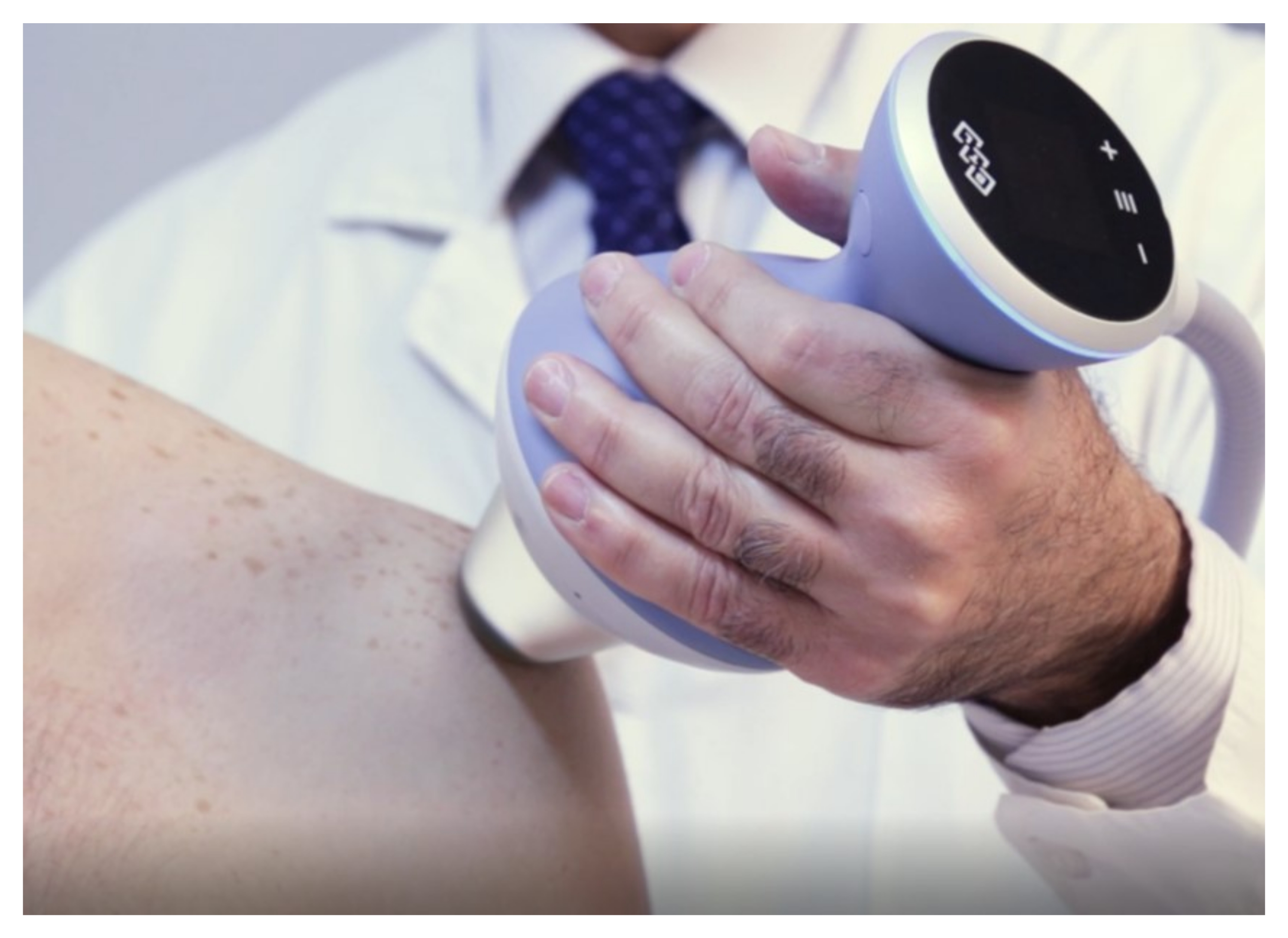
- Location of the area to be treated: Locate the area to be treated, considering anatomical landmarks and, if necessary, with the help of ultrasound images (Figure 2).
NOTE: It is very important to locate the exact area of the calcification and to focus the treatment on it4,42. The use of anatomical references and the help of ultrasound localization allows identifying the correct location. The need for a diagnostic ultrasound to locate the area is disputed, as there are studies that have not found significant differences with its use43. The application of focused waves is risky if done on large vessels and nerves or in areas close to the pleura44. Anatomical approaches that avoid these structures should be used. - Operator positioning: The use of this type of device implies the support of an applicator by the operator. Work in an ergonomic position.
NOTE: The operator must always be aware of the condition of the patient, as exceptional cases of fainting during treatment have been reported44. - Anesthesia: Do not use anesthesia. The use of local anesthesia is contraindicated because the presence of fluids alters the acoustic impedance of the tissue. In general, tolerance to treatment with this type of device is good, even when working with high energy levels.
- Contact gel: Apply adequate contact gel to the skin in the area to be treated.
- Navigation controls: Turn on the computer and access the touch screen. Select the Manual option at the top of the screen. The controls of the various treatment parameters are displayed. Use the rotary selection knob to set the parameters, including the intensity, frequency, and number of shocks.
- Intensity: Set the initial intensity of the treatment. The energy flux density (EFD) is graduated in the device used in millijoules per square millimeter of tissue (mJ/mm2).
- Increase the intensity progressively as tolerated by the patient. Start with low intensity. Reach at least an energy level above 0.40 mJ/mm2 to obtain reproducible results19,20,21,22,23,24,25,26. The mean maximum intensity used is 0.50 mJ/mm2.
- Frequency: Select the frequency to be used. The equipment has a frequency between 1-25 Hz. Use 4-6 Hz.
- Number of shocks: Select the number of shocks to be applied using the rotary selection knob. Apply at least 4,000 shocks per session.
- Modification of parameters during treatment: Modify the frequency and especially the intensity during the application. Use the display located on the applicator head to easily modify the application parameters during the treatment without having to interrupt it.
- The interval between sessions: Carry out three sessions at weekly intervals.

Figure 1: Applicator and coupling pad variations. Three different sizes of coupling pads are available. Each one allows the focus to be taken to a different depth in the tissues. Please click here to view a larger version of this figure.

Figure 2: Applicator positioning. Focused wave application in the supraspinatus tendon. Please click here to view a larger version of this figure.
3. Post-treatment protocol
- After the first session, alert the patients that in approximately 5% of cases, they may suffer an acute resorption process with intense pain. If discomfort occurs, recommend the patient to apply ice to the areas of pain in short sessions of no more than 10 min.
NOTE: Paracetamol is preferred over non-steroidal anti-inflammatory drugs. Shock waves modulate the inflammatory process, and it is better not to alter it by other means while the treatment lasts. - Immobilization: It is not necessary to immobilize the treated limb. Advise the patient to avoid extreme efforts and range of motions that may cause pain.
- Rehabilitation program
- As the sessions progress, if tolerance is good, increase the range of movement and incorporate exercises to strengthen the depressors of the humeral head.
- If symptomatic relapses do not occur, include recovery exercises for the extreme ranges of movement. The strengthening of the three portions of the deltoid and the scapular stabilizers are also indicated.
- Radiological follow-up:
- Perform the first radiographic examination at 6 weeks after the sessions and the second one at 12 weeks. In the case of observing a partial resorption process that has not been completed after 12 weeks, continue the radiological follow-up.
- If there have been no changes, opt for a new treatment module or move on to invasive procedures.
NOTE: In many patients, very important changes can be seen in the first control x-ray. In other cases, variable changes can be seen from baseline studies, and there may be no change even on long-term radiographs in other cases.
Results
A retrospective study of a series of patients with shoulder pain due to calcium deposits in the rotator cuff tendons was carried out in our institution. The inclusion criteria were Gärtner stage I and II calcifications and at least 3 months of previous conservative treatment without satisfactory results. Patients with Gärtner III calcifications, other associated pathology in the affected shoulder, previous local cortisone injection, and a history of surgery in the affected shoulder were excluded.
Discussion
This study shows encouraging results with the application of focused shock waves generated by a single-crystal piezoelectric device in a series of retrospectively evaluated patients with calcific tendinitis of the shoulder. According to the bibliographic search we carried out, this is the first study that reports results with a single-crystal piezoelectric device. Recently, Louwerens39 published a study using a piezoelectric shock wave device for the treatment of rotator cuff calcifications. Howev...
Disclosures
None
Acknowledgements
None
Materials
| Name | Company | Catalog Number | Comments |
| BTL 6000 FSWT | BTL | 09400B001107 | Focused Shock Wave Piezoelectric Source |
| Ultrasound & SWT Gel 300 mL | BTL | 237-GEL102 | Alcohol free hypoallergic gel |
References
- Gondos, B. Observations on periarthritis calcarea. The American Journal of Roentgenology Radium Therapy and Nuclear Medicine. 77 (1), 93-108 (1957).
- Suzuki, K., Potts, A., Anakwenze, O., Singh, A. Calcific tendinitis of the rotator cuff: management options. The Journal of the American Academy of Orthopaedic Surgeons. 22 (11), 707-717 (2014).
- Uhthoff, H. K., Sarkar, K., Maynard, J. A. Calcifying tendinitis: a new concept of its pathogenesis. Clinical Orthopaedics and Related Research. 118, 164-168 (1976).
- Moya, D., Ramón, S., Guiloff, L., Gerdesmeyer, L. Current knowledge on evidence-based shockwave treatments for shoulder pathology. International Journal of Surgery. 24, 171-178 (2015).
- Louwerens, J. K., Sierevelt, I. N., van Hove, R. P., vanden Bekerom, M. P., van Noort, A. Prevalence of calcific deposits within the rotator cuff tendons in adults with and without subacromial pain syndrome: clinical and radiologic analysis of 1219 patients. Journal of Shoulder and Elbow Surgery. 24 (10), 1588-1593 (2015).
- Gärtner, J., Heyer, A. Calcific tendinitis of the shoulder. Orthopade. 24 (3), 284-302 (1995).
- Ogon, P., et al. Prognostic factors in nonoperative therapy for chronic symptomatic calcific tendinitis of the shoulder. Arthritis and Rheumatism. 60 (10), 2978-2984 (2009).
- Drummond Junior, M., et al. Predictive factors for failure of conservative management in the treatment of calcific tendinitis of the shoulder. JSES International. 5 (3), 469-473 (2021).
- DePalma, A. F., Kruper, J. S. Long-term study of shoulder joints afflicted with and treated for calcific tendinitis. Clinical Orthopaedics. 20, 61-72 (1961).
- Gschwend, N., Patte, D., Zippel, J. Therapy of calcific tendinitis of the shoulder. Archiv fur Orthopadische und Unfall-Chirurgie. 73 (2), 120-135 (1972).
- Sansone, V., Maiorano, E., Galluzzo, A., Pascale, V. Calcific tendinopathy of the shoulder: clinical perspectives into the mechanisms, pathogenesis, and treatment. Orthopedic Research and Reviews. 10, 63-72 (2018).
- Ark, J. W., Flock, T. J., Flatow, E. L., Bigliani, L. U. Arthroscopic treatment of calcific tendinitis in the shoulder. Arthroscopy. 8 (2), 613-623 (2012).
- Porcellini, G., Paladini, P., Campi, F., Paganelli, M. Arthroscopic treatment of calcifying tendinitis of the shoulder: clinical and ultrasonographic follow-up findings at two to five years. Journal of Shoulder and Elbow Surgery. 13 (5), 503-508 (2004).
- Serafini, G., et al. Rotator cuff calcific tendonitis: short-term and 10-year outcomes after two-needle us-guided percutaneous treatment-nonrandomized controlled trial. Radiology. 252 (1), 157-164 (2009).
- Kim, Y. S., Lee, H. J., Kim, Y. V., Kong, C. G. Which method is more effective in the treatment of calcific tendinitis in the shoulder? Prospective randomized comparison between ultrasound-guided needling and extracorporeal shock wave therapy. Journal of Shoulder and Elbow Surgery. 23 (11), 1640-1646 (2014).
- de Witte, P. B., Kolk, A., Overes, F., Nelissen, R. G. H. H., Reijnierse, M. Rotator cuff calcific tendinitis: ultrasound-guided needling and lavage versus subacromial corticosteroids: Five-year outcomes of a randomized controlled trial. The American Journal of Sports Medicine. 45 (14), 3305-3314 (2017).
- Moya, D., et al. The role of extracorporeal shockwave treatment in musculoskeletal disorders. The Journal of Bone and Joint Surgery. American Volume. 100 (3), 251-263 (2018).
- Loske, A. M. . Medical and Biomedical Applications of Shock Waves. , (2017).
- Loske, A. M., Moya, D. Shock waves and radial pressure waves: time to put a clear nomenclature into practice. Journal of Regenerative Science. 1 (1), 4-8 (2021).
- Rompe, J. D., Zoellner, J., Nafe, B. Shock wave therapy versus conventional surgery in the treatment of calcifying tendinitis of the shoulder. Clinical Orthopaedics and Related Research. (387), 72-82 (2001).
- Rebuzzi, E., Coletti, N., Schiavetti, S., Giusto, F. Arthroscopy surgery versus shock wave therapy for chronic calcifying tendinitis of the shoulder. Journal of Orthopaedics and Traumatology. 9 (4), 179-185 (2008).
- Haake, M., Rautmann, M., Wirth, T. Assessment of the treatment costs of extracorporeal shock wave therapy versus surgical treatment for shoulder diseases. International Journal of Technology Assessment in Health Care. 17 (4), 612-617 (2001).
- Eid, J. Economic aspects in the treatment of tendinosis calcarea of the shoulder. 9th International Congress of the International Society for Musculoskeletal Shockwave Therapy. , (2006).
- Ramon, S., Moya, D., Alvarez, P., Cugat, R., Corbella, X. Efficiency in treatment of calcifying tendinopathy of the shoulder: extracorporeal shockwave therapy vs. surgery. 13th International Congress of Shoulder and Elbow Surgery. , (2016).
- Moya, D., et al. Incorrect methodology may favor ultrasound-guided needling over shock wave treatment in calcific tendinopathy of the shoulder. Journal of Shoulder and Elbow Surgery. 25 (8), 241-243 (2016).
- Albert, J. D., et al. High-energy extracorporeal shock-wave therapy for calcifying tendinitis of the rotator cuff: a randomised trial. The Journal of Bone and Joint Surgery. 89 (3), 335-341 (2007).
- Verstraelen, F. U., Inden Kleef, N. J., Jansen, L., Morrenhof, J. W. High-energy versus low-energy extracorporeal shock wave therapy for calcifying tendinitis of the shoulder: which is superior? A meta-analysis. Clinical Orthopaedics and Related Research. 472 (9), 2816-2825 (2014).
- Bannuru, R. R., Flavin, N. E., Vaysbrot, E., Harvey, W., McAlindon, T. High-energy extracorporeal shock-wave therapy for treating chronic calcific tendinitis of the shoulder: a systematic review. Annals of Internal Medicine. 160 (8), 542-549 (2014).
- Daecke, W., Kusnierczak, D., Loew, M. Extracorporeal shockwave therapy (ESWT) in tendinosis calcarea of the rotator cuff. Long-term results efficacy. Der Orthopade. 31 (7), 645-651 (2002).
- Huisstede, B. M., Gebremariam, L., vander Sande, R., Hay, E. M., Koes, B. W. Evidence for effectiveness of extracorporal shock-wave therapy (ESWT) to treat calcific and non-calcific rotator cuff tendinosis-systematic review. Manual Therapy. 16 (5), 419-433 (2011).
- Ioppolo, F., et al. Extracorporeal shock-wave therapy for supraspinatus calcifying tendinitis: a randomized clinical trial comparing two different energy levels. Physical Therapy. 92 (11), 1376-1385 (2012).
- Speed, C. A systematic review of shockwave therapies in soft tissue conditions: focusing on the evidence. British Journal of Sports Medicine. 48 (21), 1538-1542 (2014).
- Vavken, P., Holinka, J., Rompe, J. D., Dorotka, R. Focused extracorporeal shock wave therapy in calcifying tendinitis of the shoulder: a meta-analysis. Sports Health. 1 (2), 137-144 (2009).
- Cosentino, R., et al. Extracorporeal shock wave therapy for chronic calcific tendinitis of the shoulder: single blind study. Annals of the Rheumatic Diseases. 62 (3), 248-250 (2003).
- Hsu, C. J., et al. Extracorporeal shock wave therapy for calcifying tendinitis of the shoulder. Journal of Shoulder and Elbow Surgery. 17 (1), 55-59 (2008).
- Wang, C. J., et al. Shock wave therapy for calcific tendinitis of the shoulder: a prospective clinical study with two-year follow-up. American Journal of Sports Medicine. 31 (3), 425-430 (2003).
- Gerdesmeyer, L., et al. Extracorporeal shock wave therapy for the treatment of chronic calcifying tendonitis of the rotator cuff: a randomized controlled trial. JAMA. 290 (19), 2573-2580 (2003).
- Peters, J., et al. Extracorporeal shock wave therapy in calcific tendinitis of the shoulder. Skeletal Radiology. 33 (12), 712-718 (2004).
- Louwerens, J. K. G., et al. Comparing ultrasound-guided needling combined with a subacromial corticosteroid injection versus high-energy extracorporeal shockwave therapy for calcific tendinitis of the rotator cuff: A randomized controlled trial. Arthroscopy. 36 (7), 1823-1833 (2020).
- Tatari, H., Kosay, C., Baran, O., Ozcan, O., Ozer, E. Deleterious effects of local corticosteroid injections on the Achilles tendon of rats. Archives of Orthopaedic and Trauma Surgery. 121 (6), 333-337 (2001).
- Ghellioni, G. V., da Silva, L. S., Piovezan, A. P., Martins, R. O. Effect of methylprednisolone use on the rotator cuff in rats: biomechanical and histological study. Revista Brasileria de Ortopedia. 50 (3), 260-265 (2015).
- Eid, J., Moya, D. Quality standards and techniques for the application of focused shockwaves and radial pressure waves in musculoskeletal disorders. Journal of Regenerative Science. 1 (1), 9-12 (2021).
- Njawaya, M. M., et al. Ultrasound guidance does not improve the results of shock wave for plantar fasciitis or calcific achilles tendinopathy: A randomized control trial. Clinical Journal of Sport Medicine. 28 (1), 21-27 (2018).
- Moya, D., et al. Poor results and complications in the use of focused shockwaves and radial pressure waves in musculoskeletal pathology. Rehabilitation. 56 (1), 64-73 (2022).
- Bernáldez Domínguez, P., Dallo Lazzarini , I. The Sports, Ultrasound, Biologics, and Arthroscopy Protocol in the New Era of Orthopaedic Sports Injuries Treatments. Journal of Regenerative Science. 1 (1), 16 (2021).
- Bernáldez Domínguez, P., Dallo Lazzarini, I. State of the Art in Ultrasound-Guided Surgery Concept, Planning, Instruments, Classifications, Indications, and Literature Review. Journal of Regenerative Science. 2 (1), 16-21 (2022).
- Raedel, R. F., Wertenbruch, J., Dreisilker, U., Kievernagel, G., Schramm, G. Ten-year results of high-energy extracorporeal shockwave therapy (electro-hydraulic) of tendinosis calcarea of the shoulder. 11th International Congress of Medical Shockwave Treatment. , (2008).
- Lorbach, O., Kusma, M., Pape, D., Kohn, D., Dienst, M. Influence of deposit stage and failed ESWT on the surgical results of arthroscopic treatment of calcifying tendonitis of the shoulder. Knee Surgery, Sports Traumatology, Arthroscopy. 16 (5), 516-521 (2008).
Reprints and Permissions
Request permission to reuse the text or figures of this JoVE article
Request PermissionThis article has been published
Video Coming Soon
Copyright © 2025 MyJoVE Corporation. All rights reserved