
A subscription to JoVE is required to view this content. Sign in or start your free trial.
Method Article
Lumican Extraction from Amniotic Membrane and Determination of its Storage Temperature
In This Article
Summary
The present protocol describes the extraction of lumican from the amniotic membrane (AM) and their storing conditions as AM extract (AME) at -20 °C, 4 °C, and room temperature (RT) for 6, 12, 20, and 32 days to quantify its proteins and lumican concentration.
Abstract
Lumican is a small leucine-rich proteoglycan in the human amniotic membrane (AM) that promotes corneal epithelialization and the organization of collagen fibers, maintaining corneal transparency. In the present work, a method for protein extraction from AM to obtain lumican is proposed. Additionally, the stability of lumican in the AM extract (AME) stored at different temperatures and time periods is evaluated. 100 mg of AM were thawed and mechanical de-epithelialized. The de-epithelialized AM was frozen and crushed until a fine powder was obtained, which was solubilized with 2.5 mL of saline buffer with protease inhibitors and centrifuged for protein extraction. The supernatant was collected and stored at -20 °C, 4 °C, and room temperature (RT) for 6, 12, 20, and 32 days. Afterward, lumican was quantified in each AME. This technique allows an accessible and acquirable protocol for lumican extraction from AM. Lumican concentration was affected by storage time and temperature conditions. Lumican in the AME of 12 days stored at -20 °C and 4 °C was significantly higher than other AME. This lumican extraction could be useful for developing treatments and pharmaceutical solutions. Further studies are needed to determine the uses of AME lumican in re-epithelialization and wound healing process.
Introduction
One of the most used treatments for corneal affections is amniotic membrane transplantation; however, in recent years, new proposals have emerged for using various components of amniotic tissue as alternative and adjuvant treatments. Among the most studied components of AM are those obtained from the AM extract (AME)1,2,3,4,5,6,7. AM contains multiple soluble factors such as antiangiogenic proteins, interleukins (IL), tissue inhibitors of metalloproteinases (TIMPs), anti-inflammatory proteins mediated by TSG-6 that inhibit neutrophil extracellular traps, growth factors: epidermal growth factor (EGF), transforming growth factor (TGF) (alpha and beta), keratinocyte growth factor (KGF), hepatocyte growth factor (HGF), and lumican, which maintains corneal transparency by regulating collagen fibrillogenesis1,2,3,4,5,6,7,8,9.
Lumican is a small leucine-rich proteoglycan (SLRP), one of the main extracellular components of interstitial collagenase in the corneal stroma matrix, responsible for organizing collagen fibers and maintaining corneal transparency4,10,11. Proteoglycans are molecules in the extracellular matrix (ECM), which are the main ones in carrying out cell signaling and maintaining intracellular homeostasis12. ECM proteins have been reported to drive the cellular processes of proliferation, differentiation, and migration during wound healing11.
Evidence indicates the possible participation of lumican in the process of corneal re-epithelialization. Saika et al., in a study, showed that after a corneal injury, lumican could be detected in corneal keratocytes between the first 8 h and up to 3 days after injury. Presenting the highest concentration of lumican on the second and third day, this proteoglycan is subsequently undetectable on the seventh day13. These data suggest the participation of lumican in the activation of the corneal re-epithelialization process. On the other hand, in another study, it was reported that the absence of lumican delays re-epithelialization; interestingly, adding lumican could accelerate the re-epithelialization process4,11,13. Likewise, a recent study has reported that lumican can modulate the inflammatory functions of corneal limbus fibroblasts14, which suggests a role for lumican as a modulator of the inflammatory, antifibrotic and re-epithelializing response. Similarly, lumican can modulate the corneal response by interacting with signaling molecules such as Fas-FasL. Also, the absence of lumican in a knockout Lum-/- mouse model demonstrated that the lack of lumican signaling prevents adequate corneal repair15.
Primarily, this method aims to demonstrate a feasible and approachable way to extract lumican from AM. With this advantageous method of lumican extraction, it is possible to obtain similar concentrations of proteins, decreasing the processing time and making it more convenient for investigators compared to the previous studies16. Furthermore, this AME lumican could be used as an adjuvant for corneal repair and re-epithelialization processes.
Access restricted. Please log in or start a trial to view this content.
Protocol
All the experimental procedures were approved by the Institutional Review Board (Project No. CEI-2020/06/04). The AM was obtained from the Instituto de Oftalmologia Conde de Valenciana amnion bank (from deidentified human subjects), which is prepared as described by Chávez-García et al.17.
1. Preparation of the amniotic membrane extract
- Obtain 100 mg of AM from the amnion bank.
NOTE: According to a previous report, 50 mg of AM secretes a total of 10 ng/mL of lumican14. To obtain a higher concentration of lumican, use 100 mg of AM, equivalent to a total area of 32 cm2. - If the AM is frozen, thaw it at room temperature.
NOTE: Perform the following procedures under a laminar flow hood class II B. - Wash the AM in a Petri dish with 10 mL of sterile balanced salt solution (BSS, see Table of Materials) for 2 min.
- Pour the BSS into a beaker.
- Repeat step 3 and visually confirm that glycerol medium is not present in Petri dish BSS.
NOTE: Repeat step 3. as needed until glycerol medium is not present in Petri dish BSS.
- Incubate the AM with 10 mL of dispase II (1.7 IU/mL, see Table of Materials) at 37 °C, 5% CO2 for 30 min.
NOTE: Dispase II is a neutral protease with gentle activity over epithelial cells. This enzyme effectively separates the intact epidermis from the dermis and isolates intact epithelial sheets18. - After dispase incubation, perform a mechanical de-epithelialization14 with a rubber policeman (see Table of Materials). Confirm de-epithelialization by microscopy visualization.
NOTE: The process of de-epithelization is corroborated in an inverted microscope using 4x and 20x objectives. Visualize the tissue to exclude the presence of any cellular layer. - Wash the AM in a Petri dish with 10 mL of BSS for 2 min. Pour the BSS into a beaker.
- Place the de-epithelialized AM (dAM) in a 2 mL microcentrifuge tube. Immerse the dAM in liquid nitrogen for 40 min.
- Manually ground the frozen dAM for 2-3 min in a precooled mortar at -85 °C until a fine powder is obtained.
- In the mortar, solubilize the dAM powder with 2.5 mL of protease inhibitor solution (BSS with protease inhibitors).
NOTE: Every tablet of protease inhibitor consists of the following mixture of enzymes: pancreas extract (0.02 mg/mL), thermolysin (metalloprotease) (0.0005 mg/mL), chymotrypsin (0.002 mg/mL), trypsin (0.02 mg/mL), and papain (0.33 mg/mL) (see Table of Materials). - Collect the mixture with a micropipette, and clean the mortar walls with the help of a scalpel knife. Place the mixture in a 5 mL tube.
- Mix well with the vortex for 30 s.
- Homogenize the tissue mixture by centrifuging at 34 x g for 20 min at 4 °C and immediately centrifuge at 3360 x g for 20 min at 4 °C.
- The supernatant collected is the AME (Figure 1). Store 0.7 mL of each AME in different 2 mL microcentrifuge tubes for 6, 12, 20, and 33 days at the different temperature conditions of -20 °C, 4 °C, and room temperature (RT).

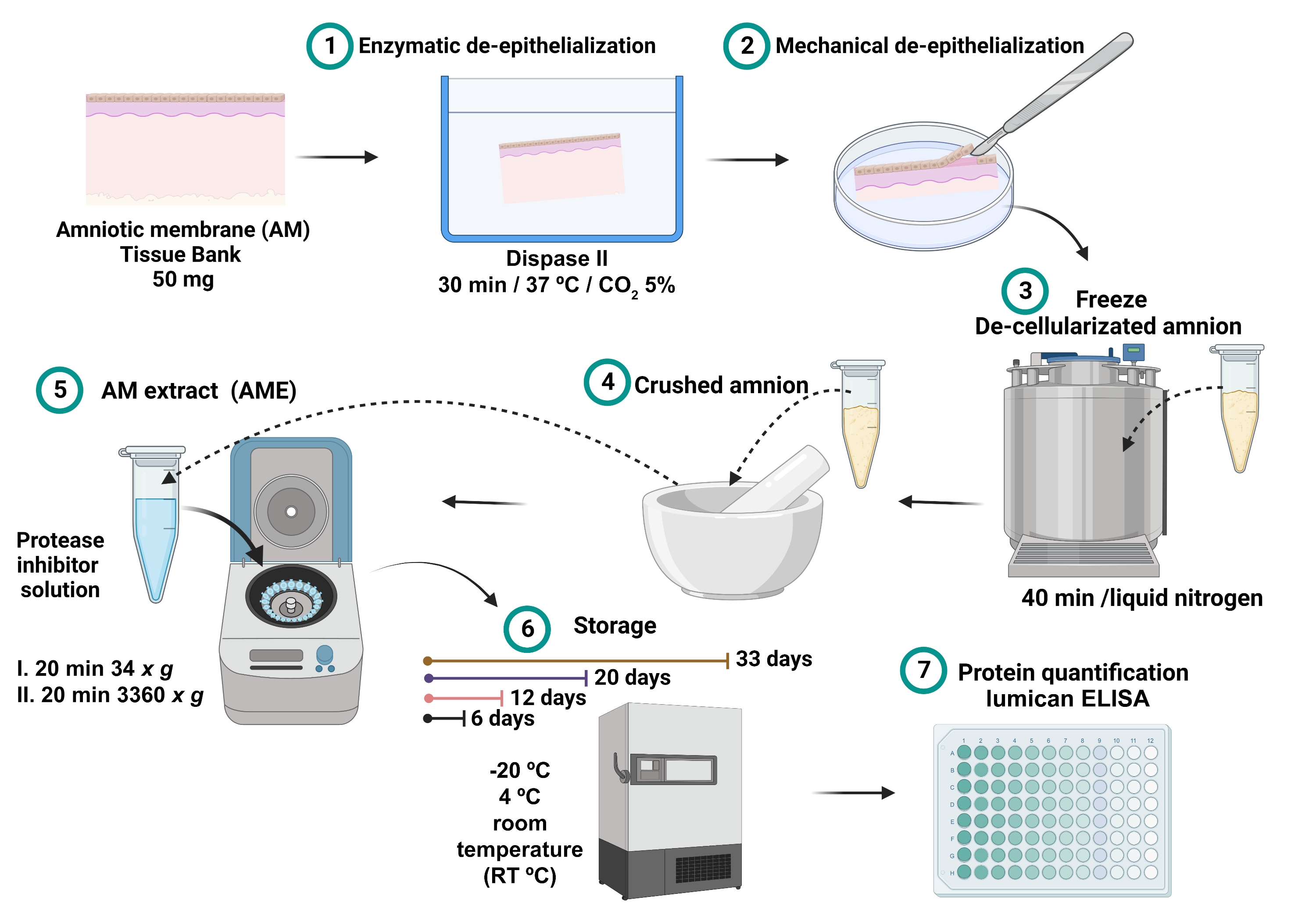
Figure 1: Process of AME preparation and lumican concentration measurement. 100 mg of AM were incubated with dispase II at 37 °C for 30 min and mechanically de-epithelialized. The de-epithelialized AM was washed and immersed in liquid nitrogen for 40 min, and then crushed until a fine powder was obtained, which was solubilized with 2.5 mL of saline buffer with protease inhibitors and centrifuged. The supernatant was collected and stored at -20 °C, 4 °C and RT for 6, 12, 20, and 32 days until total protein and lumican quantitation. Please click here to view a larger version of this figure.
2. AME protein quantification
NOTE: The quantification of total protein in the AME must be carried out immediately after obtention. Quantify proteins using Lowry protein assay and follow the manufacturer's instructions (see Table of Materials). It is recommended that all standards and samples be assayed in triplicate.
- Pipette 40 µL of each sample of AME into a 96-well microplate.
- Prepare a standard curve into the same microplate using bovine serum albumin (BSA) standard for a final BSA concentration of 0-1,500 µg/mL (0, 1, 5, 25, 125, 250, 500, 750, 1,000 and 1,500 µg/mL).
- Pipette 200 µL of the modified Lowry reagent to each well. Immediately mix it on a plate mixer for 30 s.
- Cover the microplate with aluminum foil and incubate it at RT for 10 min.
- Pipette 20 µL of 1x Folin-Ciocalteu reagent to each well. Immediately mix it on a plate mixer for 30 s.
NOTE: To prepare 1x Folin-Ciocalteu reagent, dilute 2x (2N) reagent 1:1 with ultrapure water. Prepare 1x Folin-Ciocalteu reagent on the same day of use as the diluted reagent is unstable. - Cover the microplate from light with aluminum foil and incubate it at RT for 30 min.
- Measure the absorbance of samples at 660 nm in an ELISA plate spectrometer (see Table of Materials).
NOTE: Color can be measured at wavelengths between 650 nm and 750 nm. - Average the 660 nm absorbance value of the standard blank samples and subtract it from other 660 nm values of standard and unknown samples.
- Measure the absorbance with an ELISA plate spectrometer in an endpoint mode with low shaking for 10 s.
- Use the standard curve to determine the protein concentration of each unknown sample.
- For the calculation of protein, determine the concentration from a lineal regression graph using the values of absorbance on the Y axis against the concentrations in mg/mL on the X axis of each standard BSA curve.
- Obtain the equation of linear regression and r value to calculate the protein concentration.
NOTE: Results are expressed as normalized relative concentration values of total protein relative to mg of AM (µg/mL protein/mg AM tissue).
- Obtain the equation of linear regression and r value to calculate the protein concentration.
3. Quantification of Lumican in AME
NOTE: The concentration of lumican must be measured in the AME stored at different storage conditions and time periods. Quantify lumican using sandwich ELISA and follow the manufacturer's instructions. It is recommended that all standards and samples be assayed in duplicate.
- Dilute the human lumican capture antibody (see Table of Materials) to the employed concentration in phosphate-buffered saline (PBS).
NOTE: The capture antibody vial contains 120 µg of antibody. After reconstitution with 0.5 mL of PBS, dilute the capture antibody at a working solution of 2 µg/mL.- Instantly pipette 100 µL per well of the diluted capture antibody to a 96-well microplate. Enclose the plate and incubate it overnight at RT.
- Aspirate each well and wash it by pipetting with 300 µL of wash buffer: 0.05% polyoxyethylene sorbitan monolaurate 20 in PBS, pH 7.2-7.4 (see Table of Materials) using a multi-channel pipettor. Repeat three times.
NOTE: After the last wash, remove any remaining wash buffer by everting the plate and gently tap it against paper towels. - Block plates by adding 300 µL of reagent diluent: 1% BSA in PBS, pH 7.2-7.4, 0.2 µm filtered (see Table of Materials) to each well. Incubate at RT for 1 h.
- Repeat step 2.
- Prepare a standard curve into a 96-well microplate using two-fold serial dilutions from 0-8,000 pg/mL for final concentrations of 125, 250, 500, 1,000, 2,000, 4,000 and 8,000 pg/mL. The ELISA lumican kit contains a recombinant lumican standard of 75 ng (see Table of Materials).
- Add 100 µL of samples and the standard curve in the capture antibody-coated 96-well microplate.
- Cover the microplate and incubate for 2 h at RT with low agitation in a compact rocker maintaining the velocity between 2-3 rpm.
- Repeat step 2.
- Add 100 µL of the biotinylated-detection antibody (see Table of Materials) to each well. Cover from light and incubate 2 h at RT with low agitation in a compact rocker maintaining the velocity between 2-3 rpm.
NOTE: The biotinylated-detection antibody vial contains 24 µg of antibody. After reconstitution with 1.0 mL of reagent diluent, dilute the biotinylated-detection antibody at a working solution of 400 ng/mL. - Repeat step 2.
- Add 100 µL of the working dilution of streptavidin-horseradish peroxidase (HRP, see Table of Materials) to each well. Cover the microplate from light and incubate for 20 min at RT.
NOTE: The reactive streptavidin-HRP was 40-fold concentrated. The work solution 1x of streptavidin- HRP was made with reagent diluent.
NOTE: Avoid placing the plate in direct light. - Repeat step 2.
- Finally, add 100 µL of substrate tetramethylbenzidine (TMB, see Table of Materials) solution to each well.
NOTE: Prepare TMB solution with an equal volume of stabilized hydrogen peroxide 30% solution provided in the kit.
NOTE: Prepare the solution immediately before use and maintain it at room temperature. - Incubate for 30 min at RT in a dark place.
NOTE: Avoid placing the plate in direct light. Do not aspirate the TMB solution as no further washing is needed. - Add 50 µL of 1N H2SO4 stop solution to stop the colorimetric reaction. Gently tap the plate to ensure thorough mixing.
- Immediately determine the absorbance of each well using a microplate reader set to 450 nm in an ELISA plate spectrometer.
- Measure the absorbance with an ELISA plate spectrometer in an endpoint mode with low shaking for 10 s.
- Average the 450 nm absorbance value of the standard blank samples and subtract it from other 450 nm values of standard and unknown samples.
- Use the standard curve to determine the lumican concentration of each unknown sample.
- For the calculation of lumican concentration, make a lineal regression graph using the values of absorbance on the Y axis against the concentrations in pg/mL on the X axis of each standard lumican curve.
- Obtain the equation of linear regression and r value to calculate the lumican concentration.
NOTE: The concentration of lumican was normalized with respect to the mg of tissue extracted. Results are expressed as normalized relative concentration values of lumican to mg AM (ng/mL lumican/mg AM tissue).
- Obtain the equation of linear regression and r value to calculate the lumican concentration.
Access restricted. Please log in or start a trial to view this content.
Results
Results are reported as the mean value ± standard deviation (SD). Student's t-tests and analysis of variance (ANOVA) were performed. P-values < 0.05 was considered statistically significant. Statistical analysis was performed using statistics software (see Table of Materials).
The total protein quantity in the AME was affected by time and storage conditions. The basal protein concentration was similar among all AME; the range of total protein was...
Access restricted. Please log in or start a trial to view this content.
Discussion
In this study, the presence of lumican was analyzed in the AME and its direct correlation with its stability under different storage conditions. Interestingly, when the total protein concentration in AME was quantified, protein concentration increased after storage. Evidence suggests three mechanisms that could change protein concentration in frozen storage: cold denaturation, the frozen concentration of solutes, and ice-induced partial unfolding of protein structure19. The freezing process could ...
Access restricted. Please log in or start a trial to view this content.
Disclosures
The study was funded by the Support Program for Research and Technological Innovation Projects of the Universidad Nacional Autonoma de Mexico (Grant No. PAPIIT IN203821), and Ministry of Education, Science, Technology and Innovation (Grant No. SECTEI 250/2019).
Acknowledgements
The authors have no competing financial interests.
Access restricted. Please log in or start a trial to view this content.
Materials
| Name | Company | Catalog Number | Comments |
| 1 N H2SO4 stop solution | R&D Systems | DY994 | |
| 100 μL micropipette | Eppendorf | ||
| 1000 μL micropipette | Eppendorf | ||
| 15 mm Petri dish | Symlaboratorios | ||
| 18 G Needle (1.2 mm x 40 mm) | BD Becton Dickinson | 305211 | |
| 2 mL microcentrifuge tube | Eppendorf | Z606340 | |
| 20 mL plastic syringe | BD Becton Dickinson | 302562 | |
| 20 μL micropipette | Eppendorf | ||
| 20-200 μL micropipette | Eppendorf | ||
| 5 mL microcentrifuge tube | Eppendorf | 30119401 | |
| 96-well microplate | SARSTEDT | 821581 | |
| Aluminum foil | N/A | N/A | |
| Amniotic membrane | Instituto de Oftalmologia Conde de Valenciana Amnion Bank | 100 mg | |
| Balanced salt solution | Bausch + Lomb | BSS-403802 | |
| Beaker | N/A | N/A | |
| BioRender | BioRender | figures design | |
| Compact Rocker | BioRad | 970822DD | Mod. 5202SD-BIO |
| complete, EDTA-free, Protease inhibitor cocktail tablets | Roche | 11 873 580 001 | Protease Inhibitor |
| Daiggner vortex Genie 2 | A.Daigger & Co. , INC | 22220A | |
| Dispase II | Gibco | 17105-041 | |
| ELISA plate spectrometer | Thermo Labsystems | 35401106 | Multiscan |
| Freezer | |||
| GraphPad Prism | GraphPad Software, Inc | version 9 | statistical analysis and graphic program |
| Human lumican DuoSet ELISA kit | R&D Systems | DY2846-05 | includes human Lumican capture antibody |
| Incubator | Forma Scientific | 3326 S/N 36481-7002 | |
| Inverted light Microscope | Olympus | 6A13921 | to confirm de-epithelialization Mod.CK2 |
| Laminar flow hood | Forma Scientific | 14753-567 | Mod.1184 |
| Liquid nitrogen | N/A | N/A | |
| Mortar | N/A | N/A | |
| Multi-channel pipettor | Eppendorf | ||
| Nitrogen Tank | Thermo Scientific | Mod. Biocan 20 | |
| Paper towels | N/A | N/A | |
| Phosphate-buffered saline | R&D Systems | DY006 | |
| Pierce Modified Lowry Protein Assay Kit | Thermo Scientific | 23240 | |
| Plate sealers | R&D Systems | DY992 | |
| Reagent diluent | R&D Systems | DY995 | 1% BSA in PBS, pH 7.2-7.4, 0.2 μm filtered |
| Refrigerated centrifuge | centurion scientific Ltd | 15877 | Mod. K2015R |
| Rubber policeman cell scraper | NEST | 710001 | for mechanical de-epithelialization |
| Scalpel knife | Braun | BB521 | No. 10 or 21 |
| Streptavidin-HRP 40-fold concentrated | R&D Systems | part 893975 | |
| Substrate tetramethylbenzidine (TMB) solution | R&D Systems | DY999 | |
| Toothed tweezers | Invent Germany | 6b | inox |
| Ultrapure water | PISA | ||
| Wash buffer | R&D Systems | WA126 | 0.05% Tween 20 in PBS, pH 7.2-7.4 |
References
- Jirsova, K., Jones, G. Amniotic membrane in ophthalmology: properties, preparation, storage and indications for grafting-a review. Cell and Tissue Banking. 18 (2), 193-204 (2017).
- Witherel, C., Yu, T., Concannon, M., Dampier, W., Spiller, K. Immunomodulatory effects of human cryopreserved viable amniotic membrane in a pro-inflammatory environment in vitro. Cellular and Molecular Bioengineering. 10 (5), 451-462 (2017).
- Ruiz-Cañada, C., et al. Amniotic membrane stimulates cell migration by modulating transforming growth factor-β signalling. Journal of Tissue Engineering and Regenerative Medicine. 12 (3), 808-820 (2017).
- Yeh, L., et al. Soluble lumican glycoprotein purified from human amniotic membrane promotes corneal epithelial wound healing. Investigative Opthalmology & Visual Science. 46 (2), 479(2005).
- Navas, A., et al. Anti-Inflammatory and anti-fibrotic effects of human amniotic membrane mesenchymal stem cells and their potential in corneal repair. Stem Cells Translational Medicine. 7 (12), 906-917 (2018).
- Magaña-Guerrero, F., Domínguez-López, A., Martínez-Aboytes, P., Buentello-Volante, B., Garfias, Y. Human amniotic membrane mesenchymal stem cells inhibit neutrophil extracellular traps through TSG-6. Scientific Reports. 7, 12426(2017).
- Garfias, Y., Zaga-Clavellina, V., Vadillo-Ortega, F., Osorio, M., Jimenez-Martinez, M. Amniotic membrane is an immunosuppressor of peripheral blood mononuclear cells. Immunological Investigations. 40 (2), 183-196 (2010).
- Koob, T., et al. Biological properties of dehydrated human amnion/chorion composite graft: implications for chronic wound healing. International Wound Journal. 10 (5), 493-500 (2013).
- Miyagi, H., Thomasy, S., Russell, P., Murphy, C. The role of hepatocyte growth factor in corneal wound healing. Experimental Eye Research. 166, 49-55 (2018).
- Chen, S., Mienaltowski, M., Birk, D. Regulation of corneal stroma extracellular matrix assembly. Experimental Eye Research. 133, 69-80 (2015).
- Karamanou, K., Perrot, G., Maquart, F., Brézillon, S. Lumican as a multivalent effector in wound healing. Advanced Drug Delivery Reviews. 129, 344-351 (2018).
- Theocharis, A., et al. Cell-matrix interactions: focus on proteoglycan-proteinase interplay and pharmacological targeting in cancer. FEBS Journal. 281 (22), 5023-5042 (2014).
- Saika, S., et al. Role of lumican in the corneal epithelium during wound healing. Journal of Biological Chemistry. 275 (4), 2607-2612 (2000).
- Domínguez-López, A., et al. Amniotic membrane conditioned medium (AMCM) reduces inflammatory response on human limbal myofibroblast, and the potential role of lumican. Molecular Vision. 27, 370-383 (2021).
- Vij, N., Roberts, L., Joyce, S., Chakravarti, S. Lumican regulates corneal inflammatory responses by modulating Fas-Fas Ligand signaling. Investigative Opthalmology & Visual Science. 46 (1), 88(2005).
- Mahbod, M., et al. Amniotic membrane extract preparation: What is the best method. Journal of Ophthalmic and Vision Research. 9 (3), 314-319 (2014).
- Chávez-García, C., et al. Ophthalmic indications of amniotic membrane transplantation in Mexico: an eight years Amniotic Membrane Bank experience. Cell and Tissue Banking. 17 (2), 261-268 (2015).
- Stenn, K. S., Link, R., Moellmann, G., Madri, J., Kuklinska, E. Dispase, a neutral protease from Bacillus polymyxa, is a powerful fibronectinase and type IV collagenase. Journal of Investigative Dermatology. 93 (2), 287-290 (1989).
- Bhatnagar, B. S., Bogner, R. H., Pikal, M. J. Protein stability during freezing: separation of stresses and mechanisms of protein stabilization. Pharmaceutical Development and Technology. 12 (5), 505-523 (2007).
- McClain, A. K., McCarrel, T. M. The effect of four different freezing conditions and time in frozen storage on the concentration of commonly measured growth factors and enzymes in equine platelet-rich plasma over six months. BMC Veterinary Research. 15 (1), 292(2019).
- Tamhane, A., et al. Evaluation of amniotic membrane transplantation as an adjunct to medical therapy as compared with medical therapy alone in acute ocular burns. Ophthalmology. 112 (11), 1963-1969 (2005).
- Shtein, R., et al. Autologous serum-based eye drops for treatment of ocular surface disease. Ophthalmology. 127 (1), 128-133 (2020).
- Shahriari, H., Tokhmehchi, F., Reza, M., Hashemi, N. Comparison of the effect of amniotic membrane suspension and autologous serum on alkaline corneal epithelial wound healing in the rabbit model. Cornea. 27 (10), 1148-1150 (2008).
- Schuerch, K., Baeriswyl, A., Frueh, B., Tappeiner, C. Efficacy of amniotic membrane transplantation for the treatment of corneal ulcers. Cornea. 39 (4), 479-483 (2019).
- Chen, H., et al. Amniotic membrane transplantation for persistent corneal ulcers and perforations in acute fungal keratitis. Cornea. 25 (5), 564-572 (2006).
- Guo, Q., et al. A comparison of the effectiveness between amniotic membrane homogenate and transplanted amniotic membrane in healing corneal damage in a rabbit model. Acta Ophthalmologica. 89 (4), 315-319 (2011).
- Sabater, A., Perez, V. Amniotic membrane use for management of corneal limbal stem cell deficiency. Current Opinion in Ophthalmology. 28 (4), 363-369 (2017).
- Ahmad, T., et al. Autolysis of bovine skin, its endogenous proteases, protease inhibitors and their effects on quality characteristics of extracted gelatin. Food Chemistry. 265, 1-8 (2018).
- Mullegama, S. V., et al. Nucleic acid extraction from human biological samples. Methods in Molecular Biology. 1897, 359-383 (2019).
- Skog, M., et al. The effect of enzymatic digestion on cultured epithelial autografts. Cell Transplantation. 28 (5), 638-644 (2019).
Access restricted. Please log in or start a trial to view this content.
Reprints and Permissions
Request permission to reuse the text or figures of this JoVE article
Request PermissionThis article has been published
Video Coming Soon
Copyright © 2025 MyJoVE Corporation. All rights reserved