A subscription to JoVE is required to view this content. Sign in or start your free trial.
Electroencephalography Network Indices as Biomarkers of Upper Limb Impairment in Chronic Stroke
* These authors contributed equally
In This Article
Summary
The experimental protocol demonstrates the paradigm for acquiring and analyzing electroencephalography (EEG) signals during upper limb movement in individuals with stroke. The alteration of the functional network of low-beta EEG frequency bands was observed during the movement of the impaired upper limb and was associated with the degree of motor impairment.
Abstract
Alteration of electroencephalography (EEG) signals during task-specific movement of the impaired limb has been reported as a potential biomarker for the severity of motor impairment and for the prediction of motor recovery in individuals with stroke. When implementing EEG experiments, detailed paradigms and well-organized experiment protocols are required to obtain robust and interpretable results. In this protocol, we illustrate a task-specific paradigm with upper limb movement and methods and techniques needed for the acquisition and analysis of EEG data. The paradigm consists of 1 min of rest followed by 10 trials comprising alternating 5 s and 3 s of resting and task (hand extension)-states, respectively, over 4 sessions. EEG signals were acquired using 32 Ag/AgCl scalp electrodes at a sampling rate of 1,000 Hz. Event-related spectral perturbation analysis associated with limb movement and functional network analyses at the global level in the low-beta (12-20 Hz) frequency band were performed. Representative results showed an alteration of the functional network of low-beta EEG frequency bands during movement of the impaired upper limb, and the altered functional network was associated with the degree of motor impairment in chronic stroke patients. The results demonstrate the feasibility of the experimental paradigm in EEG measurements during upper limb movement in individuals with stroke. Further research using this paradigm is needed to determine the potential value of EEG signals as biomarkers of motor impairment and recovery.
Introduction
Upper limb motor impairment is one of the most common consequences of stroke and is related to limitations in activities of daily living1,2. Alpha (8-13 Hz) and beta (13-30 Hz) band rhythms are known to be closely associated with movements. In particular, studies have shown that altered neural activity in the alpha and lower beta (12-20 Hz) frequency bands during movement of an impaired limb is correlated with the degree of motor impairment in individuals with stroke3,4,5. Based on these findings, electroencephalograp....
Protocol
All experimental procedures were reviewed and approved by the Institutional Review Board of Seoul National University Bundang Hospital. For the experiments in this study, 34 participants with stroke were recruited. Signed informed consent was obtained from all participants. A signed informed consent was obtained from a legal representative if a participant met the criteria but could not sign the consent form because of disability.
1. Experimental setup
- Patient recruitment
- Perform the screening process using the following inclusion criteria:
Aged 18 to 85 years with the presence of impaired up....
- Perform the screening process using the following inclusion criteria:
Results
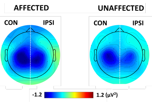
Figure 7 presents the topographical low-beta ERD maps of each hand-movement task. A significantly strong low-beta ERD was observed in the contralesional hemisphere compared with the ipsilesional hemisphere for both the affected and unaffected hand-movement tasks.

Figure 7: Mean topograp.......
Discussion
This study has introduced an EEG experiment for measuring upper limb movement-related neuronal activities in individuals with stroke. The experimental paradigm and methods of acquisition and analysis of EEG were applied to determine the ERD patterns in the ipsilesional and contralesional motor cortex.
The results of the ERSP maps (Figure 7) demonstrated the difference in the degree of neuronal activation when moving the impaired and unaffected hands. The results .......
Disclosures
MS, NJP, WSK, and HJH have a patent-pending entitled "Method for providing information related to motor impairment and devices used therefor", Number 10-2022-0007841.
Acknowledgements
This work was supported by the National Research Foundation of Korea (NRF) grant funded by the Korea government (MSIT) (No. NRF-2022R1A2C1006046), by the Original Technology Research Program for Brain Science through the National Research Foundation of Korea (NRF) funded by the Ministry of Education, Science and Technology (2019M3C7A1031995), by National Research Foundation of Korea (NRF) grant funded by the Korea government (MSIT) (No. NRF-2022R1A6A3A13053491), and by the MSIT(Ministry of Science and ICT), Korea, under the ITRC (Information Technology Research Center) support program (IITP-2023-RS-2023-00258971) supervised by the IITP (Institute for Information &....
Materials
| Name | Company | Catalog Number | Comments |
| actiCAP | Easycap, GmbH Ltd., Herrsching, Germany | CAC-32-SAMW-56 | Textile EEG cap platform to accommodate EEG electrodes |
| Brain Vision Recorder (Software) | Brain Products GmBH Ltd., Munich, Germany | - | Software used to record EEG signal |
| Curry 7 (Software) | Compumedics, Australia | - | Software used in preprocessing of EEG data |
| LiveAmp | Brain Products, GmbH Ltd., Gilching, Germany | LA-055606-0348 | EEG system (amplifier) used for the measurement |
| MATLAB R2019a (Software) | MathWorks Inc., Natick, MA, USA | - | Software used to run the experimental stimulus and analyze the EEG data |
| Recording PC | Lenovo Group Limited, Hong Kong, China | Model: X58K | Intel Core i7-7700HQ CPU@2.80 GHz, RAM 8 GB /EEG data recording using Brain Vision Recorder |
| Sensor&Trigger Extension(STE) | Brain Products GmBH Ltd., Munich, Germany | STE-055604-0162 | Adds physioloigcal signals to the EEG amplifier |
| Splitter box | Brain Products GmBH Ltd., Munich, Germany | BP-135-1600 | Connects Ag/AgCl electrodes to the EEG amplifier |
| Stimulation PC | Hansung Corporation, Seoul, Korea | Model: ThinkPad P71 | Intel Core i7-8750H CPU@2.20 GHz, RAM 8 GB Presenting stimulation screen using MATLAB |
| TriggerBox | Brain Products GmBH Ltd., Munich, Germany | BP-245-1010 | Receives trigger signal from PC and relay it to EEG recording system |
References
- Faria-Fortini, I., Michaelsen, S. M., Cassiano, J. G., Teixeira-Salmela, L. F. Upper extremity function in stroke subjects: Relationships between the international classification of functioning, disability, and health domains. Journal of Hand Therapy. 24 (3), 257-265 (2011).
- Veerbeek, J. M., Kwakkel, G., van Wegen, E. E., Ket, J. C., Heymans, M.....
Reprints and Permissions
Request permission to reuse the text or figures of this JoVE article
Request PermissionThis article has been published
Video Coming Soon
Copyright © 2025 MyJoVE Corporation. All rights reserved