
Summary
Abstract
Introduction
Protocol
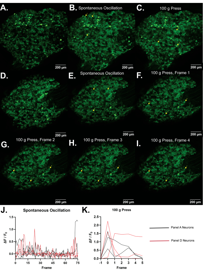
Representative Results
Discussion
Acknowledgements
Materials
References
Neuroscience
في الجسم الحي تصوير الكالسيوم للمجموعات العصبية في شبكات الخلايا العصبية الحسية الأولية في العقد الجذرية الظهرية السليمة
يصف هذا البروتوكول التعرض الجراحي لعقدة الجذر الظهري (DRG) متبوعا ب GCaMP3 (مؤشر Ca2+ المشفر وراثيا; بروتين الفلورسنت الأخضر - كالمودولين - M13 بروتين 3) تصوير Ca2+ للمجموعات العصبية باستخدام الفئران Pirt-GCaMP3 أثناء تطبيق مجموعة متنوعة من المحفزات على المخلب الخلفي المماثل.
يمكن استخدام تصوير Ca 2+ كوكيل للنشاط الخلوي ، بما في ذلك إمكانات الفعل وآليات الإشارات المختلفة التي تتضمن دخول Ca 2+ إلى السيتوبلازم أو إطلاق مخازن Ca2+ داخل الخلايا. يوفر تصوير Ca2+ القائم على Pirt-GCaMP3 للخلايا العصبية الحسية الأولية لعقدة الجذر الظهرية (DRG) في الفئران ميزة القياس المتزامن لعدد كبير من الخلايا. يمكن مراقبة ما يصل إلى 1800 خلية عصبية ، مما يسمح بدراسة الشبكات العصبية والعمليات الحسية الجسدية كمجموعة في سياقها الفسيولوجي الطبيعي على مستوى السكان في الجسم الحي. يسمح العدد الكبير من الخلايا العصبية التي تتم مراقبتها باكتشاف أنماط النشاط التي يصعب اكتشافها باستخدام طرق أخرى. يمكن تطبيق المحفزات على مؤخرة الفأر ، مما يسمح بدراسة التأثيرات المباشرة للمنبهات على مجموعة الخلايا العصبية DRG. يشير عدد الخلايا العصبية التي تنتج عابرات Ca 2+ بالإضافة إلى سعة العابرين Ca2+ إلى الحساسية لطرائق حسية محددة. يوفر قطر الخلايا العصبية دليلا على أنواع الألياف المنشطة (الميكانيكي غير الضار مقابل ألياف الألم الضارة ، ألياف Aβ و Aδ و C). يمكن تسمية الخلايا العصبية التي تعبر عن مستقبلات محددة وراثيا باستخدام td-Tomato و Cre recombinases المحددة مع Pirt-GCaMP. لذلك ، يوفر تصوير Pirt-GCaMP3 Ca2+ ل DRG أداة ونموذجا قويا لتحليل طرائق حسية محددة وأنواع فرعية من الخلايا العصبية تعمل كمجموعة على المستوى السكاني لدراسة الألم والحكة واللمس والإشارات الحسية الجسدية الأخرى.
الخلايا العصبية الحسية الأولية تعصب الجلد مباشرة وتحمل المعلومات الحسية الجسدية مرة أخرى إلى الجهاز العصبي المركزي 1,2. العقد الجذرية الظهرية (DRGs) هي مجموعات جسم الخلية من 10000-15000 خلية عصبية حسية أولية 3,4. تقدم الخلايا العصبية DRG أحجاما متنوعة ومستويات الميالين وأنماط التعبير الجيني والمستقبلات. تشمل الخلايا العصبية ذات القطر الأصغر الخلايا العصبية المستشعرة للألم وتستجيب الخلايا العصبية ذات القطر الأكبر عادة للمنبهات الميكانيكية غير المؤلمة 5,6. يمكن للاضطرابات في الخلايا العصبية الحسي....
تم تنفيذ جميع الإجراءات الموضحة هنا وفقا لبروتوكول معتمد من قبل اللجنة المؤسسية لرعاية واستخدام الحيوان التابعة لمركز العلوم الصحية بجامعة تكساس في سان أنطونيو.
ملاحظة: بمجرد البدء ، يجب إكمال جراحة الحيوانات (الخطوة 1) والتصوير (الخطوة 2) بطريقة مستمرة. قد يتم إجراء تحليل ا?.......
الألم المستمر موجود في مجموعة واسعة من الاضطرابات ، مما يضعف و / أو يقلل من نوعية الحياة لحوالي 8٪ من الناس29. تكتشف الخلايا العصبية الحسية الأولية المنبهات الضارة على الجلد ، وتساهم اللدونة في الألم المستمر8. في حين يمكن دراسة الخلايا العصبية في زراعة الخلايا والن?.......
| Name | Company | Catalog Number | Comments |
| Anased Injection (Xylazine) | Covetrus, Akorn | 33197 | |
| C Epiplan-Apochromat 10x/0.4 DIC | Cal Zeiss | 422642-9900-000 | |
| Cotton Tipped Applicators | McKesson | 24-106-1S | |
| Curved Hemostat | Fine Science Tools | 13007-12 | |
| DC Temperature Controller | FHC | 40-90-8D | |
| DC Temperature Controller Heating Pad | FHC | 40-90-2-05 | |
| Dumont Ceramic Coated Forceps | Fine Science Tools | 11252-50 | |
| FHC DC Temperature Controller | FHC | 40-90-8D | |
| Fluriso (Isoflurane) | MWI Animal Health, Piramal Group | 501017 | |
| Friedman-Pearson Rongeurs | Fine Science Tools | 16221-14 | |
| GelFoam | Pfizer | 09-0353-01 | |
| Ketaset (Ketamine) | Zoetis | KET-00002R2 | |
| Luminescent Green Stage Tape | JSITON/ Amazon | B803YW8ZWL | |
| Matrx VIP 3000 Isoflurane Vaporizer | Midmark | 91305430 | |
| Micro dissecting scissors | Roboz | RS-5882 | |
| Micro dissecting spring scissors | Fine Science Tools | 15023-10 | |
| Micro dissecting spring scissors | Roboz | RS-5677 | |
| Mini Rectal Thermistor Probe | FHC | 40-90-5D-02 | |
| Operating scissors | Roboz | RS-6812 | |
| Pirt-GCaMP3 C57BL/6J mice | Johns Hopkins University | N/A | Either sex can be imaged equally well. Mice should be at least 8 weeks old due to weak or intermittent Pirt promoter expression in younger mice. |
| SMALGO small animal algometer | Bioseb In vivo Research Instruments | BIO-SMALGO | |
| Stereotaxic frame | Kopf Model 923-B | 923-B | |
| td-Tomato C57BL/6J mice | Jackson Laboratory | 7909 | |
| Top Plate, 6 in x 10 in | Newport | 290-TP | |
| TrpV1-Cre C57BL/6J mice | Jackson Laboratory | 17769 | |
| Zeiss LSM 800 confocal microscope | Cal Zeiss | LSM800 | |
| Zeiss Zen 2.6 Blue Edition Software | Cal Zeiss | Zen (Blue Edition) 2.6 |
- Rivero-Melián, C., Grant, G. Distribution of lumbar dorsal root fibers in the lower thoracic and lumbosacral spinal cord of the rat studied with choleragenoid horseradish peroxidase conjugate. The Journal of Comparative Neurology. 299 (4), 470-481 (1990).
- Wessels, W. J., Marani, E. A rostrocaudal somatotopic organization in the brachial dorsal root ganglia of neonatal rats. Clinical Neurology and Neurosurgery. 95, 3-11 (1993).
- Schmalbruch, H. The number of neurons in dorsal root ganglia L4-L6 of the rat. The Anatomical Record. 219 (3), 315-322 (1987).
- Sørensen, B., Tandrup, T., Koltzenburg, M., Jakobsen, J. No further loss of dorsal root ganglion cells after axotomy in p75 neurotrophin receptor knockout mice. The Journal of Comparative Neurology. 459 (3), 242-250 (2003).
- Basbaum, A. I., Woolf, C. J. Pain. Current Biology. 9 (12), 429-431 (1999).
- Liu, Y., Ma, Q. Generation of somatic sensory neuron diversity and implications on sensory coding. Current Opinion in Neurobiology. 21 (1), 52-60 (2011).
- Basbaum, A. I., Bautista, D. M., Scherrer, G., Julius, D. Cellular and molecular mechanisms of pain. Cell. 139 (2), 267-284 (2009).
- Stucky, C. L., Mikesell, A. R. Cutaneous pain in disorders affecting peripheral nerves. Neuroscience Letters. 765, 136233 (2021).
- Iseppon, F., Linley, J. E., Wood, J. N. Calcium imaging for analgesic drug discovery. Neurobiology of Pain. 11, 100083 (2022).
- Chen, Z., et al. Adjacent intact nociceptive neurons drive the acute outburst of pain following peripheral axotomy. Scientific Reports. 9 (1), 7651 (2019).
- Chisholm, K. I., Khovanov, N., Lopes, D. M., La Russa, F., McMahon, S. B. Large scale in vivo recording of sensory neuron activity with GCaMP6. eNeuro. 5 (1), (2018).
- Emery, E. C., et al. In vivo characterization of distinct modality-specific subsets of somatosensory neurons using GCaMP. Science Advances. 2 (11), 1600990 (2016).
- Ishida, H., et al. In vivo calcium imaging visualizes incision-induced primary afferent sensitization and its amelioration by capsaicin pretreatment. The Journal of Neuroscience. 41 (41), 8494-8507 (2021).
- Kim, Y. S., et al. Coupled activation of primary sensory neurons contributes to chronic pain. Neuron. 91 (5), 1085-1096 (2016).
- MacDonald, D. I., et al. Silent cold-sensing neurons contribute to cold allodynia in neuropathic pain. Brain. 144 (6), 1711-1726 (2021).
- Wang, F., et al. Sensory afferents use different coding strategies for heat and cold. Cell Reports. 23 (7), 2001-2013 (2018).
- Kucharczyk, M. W., et al. The impact of bone cancer on the peripheral encoding of mechanical pressure stimuli. Pain. 161 (8), 1894-1905 (2020).
- Kim, A. Y., et al. a phosphoinositide-binding protein, functions as a regulatory subunit of TRPV1. Cell. 133 (3), 475-485 (2008).
- Kim, Y. S., et al. Central terminal sensitization of TRPV1 by descending serotonergic facilitation modulates chronic pain. Neuron. 81 (4), 873-887 (2014).
- Tian, L., et al. Imaging neural activity in worms, flies and mice with improved GCaMP calcium indicators. Nature Methods. 6 (12), 875-881 (2009).
- Thévenaz, P., Ruttimann, U. E., Unser, M. A pyramid approach to subpixel registration based on intensity. IEEE Transactions on Image Processing. 7 (1), 27-41 (1998).
- Mahadevan, A. S., et al. cytoNet: Spatiotemporal network analysis of cell communities. PLoS Computational Biology. 18 (6), 1009846 (2022).
- Barretto, R. P., et al. The neural representation of taste quality at the periphery. Nature. 517 (7534), 373-376 (2015).
- Leijon, S. C. M., et al. Oral thermosensing by murine trigeminal neurons: modulation by capsaicin, menthol and mustard oil. The Journal of Physiology. 597 (7), 2045-2061 (2019).
- Sekiguchi, K. J., et al. Imaging large-scale cellular activity in spinal cord of freely behaving mice. Nature Communications. 7, 11450 (2016).
- Wu, A., Dvoryanchikov, G., Pereira, E., Chaudhari, N., Roper, S. D. Breadth of tuning in taste afferent neurons varies with stimulus strength. Nature Communications. 6, 8171 (2015).
- Ran, C., Hoon, M. A., Chen, X. The coding of cutaneous temperature in the spinal cord. Nature Neuroscience. 19 (9), 1201-1209 (2016).
- Yarmolinsky, D. A., et al. Coding and plasticity in the mammalian thermosensory system. Neuron. 92 (5), 1079-1092 (2016).
- Torrance, N., Smith, B. H., Bennett, M. I., Lee, A. J. The epidemiology of chronic pain of predominantly neuropathic origin. Results from a general population survey. The Journal of Pain. 7 (4), 281-289 (2006).
- Shannonhouse, J., et al. Meclizine and metabotropic glutamate receptor agonists attenuate severe pain and Ca(2+) activity of primary sensory neurons in chemotherapy-induced peripheral neuropathy. The Journal of Neuroscience. 42 (31), 6020-6037 (2022).
- Luiz, A. P., et al. Cold sensing by Na(V)1.8-positive and Na(V)1.8-negative sensory neurons. Proceedings of the National Academy of Sciences of the United States of America. 116 (9), 3811-3816 (2019).
- Hartung, J. E., Gold, M. S. GCaMP as an indirect measure of electrical activity in rat trigeminal ganglion neurons. Cell Calcium. 89, 102225 (2020).
- Chung, M. K., Wang, S., Oh, S. L., Kim, Y. S. Acute and chronic pain from facial skin and oral mucosa: Unique neurobiology and challenging treatment. International Journal of Molecular Sciences. 22 (11), 5810 (2021).
- Chan, S. L., Mayne, M., Holden, C. P., Geiger, J. D., Mattson, M. P. Presenilin-1 mutations increase levels of ryanodine receptors and calcium release in PC12 cells and cortical neurons. The Journal of Biological Chemistry. 275 (24), 18195-18200 (2000).
- Sierra, D. A., Popov, S., Wilkie, T. M. Regulators of G-protein signaling in receptor complexes. Trends in Cardiovascular Medicine. 10 (6), 263-268 (2000).
- Yoshihara, K., et al. Astrocytic Ca(2+) responses in the spinal dorsal horn by noxious stimuli to the skin. Journal of Pharmacological Sciences. 137 (1), 101-104 (2018).
- Tan, C. H., McNaughton, P. A. The TRPM2 ion channel is required for sensitivity to warmth. Nature. 536 (7617), 460-463 (2016).
- Akemann, W., Mutoh, H., Perron, A., Rossier, J., Knöpfel, T. Imaging brain electric signals with genetically targeted voltage-sensitive fluorescent proteins. Nature Methods. 7 (8), 643-649 (2010).
- Gong, Y., et al. High-speed recording of neural spikes in awake mice and flies with a fluorescent voltage sensor. Science. 350 (6266), 1361-1366 (2015).
- Grewe, B. F., Langer, D., Kasper, H., Kampa, B. M., Helmchen, F. High-speed in vivo calcium imaging reveals neuronal network activity with near-millisecond precision. Nature Methods. 7 (5), 399-405 (2010).
- Harada, K., et al. Red fluorescent protein-based cAMP indicator applicable to optogenetics and in vivo imaging. Scientific Reports. 7 (1), 7351 (2017).
ABOUT JoVE
Copyright © 2024 MyJoVE Corporation. All rights reserved