
Summary
Abstract
Introduction
Protocol
Representative Results
Discussion
Acknowledgements
Materials
References
Neuroscience
Im lebenden Organismus Calcium-Bildgebung neuronaler Ensembles in Netzwerken primärer sensorischer Neurone in intakten dorsalen Wurzelganglien
Dieses Protokoll beschreibt die chirurgische Exposition des dorsalen Wurzelganglions (DRG) gefolgt von GCaMP3 (genetisch kodierter Ca2+ Indikator; Grün fluoreszierendes Protein-Calmodulin-M13 Protein 3) Ca2+ Bildgebung der neuronalen Ensembles mit Pirt-GCaMP3 Mäusen unter Anwendung einer Vielzahl von Stimuli auf die ipsilaterale Hinterpfote.
Die Ca 2+-Bildgebung kann als Proxy für die zelluläre Aktivität verwendet werden, einschließlich Aktionspotentialen und verschiedener Signalmechanismen, die den Eintritt von Ca 2+ in das Zytoplasma oder die Freisetzung intrazellulärer Ca 2+-Speicher beinhalten. Die Pirt-GCaMP3-basierte Ca2+ Bildgebung von primären sensorischen Neuronen des dorsalen Wurzelganglions (DRG) in Mäusen bietet den Vorteil der gleichzeitigen Messung einer großen Anzahl von Zellen. Bis zu 1.800 Neuronen können überwacht werden, so dass neuronale Netzwerke und somatosensorische Prozesse als Ensemble in ihrem normalen physiologischen Kontext auf Populationsebene in vivo untersucht werden können. Die große Anzahl der überwachten Neuronen ermöglicht die Erkennung von Aktivitätsmustern, die mit anderen Methoden nur schwer zu erkennen wären. Stimuli können auf die Hinterpfote der Maus angewendet werden, so dass die direkten Auswirkungen von Stimuli auf das DRG-Neuronenensemble untersucht werden können. Die Anzahl der Neuronen, die Ca 2+-Transienten produzieren, sowie die Amplitude der Ca2+-Transienten weisen auf eine Sensitivität gegenüber bestimmten sensorischen Modalitäten hin. Der Durchmesser der Neuronen gibt Aufschluss über aktivierte Fasertypen (nicht-schädliche Mechano- vs. schädliche Schmerzfasern, Aβ-, Aδ- und C-Fasern). Neurone, die spezifische Rezeptoren exprimieren, können genetisch mit td-Tomato und spezifischen Cre-Rekombinasen zusammen mit Pirt-GCaMP markiert werden. Daher bietet die Pirt-GCaMP3 Ca2+ Bildgebung von DRG ein leistungsfähiges Werkzeug und Modell für die Analyse spezifischer sensorischer Modalitäten und Neuronensubtypen, die als Ensemble auf Populationsebene fungieren, um Schmerz, Juckreiz, Berührung und andere somatosensorische Signale zu untersuchen.
Primäre sensorische Neuronen innervieren direkt die Haut und leiten somatosensorische Informationen zurück an das zentrale Nervensystem 1,2. Dorsale Wurzelganglien (DRGs) sind Zellkörpercluster von 10.000-15.000 primären sensorischen Neuronen 3,4. DRG-Neuronen weisen unterschiedliche Größe, Myelinisierungsgrade sowie Gen- und Rezeptorexpressionsmuster auf. Neuronen mit kleinerem Durchmesser umfassen schmerzempfindliche Neuronen, und Neuronen mit größerem Durchmesser reagieren typischerweise auf nicht-schmerzhafte mechanische Reize
Alle hier beschriebenen Verfahren wurden in Übereinstimmung mit einem Protokoll durchgeführt, das vom Institutional Animal Care and Use Committee des University of Texas Health Science Center in San Antonio genehmigt wurde.
HINWEIS: Einmal begonnen, müssen die Tierchirurgie (Schritt 1) und die Bildgebung (Schritt 2) kontinuierlich abgeschlossen werden. Die Datenanalyse (Schritt 3) kann zu einem späteren Zeitpunkt durchgeführt werden.
1. Operation und Sich.......
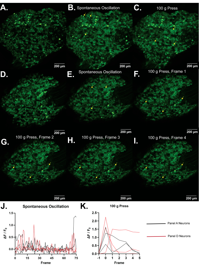
Abbildung 4: Repräsentative Bilder von L5 dorsalen Wurzelganglien von Pirt-GCaMP3 Mäusen. (A,D) Hochauflösende Einzelbildscans von L5 dorsalen Wurzelganglien von Pirt-GCaMP3 Mäusen werden gezeigt. (B,E) . Durchschnittliche Intensitätsprojektionen von 15 Bildern von Pirt-GCaMP3 L5 DRG-Ganglien a.......
Anhaltende Schmerzen treten bei einer Vielzahl von Erkrankungen auf und beeinträchtigen und/oder verringern die Lebensqualität von etwa 8 % der Menschen29. Primäre sensorische Neuronen erkennen schädliche Reize auf der Haut, und ihre Plastizität trägt zu anhaltenden Schmerzen bei8. Während Neuronen in Zellkulturen und Explantaten untersucht werden können, werden sie dadurch aus ihrem normalen physiologischen Kontext entfernt. Die chirurgische Exposition des DRG, gef.......
Diese Arbeit wurde durch die National Institutes of Health Grants R01DE026677 und R01DE031477 (an Y.S.K.), UTHSCSA Startup Fund (Y.S.K.) und einen Rising STAR Award der University of Texas System (Y.S.K.) unterstützt.
....| Name | Company | Catalog Number | Comments |
| Anased Injection (Xylazine) | Covetrus, Akorn | 33197 | |
| C Epiplan-Apochromat 10x/0.4 DIC | Cal Zeiss | 422642-9900-000 | |
| Cotton Tipped Applicators | McKesson | 24-106-1S | |
| Curved Hemostat | Fine Science Tools | 13007-12 | |
| DC Temperature Controller | FHC | 40-90-8D | |
| DC Temperature Controller Heating Pad | FHC | 40-90-2-05 | |
| Dumont Ceramic Coated Forceps | Fine Science Tools | 11252-50 | |
| FHC DC Temperature Controller | FHC | 40-90-8D | |
| Fluriso (Isoflurane) | MWI Animal Health, Piramal Group | 501017 | |
| Friedman-Pearson Rongeurs | Fine Science Tools | 16221-14 | |
| GelFoam | Pfizer | 09-0353-01 | |
| Ketaset (Ketamine) | Zoetis | KET-00002R2 | |
| Luminescent Green Stage Tape | JSITON/ Amazon | B803YW8ZWL | |
| Matrx VIP 3000 Isoflurane Vaporizer | Midmark | 91305430 | |
| Micro dissecting scissors | Roboz | RS-5882 | |
| Micro dissecting spring scissors | Fine Science Tools | 15023-10 | |
| Micro dissecting spring scissors | Roboz | RS-5677 | |
| Mini Rectal Thermistor Probe | FHC | 40-90-5D-02 | |
| Operating scissors | Roboz | RS-6812 | |
| Pirt-GCaMP3 C57BL/6J mice | Johns Hopkins University | N/A | Either sex can be imaged equally well. Mice should be at least 8 weeks old due to weak or intermittent Pirt promoter expression in younger mice. |
| SMALGO small animal algometer | Bioseb In vivo Research Instruments | BIO-SMALGO | |
| Stereotaxic frame | Kopf Model 923-B | 923-B | |
| td-Tomato C57BL/6J mice | Jackson Laboratory | 7909 | |
| Top Plate, 6 in x 10 in | Newport | 290-TP | |
| TrpV1-Cre C57BL/6J mice | Jackson Laboratory | 17769 | |
| Zeiss LSM 800 confocal microscope | Cal Zeiss | LSM800 | |
| Zeiss Zen 2.6 Blue Edition Software | Cal Zeiss | Zen (Blue Edition) 2.6 |
- Rivero-Melián, C., Grant, G. Distribution of lumbar dorsal root fibers in the lower thoracic and lumbosacral spinal cord of the rat studied with choleragenoid horseradish peroxidase conjugate. The Journal of Comparative Neurology. 299 (4), 470-481 (1990).
- Wessels, W. J., Marani, E. A rostrocaudal somatotopic organization in the brachial dorsal root ganglia of neonatal rats. Clinical Neurology and Neurosurgery. 95, 3-11 (1993).
- Schmalbruch, H. The number of neurons in dorsal root ganglia L4-L6 of the rat. The Anatomical Record. 219 (3), 315-322 (1987).
- Sørensen, B., Tandrup, T., Koltzenburg, M., Jakobsen, J. No further loss of dorsal root ganglion cells after axotomy in p75 neurotrophin receptor knockout mice. The Journal of Comparative Neurology. 459 (3), 242-250 (2003).
- Basbaum, A. I., Woolf, C. J. Pain. Current Biology. 9 (12), 429-431 (1999).
- Liu, Y., Ma, Q. Generation of somatic sensory neuron diversity and implications on sensory coding. Current Opinion in Neurobiology. 21 (1), 52-60 (2011).
- Basbaum, A. I., Bautista, D. M., Scherrer, G., Julius, D. Cellular and molecular mechanisms of pain. Cell. 139 (2), 267-284 (2009).
- Stucky, C. L., Mikesell, A. R. Cutaneous pain in disorders affecting peripheral nerves. Neuroscience Letters. 765, 136233 (2021).
- Iseppon, F., Linley, J. E., Wood, J. N. Calcium imaging for analgesic drug discovery. Neurobiology of Pain. 11, 100083 (2022).
- Chen, Z., et al. Adjacent intact nociceptive neurons drive the acute outburst of pain following peripheral axotomy. Scientific Reports. 9 (1), 7651 (2019).
- Chisholm, K. I., Khovanov, N., Lopes, D. M., La Russa, F., McMahon, S. B. Large scale in vivo recording of sensory neuron activity with GCaMP6. eNeuro. 5 (1), (2018).
- Emery, E. C., et al. In vivo characterization of distinct modality-specific subsets of somatosensory neurons using GCaMP. Science Advances. 2 (11), 1600990 (2016).
- Ishida, H., et al. In vivo calcium imaging visualizes incision-induced primary afferent sensitization and its amelioration by capsaicin pretreatment. The Journal of Neuroscience. 41 (41), 8494-8507 (2021).
- Kim, Y. S., et al. Coupled activation of primary sensory neurons contributes to chronic pain. Neuron. 91 (5), 1085-1096 (2016).
- MacDonald, D. I., et al. Silent cold-sensing neurons contribute to cold allodynia in neuropathic pain. Brain. 144 (6), 1711-1726 (2021).
- Wang, F., et al. Sensory afferents use different coding strategies for heat and cold. Cell Reports. 23 (7), 2001-2013 (2018).
- Kucharczyk, M. W., et al. The impact of bone cancer on the peripheral encoding of mechanical pressure stimuli. Pain. 161 (8), 1894-1905 (2020).
- Kim, A. Y., et al. a phosphoinositide-binding protein, functions as a regulatory subunit of TRPV1. Cell. 133 (3), 475-485 (2008).
- Kim, Y. S., et al. Central terminal sensitization of TRPV1 by descending serotonergic facilitation modulates chronic pain. Neuron. 81 (4), 873-887 (2014).
- Tian, L., et al. Imaging neural activity in worms, flies and mice with improved GCaMP calcium indicators. Nature Methods. 6 (12), 875-881 (2009).
- Thévenaz, P., Ruttimann, U. E., Unser, M. A pyramid approach to subpixel registration based on intensity. IEEE Transactions on Image Processing. 7 (1), 27-41 (1998).
- Mahadevan, A. S., et al. cytoNet: Spatiotemporal network analysis of cell communities. PLoS Computational Biology. 18 (6), 1009846 (2022).
- Barretto, R. P., et al. The neural representation of taste quality at the periphery. Nature. 517 (7534), 373-376 (2015).
- Leijon, S. C. M., et al. Oral thermosensing by murine trigeminal neurons: modulation by capsaicin, menthol and mustard oil. The Journal of Physiology. 597 (7), 2045-2061 (2019).
- Sekiguchi, K. J., et al. Imaging large-scale cellular activity in spinal cord of freely behaving mice. Nature Communications. 7, 11450 (2016).
- Wu, A., Dvoryanchikov, G., Pereira, E., Chaudhari, N., Roper, S. D. Breadth of tuning in taste afferent neurons varies with stimulus strength. Nature Communications. 6, 8171 (2015).
- Ran, C., Hoon, M. A., Chen, X. The coding of cutaneous temperature in the spinal cord. Nature Neuroscience. 19 (9), 1201-1209 (2016).
- Yarmolinsky, D. A., et al. Coding and plasticity in the mammalian thermosensory system. Neuron. 92 (5), 1079-1092 (2016).
- Torrance, N., Smith, B. H., Bennett, M. I., Lee, A. J. The epidemiology of chronic pain of predominantly neuropathic origin. Results from a general population survey. The Journal of Pain. 7 (4), 281-289 (2006).
- Shannonhouse, J., et al. Meclizine and metabotropic glutamate receptor agonists attenuate severe pain and Ca(2+) activity of primary sensory neurons in chemotherapy-induced peripheral neuropathy. The Journal of Neuroscience. 42 (31), 6020-6037 (2022).
- Luiz, A. P., et al. Cold sensing by Na(V)1.8-positive and Na(V)1.8-negative sensory neurons. Proceedings of the National Academy of Sciences of the United States of America. 116 (9), 3811-3816 (2019).
- Hartung, J. E., Gold, M. S. GCaMP as an indirect measure of electrical activity in rat trigeminal ganglion neurons. Cell Calcium. 89, 102225 (2020).
- Chung, M. K., Wang, S., Oh, S. L., Kim, Y. S. Acute and chronic pain from facial skin and oral mucosa: Unique neurobiology and challenging treatment. International Journal of Molecular Sciences. 22 (11), 5810 (2021).
- Chan, S. L., Mayne, M., Holden, C. P., Geiger, J. D., Mattson, M. P. Presenilin-1 mutations increase levels of ryanodine receptors and calcium release in PC12 cells and cortical neurons. The Journal of Biological Chemistry. 275 (24), 18195-18200 (2000).
- Sierra, D. A., Popov, S., Wilkie, T. M. Regulators of G-protein signaling in receptor complexes. Trends in Cardiovascular Medicine. 10 (6), 263-268 (2000).
- Yoshihara, K., et al. Astrocytic Ca(2+) responses in the spinal dorsal horn by noxious stimuli to the skin. Journal of Pharmacological Sciences. 137 (1), 101-104 (2018).
- Tan, C. H., McNaughton, P. A. The TRPM2 ion channel is required for sensitivity to warmth. Nature. 536 (7617), 460-463 (2016).
- Akemann, W., Mutoh, H., Perron, A., Rossier, J., Knöpfel, T. Imaging brain electric signals with genetically targeted voltage-sensitive fluorescent proteins. Nature Methods. 7 (8), 643-649 (2010).
- Gong, Y., et al. High-speed recording of neural spikes in awake mice and flies with a fluorescent voltage sensor. Science. 350 (6266), 1361-1366 (2015).
- Grewe, B. F., Langer, D., Kasper, H., Kampa, B. M., Helmchen, F. High-speed in vivo calcium imaging reveals neuronal network activity with near-millisecond precision. Nature Methods. 7 (5), 399-405 (2010).
- Harada, K., et al. Red fluorescent protein-based cAMP indicator applicable to optogenetics and in vivo imaging. Scientific Reports. 7 (1), 7351 (2017).
ABOUT JoVE
Copyright © 2024 MyJoVE Corporation. All rights reserved