
Summary
Abstract
Introduction
Protocol
Representative Results
Discussion
Acknowledgements
Materials
References
Neuroscience
In vivo Imaging del calcio di insiemi neuronali in reti di neuroni sensoriali primari nei gangli della radice dorsale intatti
Questo protocollo descrive l'esposizione chirurgica del ganglio della radice dorsale (DRG) seguito da GCaMP3 (indicatore Ca2+ codificato geneticamente; Green Fluorescent Protein-Calmodulin-M13 Protein 3) Ca2+ imaging degli insiemi neuronali utilizzando topi Pirt-GCaMP3 mentre si applica una varietà di stimoli alla zampa posteriore omolaterale.
L'imaging di Ca 2+ può essere utilizzato come proxy per l'attività cellulare, compresi i potenziali d'azione e vari meccanismi di segnalazione che coinvolgono l'ingresso di Ca 2+ nel citoplasma o il rilascio di riserve intracellulari di Ca 2+. L'imaging Ca2+ basato su Pirt-GCaMP3 dei neuroni sensoriali primari del ganglio della radice dorsale (DRG) nei topi offre il vantaggio della misurazione simultanea di un gran numero di cellule. È possibile monitorare fino a 1.800 neuroni, consentendo di studiare le reti neuronali e i processi somatosensoriali come un insieme nel loro normale contesto fisiologico a livello di popolazione in vivo. Il gran numero di neuroni monitorati consente di rilevare modelli di attività che sarebbero difficili da rilevare utilizzando altri metodi. Gli stimoli possono essere applicati alla zampa posteriore del topo, consentendo di studiare gli effetti diretti degli stimoli sull'insieme dei neuroni DRG. Il numero di neuroni che producono transitori Ca 2+ e l'ampiezza dei transitori Ca2+ indicano sensibilità a specifiche modalità sensoriali. Il diametro dei neuroni fornisce prove di tipi di fibre attivate (meccano non nocivo vs fibre del dolore nocivo, fibre Aβ, Aδ e C). I neuroni che esprimono recettori specifici possono essere geneticamente marcati con td-Tomato e specifiche ricombinasi Cre insieme a Pirt-GCaMP. Pertanto, l'imaging Pirt-GCaMP3 Ca2+ di DRG fornisce un potente strumento e modello per l'analisi di specifiche modalità sensoriali e sottotipi di neuroni che agiscono come un insieme a livello di popolazione per studiare dolore, prurito, tatto e altri segnali somatosensoriali.
I neuroni sensoriali primari innervano direttamente la pelle e trasportano le informazioni somatosensoriali al sistema nervoso centrale 1,2. I gangli delle radici dorsali (DRG) sono gruppi di corpi cellulari di 10.000-15.000 neuroni sensoriali primari 3,4. I neuroni DRG presentano diverse dimensioni, livelli di mielinizzazione e modelli di espressione genica e recettoriale. I neuroni di diametro più piccolo includono neuroni sensibili al dolore e neuroni di diametro maggiore rispondono tipicamente a stimoli meccanici non dolorosi
Tutte le procedure qui descritte sono state eseguite in conformità con un protocollo approvato dall'Institutional Animal Care and Use Committee dell'Università del Texas Health Science Center di San Antonio.
NOTA: Una volta iniziato, la chirurgia animale (fase 1) e l'imaging (fase 2) devono essere completati in modo continuo. L'analisi dei dati (fase 3) può essere eseguita successivamente.
1. Chirurgia e fissaggio dell'animale per l'imaging DRG L5 del lato .......
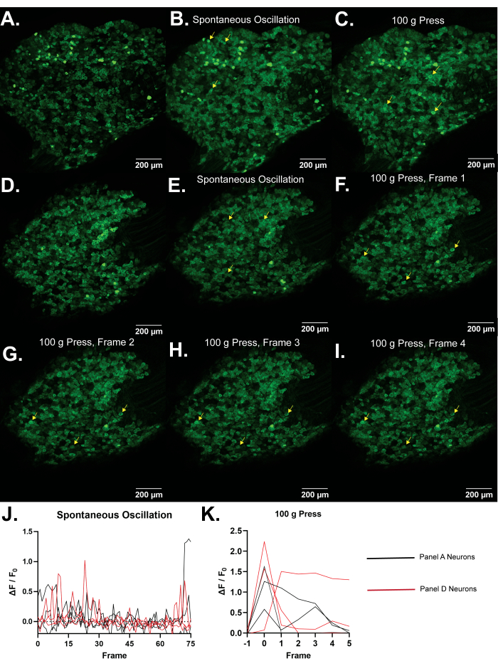
Figura 4: Immagini rappresentative dei gangli della radice dorsale L5 dei topi Pirt-GCaMP3. (A,D) Vengono mostrate scansioni ad alta risoluzione a fotogramma singolo dei gangli della radice dorsale L5 dei topi Pirt-GCaMP3. (B,E) . Proiezioni di intensità media di 15 fotogrammi di gangli Pirt-GCaMP3.......
Il dolore persistente è presente in una vasta gamma di disturbi, debilitanti e / o riducendo la qualità della vita per circa l'8% delle persone29. I neuroni sensoriali primari rilevano stimoli nocivi sulla pelle e la loro plasticità contribuisce al dolore persistente8. Mentre i neuroni possono essere studiati in coltura cellulare ed espianti, così facendo li rimuove dal loro normale contesto fisiologico. L'esposizione chirurgica del DRG, seguita dall'imaging Pirt-GCaMP3.......
Questo lavoro è stato supportato dal National Institutes of Health Grants R01DE026677 e R01DE031477 (a Y.S.K.), dal fondo di avvio UTHSCSA (Y.S.K.) e da un Rising STAR Award del sistema dell'Università del Texas (Y.S.K.).
....| Name | Company | Catalog Number | Comments |
| Anased Injection (Xylazine) | Covetrus, Akorn | 33197 | |
| C Epiplan-Apochromat 10x/0.4 DIC | Cal Zeiss | 422642-9900-000 | |
| Cotton Tipped Applicators | McKesson | 24-106-1S | |
| Curved Hemostat | Fine Science Tools | 13007-12 | |
| DC Temperature Controller | FHC | 40-90-8D | |
| DC Temperature Controller Heating Pad | FHC | 40-90-2-05 | |
| Dumont Ceramic Coated Forceps | Fine Science Tools | 11252-50 | |
| FHC DC Temperature Controller | FHC | 40-90-8D | |
| Fluriso (Isoflurane) | MWI Animal Health, Piramal Group | 501017 | |
| Friedman-Pearson Rongeurs | Fine Science Tools | 16221-14 | |
| GelFoam | Pfizer | 09-0353-01 | |
| Ketaset (Ketamine) | Zoetis | KET-00002R2 | |
| Luminescent Green Stage Tape | JSITON/ Amazon | B803YW8ZWL | |
| Matrx VIP 3000 Isoflurane Vaporizer | Midmark | 91305430 | |
| Micro dissecting scissors | Roboz | RS-5882 | |
| Micro dissecting spring scissors | Fine Science Tools | 15023-10 | |
| Micro dissecting spring scissors | Roboz | RS-5677 | |
| Mini Rectal Thermistor Probe | FHC | 40-90-5D-02 | |
| Operating scissors | Roboz | RS-6812 | |
| Pirt-GCaMP3 C57BL/6J mice | Johns Hopkins University | N/A | Either sex can be imaged equally well. Mice should be at least 8 weeks old due to weak or intermittent Pirt promoter expression in younger mice. |
| SMALGO small animal algometer | Bioseb In vivo Research Instruments | BIO-SMALGO | |
| Stereotaxic frame | Kopf Model 923-B | 923-B | |
| td-Tomato C57BL/6J mice | Jackson Laboratory | 7909 | |
| Top Plate, 6 in x 10 in | Newport | 290-TP | |
| TrpV1-Cre C57BL/6J mice | Jackson Laboratory | 17769 | |
| Zeiss LSM 800 confocal microscope | Cal Zeiss | LSM800 | |
| Zeiss Zen 2.6 Blue Edition Software | Cal Zeiss | Zen (Blue Edition) 2.6 |
- Rivero-Melián, C., Grant, G. Distribution of lumbar dorsal root fibers in the lower thoracic and lumbosacral spinal cord of the rat studied with choleragenoid horseradish peroxidase conjugate. The Journal of Comparative Neurology. 299 (4), 470-481 (1990).
- Wessels, W. J., Marani, E. A rostrocaudal somatotopic organization in the brachial dorsal root ganglia of neonatal rats. Clinical Neurology and Neurosurgery. 95, 3-11 (1993).
- Schmalbruch, H. The number of neurons in dorsal root ganglia L4-L6 of the rat. The Anatomical Record. 219 (3), 315-322 (1987).
- Sørensen, B., Tandrup, T., Koltzenburg, M., Jakobsen, J. No further loss of dorsal root ganglion cells after axotomy in p75 neurotrophin receptor knockout mice. The Journal of Comparative Neurology. 459 (3), 242-250 (2003).
- Basbaum, A. I., Woolf, C. J. Pain. Current Biology. 9 (12), 429-431 (1999).
- Liu, Y., Ma, Q. Generation of somatic sensory neuron diversity and implications on sensory coding. Current Opinion in Neurobiology. 21 (1), 52-60 (2011).
- Basbaum, A. I., Bautista, D. M., Scherrer, G., Julius, D. Cellular and molecular mechanisms of pain. Cell. 139 (2), 267-284 (2009).
- Stucky, C. L., Mikesell, A. R. Cutaneous pain in disorders affecting peripheral nerves. Neuroscience Letters. 765, 136233 (2021).
- Iseppon, F., Linley, J. E., Wood, J. N. Calcium imaging for analgesic drug discovery. Neurobiology of Pain. 11, 100083 (2022).
- Chen, Z., et al. Adjacent intact nociceptive neurons drive the acute outburst of pain following peripheral axotomy. Scientific Reports. 9 (1), 7651 (2019).
- Chisholm, K. I., Khovanov, N., Lopes, D. M., La Russa, F., McMahon, S. B. Large scale in vivo recording of sensory neuron activity with GCaMP6. eNeuro. 5 (1), (2018).
- Emery, E. C., et al. In vivo characterization of distinct modality-specific subsets of somatosensory neurons using GCaMP. Science Advances. 2 (11), 1600990 (2016).
- Ishida, H., et al. In vivo calcium imaging visualizes incision-induced primary afferent sensitization and its amelioration by capsaicin pretreatment. The Journal of Neuroscience. 41 (41), 8494-8507 (2021).
- Kim, Y. S., et al. Coupled activation of primary sensory neurons contributes to chronic pain. Neuron. 91 (5), 1085-1096 (2016).
- MacDonald, D. I., et al. Silent cold-sensing neurons contribute to cold allodynia in neuropathic pain. Brain. 144 (6), 1711-1726 (2021).
- Wang, F., et al. Sensory afferents use different coding strategies for heat and cold. Cell Reports. 23 (7), 2001-2013 (2018).
- Kucharczyk, M. W., et al. The impact of bone cancer on the peripheral encoding of mechanical pressure stimuli. Pain. 161 (8), 1894-1905 (2020).
- Kim, A. Y., et al. a phosphoinositide-binding protein, functions as a regulatory subunit of TRPV1. Cell. 133 (3), 475-485 (2008).
- Kim, Y. S., et al. Central terminal sensitization of TRPV1 by descending serotonergic facilitation modulates chronic pain. Neuron. 81 (4), 873-887 (2014).
- Tian, L., et al. Imaging neural activity in worms, flies and mice with improved GCaMP calcium indicators. Nature Methods. 6 (12), 875-881 (2009).
- Thévenaz, P., Ruttimann, U. E., Unser, M. A pyramid approach to subpixel registration based on intensity. IEEE Transactions on Image Processing. 7 (1), 27-41 (1998).
- Mahadevan, A. S., et al. cytoNet: Spatiotemporal network analysis of cell communities. PLoS Computational Biology. 18 (6), 1009846 (2022).
- Barretto, R. P., et al. The neural representation of taste quality at the periphery. Nature. 517 (7534), 373-376 (2015).
- Leijon, S. C. M., et al. Oral thermosensing by murine trigeminal neurons: modulation by capsaicin, menthol and mustard oil. The Journal of Physiology. 597 (7), 2045-2061 (2019).
- Sekiguchi, K. J., et al. Imaging large-scale cellular activity in spinal cord of freely behaving mice. Nature Communications. 7, 11450 (2016).
- Wu, A., Dvoryanchikov, G., Pereira, E., Chaudhari, N., Roper, S. D. Breadth of tuning in taste afferent neurons varies with stimulus strength. Nature Communications. 6, 8171 (2015).
- Ran, C., Hoon, M. A., Chen, X. The coding of cutaneous temperature in the spinal cord. Nature Neuroscience. 19 (9), 1201-1209 (2016).
- Yarmolinsky, D. A., et al. Coding and plasticity in the mammalian thermosensory system. Neuron. 92 (5), 1079-1092 (2016).
- Torrance, N., Smith, B. H., Bennett, M. I., Lee, A. J. The epidemiology of chronic pain of predominantly neuropathic origin. Results from a general population survey. The Journal of Pain. 7 (4), 281-289 (2006).
- Shannonhouse, J., et al. Meclizine and metabotropic glutamate receptor agonists attenuate severe pain and Ca(2+) activity of primary sensory neurons in chemotherapy-induced peripheral neuropathy. The Journal of Neuroscience. 42 (31), 6020-6037 (2022).
- Luiz, A. P., et al. Cold sensing by Na(V)1.8-positive and Na(V)1.8-negative sensory neurons. Proceedings of the National Academy of Sciences of the United States of America. 116 (9), 3811-3816 (2019).
- Hartung, J. E., Gold, M. S. GCaMP as an indirect measure of electrical activity in rat trigeminal ganglion neurons. Cell Calcium. 89, 102225 (2020).
- Chung, M. K., Wang, S., Oh, S. L., Kim, Y. S. Acute and chronic pain from facial skin and oral mucosa: Unique neurobiology and challenging treatment. International Journal of Molecular Sciences. 22 (11), 5810 (2021).
- Chan, S. L., Mayne, M., Holden, C. P., Geiger, J. D., Mattson, M. P. Presenilin-1 mutations increase levels of ryanodine receptors and calcium release in PC12 cells and cortical neurons. The Journal of Biological Chemistry. 275 (24), 18195-18200 (2000).
- Sierra, D. A., Popov, S., Wilkie, T. M. Regulators of G-protein signaling in receptor complexes. Trends in Cardiovascular Medicine. 10 (6), 263-268 (2000).
- Yoshihara, K., et al. Astrocytic Ca(2+) responses in the spinal dorsal horn by noxious stimuli to the skin. Journal of Pharmacological Sciences. 137 (1), 101-104 (2018).
- Tan, C. H., McNaughton, P. A. The TRPM2 ion channel is required for sensitivity to warmth. Nature. 536 (7617), 460-463 (2016).
- Akemann, W., Mutoh, H., Perron, A., Rossier, J., Knöpfel, T. Imaging brain electric signals with genetically targeted voltage-sensitive fluorescent proteins. Nature Methods. 7 (8), 643-649 (2010).
- Gong, Y., et al. High-speed recording of neural spikes in awake mice and flies with a fluorescent voltage sensor. Science. 350 (6266), 1361-1366 (2015).
- Grewe, B. F., Langer, D., Kasper, H., Kampa, B. M., Helmchen, F. High-speed in vivo calcium imaging reveals neuronal network activity with near-millisecond precision. Nature Methods. 7 (5), 399-405 (2010).
- Harada, K., et al. Red fluorescent protein-based cAMP indicator applicable to optogenetics and in vivo imaging. Scientific Reports. 7 (1), 7351 (2017).
ABOUT JoVE
Copyright © 2024 MyJoVE Corporation. All rights reserved