Bu içeriği görüntülemek için JoVE aboneliği gereklidir. Oturum açın veya ücretsiz deneme sürümünü başlatın.
Enzymatic Digestion and Manual Dissociation: A Method to Prepare C. elegans Embryos for Cell Culture
Bu Makalede
Overview
This video introduces a method to isolate and sterilely culture embryonic C. elegans cells in vitro.
Protokol
1. Embryonic Cells Dissociation
- Conduct the next steps of the procedure under sterile conditions using a laminar flow hood. While animals are gown on bacteria plates, the washes and the treatment with the lysis solution containing bleach should eliminate most if not all the bacteria. Thus using a laminar hood at this point of the procedure prevents new contamination of the egg suspension.
- Resuspend pelleted eggs in 1 ml of 2 mg/ml chitinase (stock in egg buffer pH 6.5) and transfer them to a new sterile 15 ml conical tube. Rock the tube for 10-30 min at room temperature. The exact incubation time changes according to the freshness of the enzyme and the temperature of the room and should therefore be determined for each preparation. It is recommended to start monitoring the eggs under an inverted cell-culture microscope after 10 min of incubation. Note: in our experience, low pH increases the chitinase enzymatic activity. For this reason we use egg buffer at pH 6.5 to dissolve chitinase (recipe reported above, where pH is adjusted to 6.5 using NaOH).
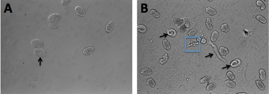
- When ~ 80% of the eggshells are digested by the chitinase treatment (Figures 1 A-B), pellet the eggs by centrifugation at 900 x g (~2,500 rpm) for 3 min. Using a P1000 pipettor and sterile tips, remove carefully the supernatant and add 3 ml of L-15 medium.
- Transfer the eggs into a 6 cm diameter plate and gently dissociate the cells using a 10 ml sterile syringe equipped with a 18 G needle. Monitor the degree of dissociation by placing a drop of suspension into a fresh plastic Petri dish and by viewing under the microscope. Do not aspirate air into the syringe during this procedure to avoid damaging cells. Continue the dissociation until ~ 80% of the cells are dissociated.
- Filter the suspension using a sterile 5 μm Millipore filter. Cell suspensions must be filtered in order to remove cell clumps, undigested eggs and hatched larvae. Filter additional 4-5 ml of fresh L-15 media through the filter to recover all the cells. Do not use excessive force during the filtration step to avoid damaging the filter and/or the cells.
2. Culturing Cells
- Pellet the dissociated cells by centrifugation at 900 x g (~2,500 rpm) for 3 min. Using a P1000 pipettor and sterile tips carefully remove all the supernatant. Resuspend the pelleted cells in complete L-15 medium and plate 1 ml/well. The amount of the medium added depends on the number of 8P plates used, the confluence of the worms on the plates, and the type of experiments that will be performed on the cells. The cell density can be determined using a hemocytometer. For patch-clamp recordings plating density of ~ 230,000 cells/cm2 is optimal.
- Keep the 24 wells plate in a plastic Tupperware container containing wet paper towels to avoid evaporation of culture medium. Store the container in a humidified incubator at 20 °C and ambient air.
- The cells are usually ready for the experiments within 24 hr when the morphological differentiation and expression of GFP markers are complete. Cells can be kept in culture for up to 2 weeks but they are usually most healthy up to 7-9 days after plating. The medium needs to be replaced once a day to maintain healthy cells.
Sonuçlar

Figure 1. C. elegans embryos before and after chitinase treatment. (A) Photograph of eggs prior to exposure to chitinase. The arrow points to the transparent and intact eggshell surrounding an embryo. (B) Eggs treated with chitinase for 10 min. The eggshells have been digested and are no longer visible around th...
Malzemeler
| Name | Company | Catalog Number | Comments |
| REAGENTS | |||
| Leibovitz's L-15 Medium (1x) Liquid | Invitrogen | 11415-064 | |
| Fetal Bovine Serum | Invitrogen | 16140-063 | |
| Penicillin-streptomycin | Sigma | P4333-100ML | |
| Chitinase from Streptomyces Griseus | Sigma | C6137-25UN | |
| Peanut Lectin | Sigma | L0881-10MG | |
| EQUIPMENT | |||
| 101-1000 μl Blue Graduated Pipet Tips | USA Scentific | 1111-2821 | |
| 10 ml Sterilized Pipet Individually Wrapped | USA Scentific | 1071-0810 | |
| Ergonomic Variable Volume (100-1000 μl) Pipettor with tip ejector | VWR International Inc. | 89079-974 | |
| Portable Pipet Aid, Drummond | VWR International Inc. | 53498-103 | |
| Transfer Plastic Pipet Sterile | VWR International Inc. | 14670-114 | |
| 15 ml Conical Tube | USA Scentific | 1475-1611 | |
| Sterile 18 gauge Needles | Becton, Dickinson and Co. | 305196 | |
| Sterile 10 ml Syringes | Becton, Dickinson and Co. | 305482 | |
| Plastic Syringe Filters Corning 0,20 μm pore size | Corning | 431224 | |
| Acrodic 25 mm Syringe filter w/5 μm versapor Membrane | VWR International Inc. | 28144-095 | |
| 60x15 mm Petri Dish Sterile | VWR International Inc. | 82050-548 | |
| 100x15 mm Petri Dish Sterile | VWR International Inc. | 82050-912 | |
| 12 mm Diameter Glass Coverslips | VWR International Inc. | 48300-560 | |
| Clear Cell Culture Plates 24 Well Flat Bottom w/lid | Thomas scientific | 6902A09 | |
| Dumont #5- Fine Forceps | Fine Science Tools | 11254-20 | |
| Centrifuge 5702 | Eppendorf | 22629883 | |
| Laminar Flow Hood | |||
| Inverted Microscope with x10 objective | |||
| Ambient air humidified Incubator |
This article has been published
Video Coming Soon
Source: Sangaletti, R. and Bianchi, L. A Method for Culturing Embryonic C. elegans Cells. J. Vis. Exp. (2013).
JoVE Hakkında
Telif Hakkı © 2020 MyJove Corporation. Tüm hakları saklıdır