
0:00
Overview
1:03
Principles of Crystal Cleavage
3:32
Observing and Analyzing Crystal Form
4:51
Observing and Analyzing Cleavage
6:04
Applications
7:16
Summary
광물의 물리적 특성 I: 결정 및 벽개
출처: 앨런 레스터 연구소 - 콜로라도 볼더 대학교
미네랄의 물리적 특성은 색상, 줄무늬, 자기 특성, 경도, 결정 성장 형태 및 결정 분열을 포함하여 다양한 측정 가능하고 식별 가능한 특성을 포함합니다. 이러한 각 특성은 광물에 특화되며, 특정 광물의 화학 적 구성 및 원자 구조와 근본적으로 관련이 있습니다.
이 실험은 결정 격자, 결정 성장 형태 및 결정 분열 내의 단위 세포라고 불리는 기본 구조 적 원자 그룹화의 대칭 반복에서 주로 줄기두 가지 특성을 검사합니다.
결정 성장 형태는 원자 수준의 대칭의 거시적 발현이며, 성장하는 결정 격자에 단위 세포(미네랄의 분자 빌딩 블록)를 추가하는 자연적인 성장 과정에 의해 생성된다. 빠른 단위 세포 추가 영역은 평면 표면, 즉 크리스탈의 면 사이의 가장자리가됩니다.
바위는 미네랄 곡물의 집합임을 인식하는 것이 중요합니다. 대부분의 바위는 폴리 미네랄 (여러 종류의 미네랄 곡물)이지만 일부는 효과적으로 단미네랄 (단일 미네랄로 구성됨)입니다. 바위는 미네랄의 조합이기 때문에 바위는 결정형태를 갖는 것으로 불리지 않습니다. 어떤 경우에는 지질학자들은 바위를 일반적인 분열이 있는 것으로 지칭하지만, 여기서이 용어는 단순히 반복적인 파괴 표면을 지칭하는 데 사용되며 원자 결정 구조의 반영이 아닙니다. 따라서 일반적으로 결정 형태와 결정 분열라는 용어는 암석 샘플이 아닌 미네랄 샘플을 참조하여 사용됩니다.
모든 미네랄은 물리적 특성을 가지고 있지만 특성과 관련된 구체적이고 쉽게 인식 할 수있는 특징이 항상 개별 결정으로 표현되는 것은 아닙니다. 예를 들어, 석영 결정은 특유의 육각형 모양을 가지고 있지만, 다른 미네랄이 자연적인 성장 형태를 차단하거나 방해하는 환경에서 결정 성장이 발생하면 육각형 모양이 형성되지 않습니다. 따라서 모든 샘플이 이러한 주요 특징을 보여주는 것은 아니기 때문에 결정 성장 또는 결정 분열 분석을 위해 적합한 샘플 그룹을 신중하게 선택하는 것이 중요합니다.
또한, 결정 분열은 비교적 테스트하기 쉽지만- 망치로 시료를 파괴함으로써 - 다른 광물은 다양한 분열 품질을 나타내며, 이러한 플라너 표면은 비정형 및 거칠기("가난한 분열"이라고 불규칙하거나 매우 매끄러운 것)("좋은" 또는 "우수 분열"이라고 불려질 수 있음). 어떤 경우에는(예를 들어 석영), 결정적 결합 강도는 모든 방향에서 균일하며, 이는 인식 할 수있는 분열 평면의 부족으로 미네랄을 초래한다.
1. 광물 샘플 그룹 설립
- 석영, halite, 방해, 가넷, 비오테이트 및/또는 muscovite : 가능한 한 많은 다음을 포함한다. 일부는 크리스탈 성장 기능 및 크리스탈 분열 기능에 대 한 다른 선택.
2. 크리스탈 형태를 관찰하고 분석
- 샘플을 관측 표면에 놓습니다.
- 모든 면을 관찰하기 위해 회전합니다. 크리스탈 면, 크리스탈 엣지(얼굴이 만나는 선), 크리스탈 정점(가장자리가 만나는 지점)을 찾습니다.
- 가능한 경우 고니오미터를 사용하여 얼굴 간 각도를 측정합니다. 이것은 단순히 특정 크리스탈 얼굴에 고니오미터의 한쪽을 놓고, 인접한 얼굴에 곤니오미터의 다른 면을 놓은 다음 각도를 읽는 것으로 수행됩니다.
- 특징적인 결정성 폴리헤드라 세트와 비교하십시오.
- 반복 단계 2.1 – 2.4 석영 (주 육각형 대피라미드 형태(그림 1)),방해 (노트 스케일노헤드론 형태(그림 2)),halite (노트 입방 결정 형태(도 3)),가넷 (노트 도데카헤드론 형태(그림 4)),및 비오티트 (참고 pseudo-hexagoal 형태)에 대한2.1)
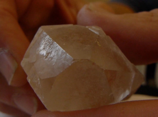
그림 1. 육각형 대피라미드 형태를 표시하는 석영.
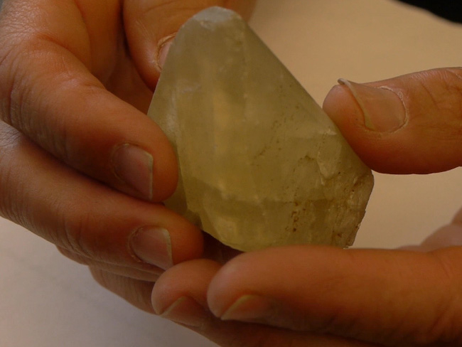
그림 2. 스케일노헤드론 형태를 표시하는 석미. 여러 결정면이 교차하여 결정 가장자리를 형성하고 가장자리의 조합은 "정점"이라고 하는 점을 형성합니다. 대칭 결정 성장 형태는 결정 격자 내의 기본 원자 구조 (단위 세포)의 반복에 의해 생성됩니다. 이 경우, 방해미 결정 성장은 스케일노헤드론으로 알려진 특정 폴리에드론을 생성한다.
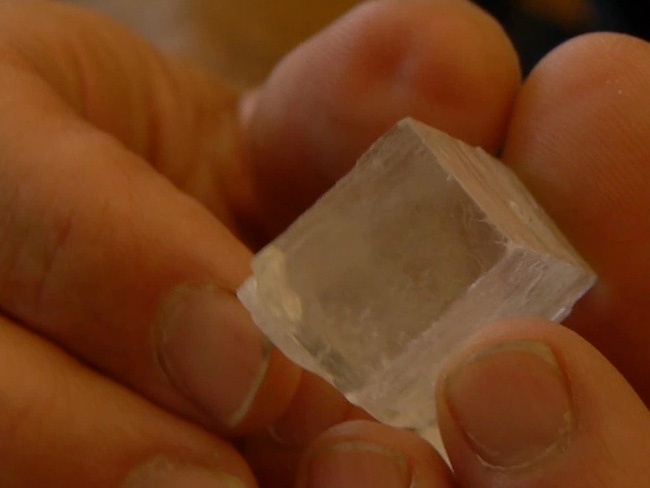
그림 3. 할라이트는 입방 크리스탈 형태를 표시합니다.
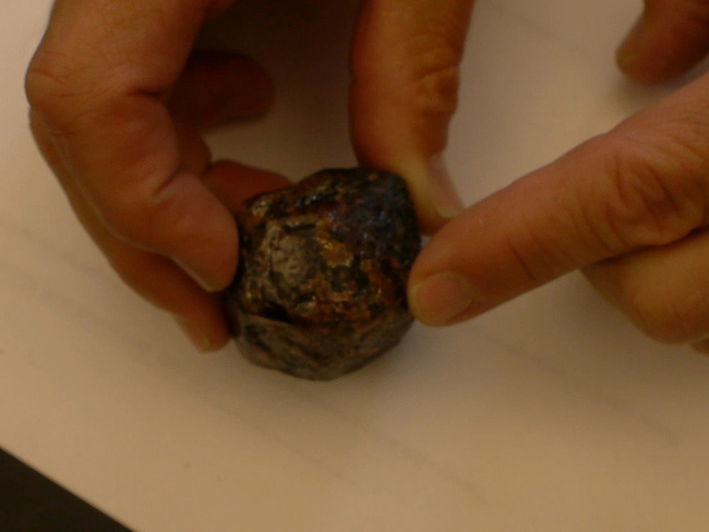
그림 4. 도데카히드론 형태를 표시하는 가넷.
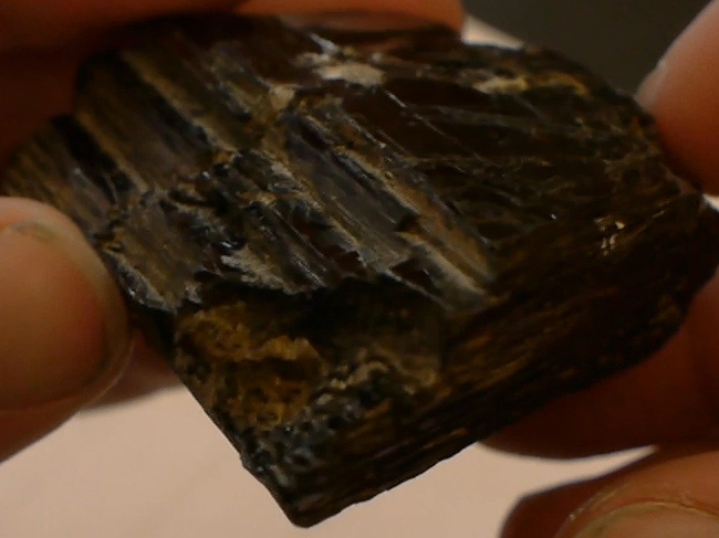
그림 5. 의사 육각형을 표시하는 비오티트.
3. 분열 관찰 및 분석
- 눈 보호에 넣어.
- 부서지는 표면에 석영 조각을 놓습니다.
- 망치를 사용하여 석영 조각을 반으로 부수습니다.
- 손 렌즈를 사용하여 절단 표면에 대한 석영의 깨진 조각을 관찰합니다. 석영에는 아무 것도 없습니다. 석영은 골절을 부출하지만 잘 정의된 골짜기표면(도 6)을나타낸다. 이것은 석영 결정 격자 (실리카 테트라 헤드랄이라고 불리는 SiO4 단)의 단위 세포가 모든 방향에서 비교적 동일한 결합 강도를 가지고 있다는 사실의 결과입니다. 이러한 결합 강도의 균일성은 바람직한 파손 평면이 없는 크리스탈을 생성합니다.
- 반복 단계 3.2 – 3.4 방해기 (마름모꼴 분열(그림 7)을표시해야한다), halite (입방 분열(그림 8)을표시해야한다), 비오타이트, 및 / 또는 muscovite (각 디스플레이 평면 골짜기(도 9).
- 손 렌즈를 사용하여 다양한 골짜기 특성을 평가합니다. 분열은 다양한 수준에서 발생할 수 있습니다. 특정 방향에서 결합 강도에 극적인 차이가 있을 때, 예를 들어, 예를 들어, SiO4 군의 시트 사이로, 이러한 시트 사이에 거의 완벽한 분열이 생성된다. 위에서 언급 했듯이 석영은 거의 총 분열부족을 나타낸다. 이러한 극단 (완벽한 분열과 분열의 부족) 사이에, 좋은 분열(예를 들어, feldspar) 및 가난한 분열 (수륙 활공 결정에 특정 얼굴)이 미네랄이 있습니다.
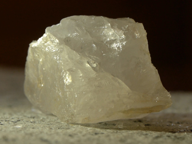
그림 6. 분열 표면없이, conchoidal 골절을 표시 석영.
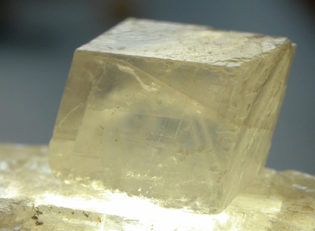
그림 7. 조개 골짜기 분열을 표시하는 석미. 대칭 파괴 및 골절 표면은 결정 격자 내의 원자 결합에서 상대적 약점의 영역에 의해 생성됩니다. 조미석 분열은 마름모꼴로 알려진 특정 다각형으로 초래한다.
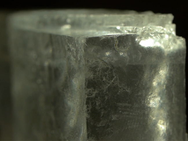
그림 8. 할라이트는 입방 분열을 표시합니다.
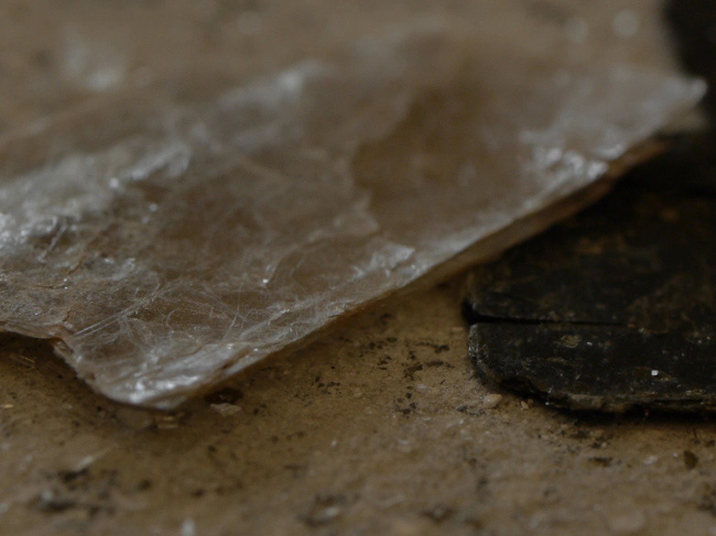
그림 9. 비염은 평면 골짜기를 표시합니다.
역사적으로, 광물의 물성을 평가하는 것은 광물 식별의 중요한 첫 번째 단계였습니다. 오늘날에도 현미경 및 현대 분석계측(예: 석유 학적 현미경 검사법, 엑스레이 회절, 엑스레이 형광 및 전자 마이크로 프로브 기술)이 부족할 때 관찰된 물리적 특성은 광물 식별을 위한 진단 도구로 여전히 매우 유용합니다. 이것은 특히 현장 지질 학연구의 경우입니다.
광물의 물리적 특성을 평가하고 관찰하는 것은 원자 수준의 구조 및 배열에 대한 거시적 특징의 중요한 의존성을 입증하는 훌륭한 수단입니다.
미네랄의 주요 물리적 특성이 항상 특정 샘플에서 발현되는 것은 아닙니다. 따라서 실제로 이러한 특성을 진단 도구로 인식하고 사용할 수 있는 것은 과학, 경험 및 공예의 조합이 필요합니다. 종종 지질학자는 핸드 렌즈를 사용하여 더 큰 암석의 매트릭스 내에서 상대적으로 작은 미네랄 크리스탈이나 곡물을 평가해야 합니다. 이러한 경우, 결정 형태와 결정 분열의 유용한 측면을 식별하는 뚜렷한 도전이 될 수 있다.
학술 적 또는 교육 환경에서 손 샘플 분석을 통한 미네랄 평가는 천연 재료의 물리적 화학에 의해 반복적 인 패턴과 특성이 부과되는 방법을 보여주는 운동입니다. 즉, 임의의 특정 미네랄의 경우 화학 적 조성 및 원자 구조에 의해 부과되는 특정 결정적 특징(예 : 결정 형태학) 및 물리적 특성(예 : 색상, 경도, 줄무늬)이 있습니다.
광물 자원 및 탐사 지질학 영역에서 손 샘플을 통한 광물 식별은 잠재적 인 광구와 경제적으로 유용한 퇴적물을 찾는 것을 목표로하는 현장 작업의 핵심 구성 요소입니다. 예를 들어, 수열 철 옥시 수산화물화(헤마타이트, 괴티트, 리모니트)와 연계하여 다양한 금속 황화물(pyrite, sphalerite, galena)의 식별은 잠재적인 Au-및 Ag가 풍부한 정맥 및 영역을 나타낼 수 있다.
역사적 지질학의 맥락에서 (지역의 깊은 시간적 역사를 해독), 광물 식별은 고대 조건의 해석을위한 무대를 설정할 수 있습니다. 예를 들어, 특정 변태미네랄(예: Al2SiO5 다형성, kyanite, andalusite, 및 실리마니트)은 고대 지각의 특정 압력 및 온도 조건의 표식입니다.
Tags
ABOUT JoVE
Copyright © 2024 MyJoVE Corporation. All rights reserved