
Eye Exam
Overview
Source: Richard Glickman-Simon, MD, Assistant Professor, Department of Public Health and Community Medicine, Tufts University School of Medicine, MA
Proper evaluation of the eyes in a general practice setting involves vision testing, orbit inspection, and ophthalmoscopic examination. Before beginning the exam, it is crucial to be familiar with the anatomy and physiology of the eye. The upper eyelid should be slightly over the iris, but it shouldn't cover the pupil when open; the lower lid lies below the iris. The sclera normally appears white or slightly buff in color. The appearance of conjunctiva, a transparent membrane covering the anterior sclera and the inner eyelids, is a sensitive indicator of ocular disorders, such as infections and inflammation. The tear-producing lacrimal gland lies above and lateral to the eyeball. Tears spread down and across the eye to drain medially into two lacrimal puncta before passing into the lacrimal sac and nasolacrimal duct to the nose.
The iris divides the anterior from the posterior chamber. Muscles of the iris control the size of the pupil, and muscles of the ciliary body behind it control the focal length of the lens. The ciliary body also produces aqueous humor, which largely determines intraocular pressure (Figure 1). Cranial nerves II and III control pupillary reaction and lens accommodation; cranial nerve III controls upper lid elevation; cranial nerves III, IV, and VI control eye movement. The six cardinal directions of gaze are controlled by six extraocular muscles (Figure 2) innervated by cranial nerves III, IV, and VI.
Visual testing is an essential part of the ophthalmological exam and is also performed as a part of cranial nerve II assessment during the neurological exam. A focused image is projected onto the retina after its light passes through the cornea, pupil, lens, and vitreous body. The projection is upside down and reversed right to left, which means that light entering from the lower temporal field of vision strikes the upper nasal quadrant of the retina. Photosensitive cells of the retina respond by generating electrical impulses, which are relayed to the optic nerve and passed to the visual cortex through the optic tracts. The right and left visual cortices process images entering from the left and right visual fields, respectively.
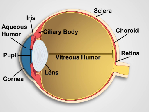
Figure 1. Anatomy of the Eye. A diagram showing a sagittal view of the human eye with the structures labeled.
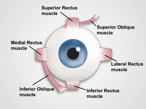
Figure 2. Muscles of the Eye. A cartoon showing a frontal view of the human eye and the extraocular muscles (labeled).
Procedure
1. Vision
Visual acuity is recorded as two numbers (e.g., 20/40 corrected). The top number indicates the distance the patient stood from the chart (20 ft), and the bottom number indicates the distance from which a person with normal vision (20/20) could see the smallest line of print accurately read by the patient (40 ft with glasses). In the US, a patient with vision of 20/200 or worse is considered legally blind.
- If available, use a well-lit, wall-mounted Snellen chart.<
Application and Summary
Many systemic and ocular pathologies have manifestations that can be identified during an eye examination. Simple visual acuity testing with a Snellen chart, or an adequate substitute, allows screening for myopia and presbyopia - impaired far and near vision, respectively. Limitations in peripheral vision raise the possibility of glaucoma, as well as other serious conditions, and should always prompt further ophthalmologic evaluation. Localized swelling of the eyelids commonly results from infected sties or nodular chala
Tags
Skip to...
Videos from this collection:

Now Playing
Eye Exam
Physical Examinations II
77.1K Views

Ophthalmoscopic Examination
Physical Examinations II
67.9K Views

Ear Exam
Physical Examinations II
55.1K Views

Nose, Sinuses, Oral Cavity and Pharynx Exam
Physical Examinations II
65.7K Views

Thyroid Exam
Physical Examinations II
105.0K Views

Lymph Node Exam
Physical Examinations II
387.3K Views

Abdominal Exam I: Inspection and Auscultation
Physical Examinations II
202.6K Views

Abdominal Exam II: Percussion
Physical Examinations II
248.2K Views

Abdominal Exam III: Palpation
Physical Examinations II
138.5K Views

Abdominal Exam IV: Acute Abdominal Pain Assessment
Physical Examinations II
67.3K Views

Male Rectal Exam
Physical Examinations II
114.4K Views

Comprehensive Breast Exam
Physical Examinations II
87.6K Views

Pelvic Exam I: Assessment of the External Genitalia
Physical Examinations II
306.9K Views

Pelvic Exam II: Speculum Exam
Physical Examinations II
150.4K Views

Pelvic Exam III: Bimanual and Rectovaginal Exam
Physical Examinations II
147.7K Views
Copyright © 2025 MyJoVE Corporation. All rights reserved