Infrared Spectroscopy
Overview
Source: Vy M. Dong and Zhiwei Chen, Department of Chemistry, University of California, Irvine, CA
This experiment will demonstrate the use of infrared (IR) spectroscopy (also known as vibrational spectroscopy) to elucidate the identity of an unknown compound by identifying the functional group(s) present. IR spectra will be obtained on an IR spectrometer using the attenuated total reflection (ATR) sampling technique with a neat sample of the unknown.
Principles
A covalent bond between two atoms can be thought of as two objects with masses m1 and m2 that are connected with a spring. Naturally, this bond stretches and compresses with a certain vibrational frequency. This frequency  is given by Equation 1, where k is the force constant of the spring, c is the speed of light, and µ is the reduced mass (Equation 2). The frequency is typically measured in wavenumbers, which are expressed in inverse centimeters (cm-1).


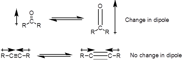
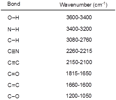
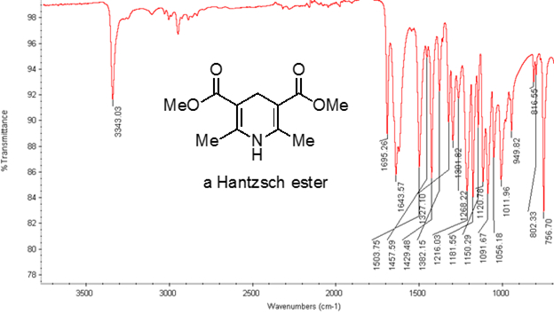
From Equation 1, the frequency is proportional to the strength of the spring and inversely proportional to the masses of the objects. Thus, C-H, N-H, and O-H bonds have higher stretching frequencies than C-C and C-O bonds, as hydrogen is a light atom. Double and triple bonds can be considered as stronger springs, so a C-O double bond has a higher stretching frequency than a C-O single bond. Infrared light is electromagnetic radiation with wavelengths ranging from 700 nm to 1 mm, which is consistent with the relative bond strengths. When a molecule absorbs infrared light with a frequency that equals the natural vibrational frequency of a covalent bond, the energy from the radiation produces an increase in the amplitude of the bond vibration. If the electronegativities (the tendency to attract electrons) of the two atoms in a covalent bond are very different, a charge separation occurs that results in a dipole moment. For example, in a C-O double bond (a carbonyl group), the electrons spend more time around the oxygen atom than the carbon atom because oxygen is more electronegative than carbon. Hence, there is a net dipole moment resulting in a partial negative charge on oxygen and a partial positive charge on carbon. On the other hand, a symmetrical alkyne does not have a net dipole moment because the two individual dipole moments on each side cancel each other. The intensity of the infrared absorption is proportional to the change in the dipole moment when the bond stretches or compresses. Hence, a carbonyl group stretch will show an intense band in the IR, and a symmetrical internal alkyne will show a small, if not invisible, band for stretching of the C-C triple bond (Figure 1). Table 1 shows some characteristic absorption frequencies. Figure 2 shows the IR spectrum of a Hantzsch ester. Notice the peak at 3,343 cm-1 for the N-H single bond and the peak at 1,695 cm-1 for the carbonyl groups. In this experiment, the ATR sampling technique is used, where the infrared light reflects off the sample that is in contact with an ATR crystal multiple times. Typically, materials with a high refractive index are used, such as germanium and zinc selenide. This method enables one to directly examine solid or liquid analytes without further preparation.

Figure 1. Diagram showing C-O double and C-C triple bond stretches and the resulting change in the dipole moment.

Table 1. Characteristic IR frequencies of covalent bonds present in organic molecules.

Figure 2. IR spectrum of a Hantzsch ester.
Procedure
- Turn on the IR spectrometer and allow it to warm up.
- Obtain an unknown sample from the instructor and record the letter and appearance of the sample.
- Collect a background spectrum.
- Using a metal spatula, place a small amount of sample under the probe.
- Twist the probe until it locks into place.
- Record the IR spectrum of the unknown sample.
- Repeat if necessary to obtain a good quality spectrum.
- Record the absorption frequencies indicative of the functional groups present.
- Clean the probe with acetone.
- Turn off the spectrometer.
- Analyze the obtained spectrum. Figure 3 shows the possible candidates for the unknown sample. State the probable identification of the unknown sample.

Figure 3. Diagram showing the possible identities of the unknown.
Results
Table 2: Appearance and observed IR frequencies of the compounds listed in Figure 3.
| Compound Number | 1 | 2 | 3 | 4 | 5 | 6 | 7 | 8 | 9 | 10 |
| Appearance | clear liquid | white solid | clear liquid | clear liquid | clear liquid | clear liquid | yellow liquid | white solid | white solid | clear liquid |
| Observed frequencies (cm-1) | 1691, 1601, 1450, 1368, 1266 |
2773, 2730, 1713, 1591, 1576 |
2940, 2867, 1717, 1422, 1347 |
3026, 2948, 2920, 1605, 1496 |
2928, 2853, 1450, 904, 852 |
3926, 3315, 2959, 2120, 1461 |
3623, 3429, 3354, 2904, 1601 |
3408, 3384, 3087, 1596, 1496 |
3226, 2966, 1598, 1474, 1238 |
3340, 2959, 2861, 1468, 1460 |
Application and Summary
In this experiment, we have demonstrated how to identify an unknown sample based on its characteristic IR spectrum. Different functional groups give different stretching frequencies, which allow the identification of the functional groups present.
As shown in this experiment, IR spectroscopy is a useful tool for the organic chemist to identify and characterize a molecule. In addition to organic chemistry, IR spectroscopy has useful applications in other areas. In the pharmaceutical industry, this technique is used for quantitative and qualitative analysis of drugs. In food science, IR spectroscopy is used to study fats and oils. Lastly, IR spectroscopy is used to measure the composition of greenhouse gases, i.e., CO2, CO, CH4, and N2O in efforts to understand global climate changes.
Skip to...
Videos from this collection:

Now Playing
Infrared Spectroscopy
Organic Chemistry II
214.1K Views

Cleaning Glassware
Organic Chemistry II
123.2K Views

Nucleophilic Substitution
Organic Chemistry II
99.2K Views

Reducing Agents
Organic Chemistry II
42.9K Views

Grignard Reaction
Organic Chemistry II
148.8K Views

n-Butyllithium Titration
Organic Chemistry II
47.7K Views

Dean-Stark Trap
Organic Chemistry II
99.8K Views

Ozonolysis of Alkenes
Organic Chemistry II
66.8K Views

Organocatalysis
Organic Chemistry II
16.6K Views

Palladium-Catalyzed Cross Coupling
Organic Chemistry II
34.2K Views

Solid Phase Synthesis
Organic Chemistry II
40.8K Views

Hydrogenation
Organic Chemistry II
49.4K Views

Polymerization
Organic Chemistry II
93.7K Views

Melting Point
Organic Chemistry II
149.6K Views

Polarimeter
Organic Chemistry II
99.8K Views
Copyright © 2025 MyJoVE Corporation. All rights reserved