
0:01
Concepts
3:53
Preparation of Donor and Recipient Strains
6:01
Conjugation
9:27
Data Analysis by Polymerase Chain Reaction (PCR)
11:29
Results
Conjugation: A Method to Transfer Ampicillin Resistance from Donor to Recipient E. coli
Source: Alexander S. Gold1, Tonya M. Colpitts1
1 Department of Microbiology, Boston University School of Medicine, National Emerging Infections Diseases Laboratories, Boston, MA
First discovered by Lederberg and Tatum in 1946, conjugation is a form of horizontal gene transfer between bacteria that relies on direct physical contact between two bacterial cells (1). Unlike other forms of gene transfer, such as transformation or transduction, conjugation is a naturally occurring process in which DNA is secreted from a donor cell to a recipient cell in a unidirectional manner. This directionality and the ability for this process to increase the genetic diversity of bacteria has given conjugation the reputation as a form of bacterial "mating," which is believed to have contributed greatly to the recent rise in antibiotic resistant bacteria (2, 3). By using selective pressures, for example the use of antibiotics, conjugation has been manipulated for use in the laboratory setting, making it a powerful tool for horizontal gene transfer between bacteria, and in some cases from bacteria to yeast, plant, and animal cells (4). Apart from applications in the laboratory, bacterium-eukaryote gene transfer by conjugation is an exciting avenue of DNA transfer with a multitude of possible biotechnical applications and naturally occurring implications (5).
Conjugation is thought to work by a "two-step mechanism" (6). First, before any DNA can be transferred, the donor cell must make direct cell-to-cell contact with the recipient. This process has been characterized best in gram-negative bacteria, the most studied of which is Escherichia coli. Cell-to-cell contact is established by the presence of a complex network of extracellular filaments on the donor known as the sex pilus, a conjugative element encoded for by the transferable gene known as the F (fertility) factor (7, 8). In addition to establishing contact between donor and recipient, several proteins are transported via the sex pilus to the recipient cytoplasm, forming a type IV secretion system (T4SS) conduit between the two cells, a necessary structure for the second step of conjugation, DNA transfer (6). By combining this function of the sex pilus with rolling circle replication of DNA, the donor cell is able to transfer DNA in the form of a transposable element, such as a plasmid or transposon, to the recipient by a "shoot and pump" model (6). In this case, the "shooting" is the transport of the pilot protein, with linked DNA, by the T4SS into the recipient cell, and the "pumping" is the active transport of DNA to the recipient, a process reliant on the T4SS and catalyzed by coupling proteins (6). The machinery used in this this process is composed of an origin of transfer sequence (oriT), which must be provided by the DNA in cis and trans genes, which encode a relaxase, mate pair formation complex, and type IV coupling protein, and can be present in cis or trans (9). This relaxase cleaves the nic site within the oriT sequence and covalently attaches to the 5' end of the transferred strand to produce the relaxosome, a single-stranded DNA-relaxase complex with other auxiliary proteins (9). Once formed, the relaxosome connects to the mating pair formation complex, via the type IV coupling protein, which allows for the transfer of the ssDNA-relaxase complex into recipient cells by the T4SS (10). Once in the cytoplasm of the recipient, the DNA can integrate into the recipient genome or exist separately in the form of a plasmid, either of which allow for the expression of its genes.
In this experiment, the widely used conjugation donor strain E. coli WM3064 was used to transfer the gene encoding for ampicillin resistance to the recipient strain E. coli J53. While both strains of the gram-negative bacteria were resistant to tetracycline, only the donor strain WM3064 had the gene for ampicillin resistance, encoded for in the pWD2-oriT shuttle vector, and was auxotrophic to diaminopimelic acid (DAP) (11-13). This experiment consisted of two main steps, the preparation of donor and recipient strains, followed by the transfer of the ampicillin resistance gene from donor to recipient by conjugation (Figure 1).
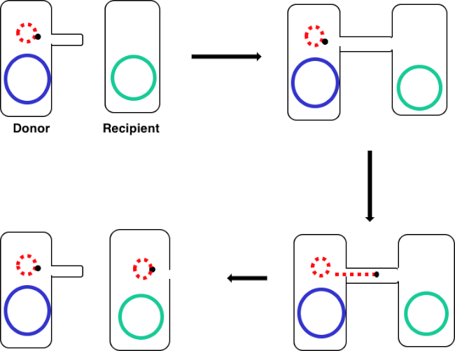
Figure 1: Conjugation schematic. This schematic shows the successful transfer of a plasmid, only one example of a transposable DNA element, from a donor cell to a recipient cell using conjugation. Upon contact with the recipient cell by the donor cell via the sex pilus, the plasmid replicates by rolling circle replication, moves through the multiprotein complex joining the two cells, and forms a new full-length plasmid in the recipient cell.
By incubating a mixture of donor and recipient cells, then successively plating these cells in the presence of tetracycline and DAP, this allowed for the successful transfer of the ampicillin resistance gene. Successively plating cells grown from this mixture in the presence of tetracycline and ampicillin, removed all donor cells due to the lack of DAP and any recipient cells that may not have gained the ampicillin resistance gene, yielding strictly recipient J53 strain bacteria that acquired ampicillin resistance (Figure 2). Once carried out, the successful transfer of the ampicillin resistance gene was confirmed by PCR. Since conjugation was successful, the J53 strain of E. coli contained pWD2-oriT and was resistant to ampicillin, and the gene encoding for this resistance is detectable by PCR. However, if unsuccessful there would have been no detection of the ampicillin resistance gene and ampicillin would still function as an effective antibiotic against the J53 strain.
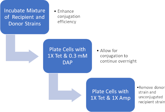
Figure 2: Protocol schematic. This schematic shows an overview of the presented protocol.
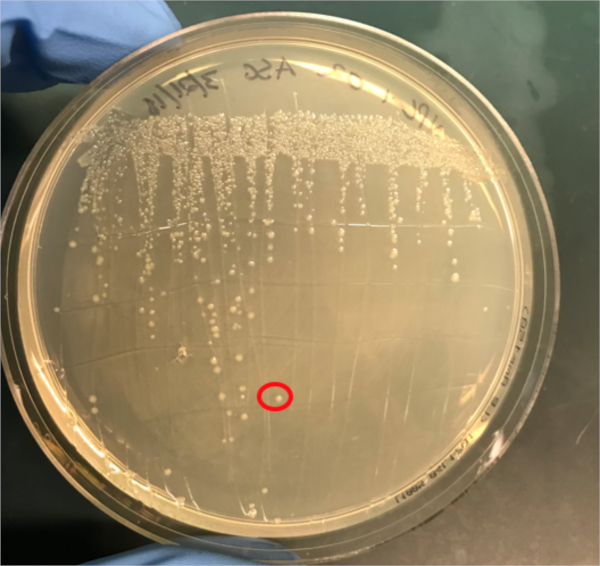
Figure 3A: The confirmation of successful conjugation by PCR. A) Freezer stocks of the conjugated and negative control samples were streaked out on agar plates and a colony was selected (red) for DNA isolation.
1. Set-Up
- Autoclave approximately 1L of Luria-Bertani medium (LB). This sterile LB will be used to make approximately 5 mL of LB containing 0.3 mM diaminopimelic acid (DAP).
- Collect the following plates: LB agar plates with 1X Tet and 0.3 mM DAP, LB agar plates with 1X Tet only, and LB agar plates with 1X Amp/Tet only.
- Ensure that some glycerol and a box of pre-sterilized plastic pipette tips are close at hand.
- Prior to starting any work involving microbes, steri
If conjugation was successful, a 500 base-pair sized band PCR product will be observed in the well in which PCR reaction 1 was loaded (Well #2 in Figure 3B), while no bands will be observed in the well in which PCR reaction 3 was loaded (Well #4 in Figure 3B). The presence of this band confirms the successful transfer of the ampicillin resistance gene, thereby conferring ampicillin resistance to the J53 strain of E. coli.
Log in or to access full content. Learn more about your institution’s access to JoVE content here
Conjugation is a naturally occurring process of horizontal gene transfer that relies on the direct cell-to-cell contact of a donor cell and a recipient cell. This process is shared among all kinds of bacteria and has been instrumental in bacterial evolution, most notably antibiotic resistance. In the lab, conjugation can be used as an effective method of gene transfer that is much less disruptive when compared to other techniques. Outside of the laboratory, the ability to transfer DNA from bacteria to eukaryotes via conj
- Lederberg J, Tatum, E.L. Gene recombination in Escherichia coli Nature. 1946;158:558.
- Holmes R.K. J, M.G. Genetics: Exchange of Genetic Information. 4th Edition ed. Baron S, editor. Galveston, TX: University of Texas Medical Branch at Galveston; 1996.
- Cruz F, Davies, J. Horizontal gene transfer and the origin of species: lessons from bacteria. Trends in Microbiology. 2000;8:128-33.
- Llosa M, Cruz, F. Bacterial conjugation: a potential tool for genomic engineering. Ressearch in Microbiology. 2005;156:1-6.
- Lacroix B, Citovsky, V. Transfer of DNA from Bacteria to Eukaryotes. mBio. 2016;7(4):1-9.
- Llosa M, et al. Bacterial conjugation: a two-step mechanism for
- DNA transport. Molecular Microbiology. 2002;45:1-8.
- Grohmann E, Muth, G., Espinosa, M. Conjugative Plasmid Transfer in Gram-Positive Bacteria. Microbiology and Molecular Biology Reviews. 2003;67:277-301.
- Firth N, Ippen-Ihler, K, Skurray, RA. Structure and function of the F factor and mechanism of conjugation. Escherichia coli and salmonella: cellular and molecular biology. 1996;2:2377-401.
- Smillie C, Garcillan-Barcia MP, Francia MV, Rocha EPC, De La Cruz F. Mobility of Plasmids. Microbiology and Molecular Biology Reviews. 2010;74(3):434-52.
- Cascales E. Definition of a Bacterial Type IV Secretion Pathway for a DNA Substrate. 2004;304(5674):1170-3.
- Wang P, Yu Z, Li B, Cai X, Zeng Z, Chen X, et al. Development of an efficient conjugation-based genetic manipulation system for Pseudoalteromonas. Microbial Cell Factories. 2015;14(1):11.
- Yi H, Cho YJ, Yong D, Chun J. Genome Sequence of Escherichia coli J53, a Reference Strain for Genetic Studies. Journal of Bacteriology. 2012;194(14):3742-3.
- Baumann RLB, E. H.; Wiseman, J. S.; Vaal, M.; Nichols, J. S. Inhibition of Escherichia coli Growth and Diaminopimelic Acid Epimerase by 3-Chlorodiaminopimelic Acid. Antimicrobial Agents and Chemotherapy 1988;32:1119-23.
- Rocha D, Santos, CS, Pacheco LG. Bacterial reference genes for gene expression studies by RT-qPCR: survey and analysis. Antonie Van Leeuwenhoek. 2015;108:685-93.
ABOUT JoVE
Copyright © 2024 MyJoVE Corporation. All rights reserved