
Imaging In-Stent Restenosis: An Inexpensive, Reliable, and Rapid Preclinical Model
September 14th, 2009
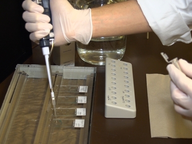
•This video demonstrates how to use a preclinical inexpensive and reliable model to study pathobiological and pathophysiological processes of in-stent restenosis development. Longitudinal in vivo monitoring using OCT (Optical Coherence Tomography) and analysis of OCT images are also demonstrated.
Tags
Related Videos

Non-plasma Bonding of PDMS for Inexpensive Fabrication of Microfluidic Devices

Assembly, Loading, and Alignment of an Analytical Ultracentrifuge Sample Cell

In vivo Imaging and Therapeutic Treatments in an Orthotopic Mouse Model of Ovarian Cancer

3D Printing of Preclinical X-ray Computed Tomographic Data Sets

An Improved Method for Accurate and Rapid Measurement of Flight Performance in Drosophila

Rapid Analysis and Exploration of Fluorescence Microscopy Images
A Rapid Automated Protocol for Muscle Fiber Population Analysis in Rat Muscle Cross Sections Using Myosin Heavy Chain Immunohistochemistry

Benefits of Cardiac Resynchronization Therapy in an Asynchronous Heart Failure Model Induced by Left Bundle Branch Ablation and Rapid Pacing

High-throughput Measurement of Gut Transit Time Using Larval Zebrafish

Mass Spectrometry Analysis to Identify Ubiquitylation of EYFP-tagged CENP-A (EYFP-CENP-A)
ABOUT JoVE
Copyright © 2024 MyJoVE Corporation. All rights reserved