
Measuring Mast Cell Exocytosis via Neuropeptide Y Monomeric Red Fluorescent Protein Release
Transcript
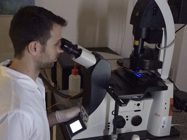
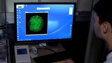
Take transfected mast cells expressing NPY-mRFP, a fusion protein containing neuropeptide Y and monomeric red fluorescent protein, in their cytoplasmic secretory granules.
Remove the media. Pipette calcium-containing buffer with an activating reagent, a calcium ionophore, and incubate.
These ionophores induce calcium influx, increasing the intracellular calcium concentration and mobilizing secretory granules to the plasma membrane.
The calcium binds to calcium-binding proteins on the secretory granule membrane.
Further, the secretory granules fuse with the plasma membrane mediated by membrane-bound proteins, resulting in exocytosis and release of contents, including NPY-mRFP.
Post-incubation, collect and transfer the released NPY-mRFP-containing supernatant to a black multi-well plate.
Add a non-ionic detergent to lyse the cells and release the remaining intracellular NPY-mRFP.
Collect and transfer the cell lysate to the multi-well plate.
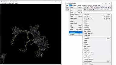
Using a fluorescence plate reader, measure the fluorescence of the cell supernatant and lysate to quantify the percentage of NPY-mRFP released and determine the extent of mast cell exocytosis.
ABOUT JoVE
Copyright © 2024 MyJoVE Corporation. All rights reserved