
Using 23Na Magnetic Resonance Imaging to Measure Sodium in Muscle Tissues
Transcript
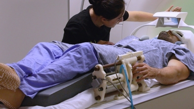
Place the patient's lower leg within the radiofrequency coil of the magnetic resonance imaging or MRI machine.
The coil contains various sodium concentrations as standards to measure tissue sodium accumulation.
Initiate a sodium-MRI scan.
The coil detects fluctuations in the MR signal intensity emitted from the tissue, influenced by the distribution and concentration of sodium ions.
Obtain a total tissue sodium map displaying areas with varying color intensities corresponding to the sodium levels.
Compare these intensities with standard sodium concentrations, which aid in measuring total tissue sodium levels.
Bright colors indicate higher levels, while darker colors indicate lower levels.
Next, capture signals corresponding to the fat and water content of the tissue to generate images of the non-muscle regions.
Subtract non-muscle regions from the total tissue sodium map to generate a refined image and measure muscle tissue sodium content.
Related Videos
Automated Segmentation of Cortical Grey Matter from T1-Weighted MRI Images (Video) | JoVE
Functional MRI in Conjunction with a Novel MRI-compatible Hand-induced Robotic Device to Evaluate Rehabilitation of Individuals Recovering from Hand Grip Deficits (Video) | JoVE
Concurrent EEG and Functional MRI Recording and Integration Analysis for Dynamic Cortical Activity Imaging (Video) | JoVE
ABOUT JoVE
Copyright © 2024 MyJoVE Corporation. All rights reserved