A subscription to JoVE is required to view this content. Sign in or start your free trial.
Method Article
تطور السيليكا طلاءات الجسيمات النانوية البوليستر على السطوح المعرضة لأشعة الشمس
In This Article
Summary
نوعين من الأسطح والصلب المغلفة البوليستر والبوليستر المغلفة بطبقة من جزيئات النانو السيليكا، وتمت دراسة. تعرضت كل السطوح لأشعة الشمس، والتي وجدت لتسبب تغيرات كبيرة في الكيمياء والنانو تضاريس السطح.
Abstract
تآكل الأسطح المعدنية هو السائد في البيئة ويشكل مصدر قلق كبير في العديد من المجالات، بما في ذلك الجيش، والنقل، والطيران، والبناء والصناعات الغذائية، وغيرها. وقد استخدمت على نطاق واسع البوليستر والطلاء تحتوي على كل من البوليستر والجسيمات النانوية السيليكا (شافي 2 NPS) لحماية الطبقات التحتية الصلب من الصدأ. في هذه الدراسة، ونحن تستخدم الأشعة السينية الضوئية الطيفي، الموهن الانعكاس الكلي الأشعة تحت الحمراء الطيفي الجزئي، والقياسات زاوية الاتصال المياه، والتنميط البصرية ومجهر القوة الذرية لتوفير نظرة ثاقبة كيف التعرض لأشعة الشمس يمكن أن يسبب تغيرات في الجزئي وسلامة النانوية من الطلاء. تم الكشف عن أي تغيير كبير في السطح الصغيرة الطبوغرافيا باستخدام profilometry البصرية، ومع ذلك، تم الكشف عن التغييرات النانو ذات دلالة إحصائية على السطح باستخدام مجهر القوة الذرية. تحليل الضوئية الطيفي للأشعة السينية والموهن التفكير الكلي الصغرى الأشعة تحت الحمراءكشفت بيانات التحليل الطيفي أن تدهور المجموعات استر وقعت خلال التعرض للأشعة فوق البنفسجية لتشكيل سجع ·، -H 2 C ·، -O ·، -co · المتطرفين. خلال عملية التحلل، وأنتجت أيضا أول أكسيد الكربون وثاني أكسيد الكربون 2.
Introduction
Environmental corrosion of metals in the environment is both prevalent and costly1-3. A recent study conducted by the Australasian Corrosion Association (ACA) reported that corrosion of metals resulted in a yearly cost of $982 million, which was directly associated with the degradation of assets and infrastructure through metallic corrosion within the water industry4. From an international perspective, the World Corrosion Organization estimated that metallic corrosion was responsible for a direct cost of $3.3 trillion, over 3% of the world's GDP5. The process of galvanizing as a corrosion preventative method has been widely used to increase the lifespan of steel material6. In humid and subtropical climates, however, water tends to condense into small pockets or grooves within the surface of the galvanized steel, leading to the acceleration of corrosion rates through pit corrosion7,8. Thermosetting polymer coatings based on polyesters have been developed to coat the galvanized steel substrata increasing their ability to withstand humid weathering conditions for items such as satellite dishes, garden furniture, air-conditioning units or agricultural construction equipment9-11. Unfortunately polymer coatings on steel surfaces have been found to be considerably adversely affected by the presence of high levels of ultraviolet (uv) radiation12-14. Coatings comprised of silica nanoparticles (SiO2) spread over a polymer layer have been widely used with a view to increasing their corrosion-, wear-, tear- and degradation-resistance15,16. The tendency of the protective polymeric coatings to form pores and cracks can be reduced by incorporating nanoparticles (NPs), which contribute to the passive obstruction of corrosion initiation17,18. Also, the mechanical stability of the protective polymeric layer can be improved by NPs inclusion. However, these coatings act as passive physical barriers and, in comparison to the galvanization approach, cannot inhibit corrosion propagation actively.
An in-depth understanding of the effect that high-levels of ultraviolet light exposure under humid conditions upon these metal coatings is yet to be obtained. In this paper, a wide range of surface analytical techniques, including X-ray photoelectron spectroscopy (XPS), attenuated total reflection infrared micro-spectroscopy (ATR IR), contact angle goniometry, optical profiling and atomic force microscopy (AFM) will be employed to examine the changes in the surface of steel coatings prepared from polyester- and silica nanoparticle-coated polyester (silica nanoparticles/polyester) after exposure to sunlight. Furthermore, the aim of this work is to give a concise, practical overview of the overall characterization techniques to examine weathered samples.
Protocol
1. عينات الصلب
- الحصول على عينات من الصلب 1 مم سمك من مورد تجاري.
والمغلفة العينات مع أي من البوليستر أو البوليستر المغلفة مع الجسيمات النانوية السيليكا: ملاحظة. - كشف العينات لأشعة الشمس في روكهامبتون، كوينزلاند، أستراليا: جمع عينات بعد فترات مدتها سنة واحدة وخمس سنوات على مدى فترة الإجمالية لمدة 5 سنوات. قطع الألواح عينة إلى أقراص مستديرة من 1 سم القطر باستخدام ثقب الناخس.
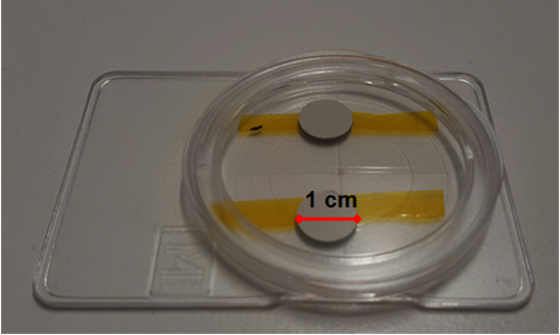
- قبل تطفو على السطح التوصيف، شطف العينات مع الماء المقطر المزدوج، ثم الجافة باستخدام غاز النيتروجين (99.99٪). إبقاء جميع العينات في حاويات محكمة الغلق لمنع أي ملوثات الهواء التكثيف على السطح (الشكل 1).

الشكل 1. إعداد الأقراص المعدنية مع طلاء القائم على البوليستر. تم تخزين العينات في حاويات حتى المطلوبة.ام / ملفات / ftp_upload / 54309 / 54309fig1large.jpg "الهدف =" _ فارغة "> الرجاء انقر هنا لعرض نسخة أكبر من هذا الرقم.
2. الكيميائية وفيزيائية توصيف السطوح
- تحليل الكيمياء السطحية باستخدام الأشعة السينية الضوئية الطيفي.
- أداء الأشعة السينية الضوئية الطيفي (XPS) باستخدام مصدر أحادي اللون الأشعة السينية (آل Kα، hν = 1486.6 فولت) التي تعمل في 150 W.
ملاحظة: حجم بقعة من استخدام شعاع الأشعة السينية هو 400 ميكرون في القطر. - عينات الحمل على لوحة عينة. وضع لوحة عينة في فراغ الغرفة من XPS ثم ضخ الغرفة. انتظر الفراغ في الغرفة للوصول إلى ~ 1 × 10 -9 م بار.
- في البرنامج الضوئية الطيفي، اضغط على خيار "الفيضانات بندقية" لإغراق العينات مع الإلكترونات ذات الطاقة المنخفضة للتصدي لشحن السطح.
- اضغط على "إدراج"> "نقطة"> "نقطة" لإدراج بوين تحليلت.
ملاحظة: سوف يكون هذا الموقع الذي يتم تنفيذ تحليل. تمكين وظيفة ارتفاع السيارات للحصول على أفضل ارتفاع لاقتناء. - اضغط على "إدراج"> "الطيف"> "متعدد الطيف" لإضافة المسح إلى هذه النقطة.
ملاحظة: سيتم فتح نافذة مع الجدول الدوري. تحديد عنصر عن طريق النقر عليه لتسليط الضوء عليه. - بعد إعداد التجارب، اضغط على "تشغيل" القيادة والمضي قدما في اجراء الفحوصات.
- اضغط على "الذروة صالح" القيادة ثم اضغط على "أضف الذروة" و "تناسب جميع مستوى" أوامر لحل أنواع متميزة كيميائيا في عالية الدقة الأطياف.
ملاحظة: هذه الخطوة سوف اكتساب الخوارزمية شيرلي لإزالة الخلفية والتمويه، Lorentzian المناسب لdeconvolute أطياف 19. - تحديد كافة عالية الدقة ومسح الأطياف. اضغط على "المسؤول التحول" الخيار لتصحيح الأطياف باستخدام hydrocaعنصر rbon من (طاقة الربط 285.0 فولت) C 1S ذروة كمرجع.
- بعد تصحيح الشحن، اضغط خيار "تصدير" لتوليد جدول البيانات للتركيز الذري النسبي للعناصر على أساس منطقة الذروة.
- أداء الأشعة السينية الضوئية الطيفي (XPS) باستخدام مصدر أحادي اللون الأشعة السينية (آل Kα، hν = 1486.6 فولت) التي تعمل في 150 W.
- كيمياء السطوح
ملاحظة: تحليل الكيمياء السطحية باستخدام مخفف الانعكاس الكلي الأشعة تحت الحمراء الصغيرة الطيفي (ATR-IR) على الأشعة تحت الحمراء (IR) الطيفي خط الأشعة في السنكروترون الاسترالية على النحو التالي:- عينات الحمل على مرحلة المجهر. فتح "بدء فيديو القياس بمساعدة" أو خيار "بدء القياس دون 3D". تشغيل وضع "VIS" على. استخدام الهدف للتركيز على سطح العينة. اضغط على "لقطة / نظرة عامة" إلى اتخاذ الصور المطلوبة.
ملاحظة: 0.5 مم الكاف 2 لوحة يمكن استخدامها كخلفية. - تغيير الهدف ATR للعينة. بعناية نقل مرحلة لوضع الالماني 45 درجة متعددة التأملالكريستال manium (معامل الانكسار من 4) 1-2 مم فوق السطوح. انقر بزر الماوس الأيمن على النافذة لايف فيديو. اضغط على "ابدأ القياس"> "تغيير قياس المعلمات". اختر الخيار "أبدا استخدام BG القائمة لجميع المواقف".
ملاحظة: هذا اختيار عدم اتخاذ أطياف الخلفية لكل نقطة قياس. - رسم خريطة على شاشة الفيديو لاختيار مجال الاهتمام. اضغط على مربع فتحة الأحمر واختيار "الفتحة"> "تغيير الفتحة". تغيير إعدادات الفعلية "حافة السكين فتحة" لX = 20 ميكرون و Y = 20 ميكرون.
- انقر بزر الماوس الأيمن على مربع فتحة بحجم حديثا وانتقل إلى "الفتحة"> "تعيين كافة بؤر لبؤر المحدد". الصحافة رمز "قياس" لبدء المسح الضوئي. حفظ البيانات.
ملاحظة: معامل الانكسار قه الكريستال هو 4، وبالتالي فإن وجود فتحة 20 ميكرومتر × 20 ميكرون تحديد حجم البقعة من 5 ميكرون × 5 ميكرون. ثيوالصورة خطوة تسمح بإعداد الخرائط FTIR مع وجود فتحة 20 من 20 ميكرون، والتي تتطابق مع 5 ميكرون من 5 ميكرون بقعة من خلال وضوح الشمس عبر مجموعة متجه مموج موجه الحد الأقصى ل4،000-850 سم - 1. - فتح الملف الرئيسي باستخدام برنامج التحليل الطيفي. اختيار ذروة الفائدة على أطياف الأشعة تحت الحمراء. انقر بزر الماوس الأيمن على قمة الفائدة. اختيار "تكامل"> "التكامل". وسوف تسمح خلق 2D خرائط ملونة كاذبة
- عينات الحمل على مرحلة المجهر. فتح "بدء فيديو القياس بمساعدة" أو خيار "بدء القياس دون 3D". تشغيل وضع "VIS" على. استخدام الهدف للتركيز على سطح العينة. اضغط على "لقطة / نظرة عامة" إلى اتخاذ الصور المطلوبة.
- قياسات بلل السطح
ملاحظة: إجراء قياس بلل باستخدام مقياس الزوايا زاوية الاتصال مجهزة nanodispenser 19.- ضع العينة على المسرح. ضبط الموقف من الجمعية محقنة مكروية بحيث يظهر الجزء السفلي من الإبرة نحو ربع من الطريق في لايف نافذة الشاشة الفيديو.
- رفع العينة باستخدام ض محور حتى المسافة بين العينة والسطح حوالي 5 ملم. نقل حقنة لأسفل حتى قطرات من دوبالمقطر لو المياه تلامس السطح. نقل حقنة تصل إلى موقعها الأصلي.
- اضغط على "تشغيل" الأمر لتسجيل قطرات الماء التي تؤثر على السطح لمدة 20 ثانية باستخدام كاميرا CCD أحادية اللون والتي تتكامل مع الأجهزة.
- اضغط على "إيقاف" القيادة للحصول على سلسلة من الصور.
- اضغط على "الاتصال زاوية" الأمر لقياس الزوايا الاتصال من الصور المكتسبة. تكرار القياسات زاوية الاتصال في ثلاثة مواقع عشوائية لكل عينة.
3. التصور من طبوغرافية السطح
- قياس التنميط البصري.
ملاحظة: يتم تشغيل الأداة في ظل وضع المسح التداخل الرأسي الضوء الأبيض.- مكان عينات على مسرح المجهر.
ملاحظة: تأكد من وجود فجوة كافية (على سبيل المثال،> 15 ملم) بين عدسة موضوعية والمسرح. - التركيز على السطح باستخدام5 × أهداف عن طريق التحكم ض محور حتى تظهر هامش على الشاشة. الصحافة القيادة "تلقائي" لتحسين كثافة. اضغط على "قياس" القيادة لبدء المسح الضوئي. حفظ الملفات الرئيسية.
- كرر الخطوة 3.1.2 لمدة 20 × و 50 × الأهداف.
- قبل تحليل خشونة الإحصائية، اضغط على "إزالة الميل" خيار لإزالة التموج السطح. الصحافة خيار "كونتور" لتحليل خشونة. انقر على خيار "3Di" لتوليد صور ثلاثية الأبعاد من الملفات التنميط البصرية باستخدام برنامج متوافق 20.
- مكان عينات على مسرح المجهر.
- مجهر القوة الذرية
- مكان عينات على الأقراص الصلب. إدراج الأقراص الصلب إلى حامل المغناطيسي.
- القيام بمسح AFM في وضع 21 التنصت. الفوسفور تحميل ميكانيكيا مخدر تحقيقات السيليكون مع ثابت ربيع 0.9 نيوتن / متر، طرف انحناء مع دائرة نصف قطرها 8 نانومتر، وتردد صدى ~20 كيلو هرتز للتصوير السطح. <لى> يدويا ضبط انعكاس الليزر على تعزية. اختيار "السيارات اللحن" القيادة ثم اضغط على "اللحن" الأمر لضبط ناتئ AFM للوصول إلى تردد صدى الأمثل ذكرت من قبل الشركة المصنعة.
- التركيز على السطح. نقل نصائح قريبة من سطح العينة. انقر على إشراك الأمر لإشراك نصائح AFM على الأسطح.
- اكتب "1 هرتز" في مربع سرعة المسح الضوئي. اختيار المناطق المسح. اضغط على "تشغيل" الأمر لإجراء المسح الضوئي. تكرار المسح على الأقل لمدة عشر مناطق من كل خمس عينات من كل حالة.
- اختيار خيار التسوية لمعالجة البيانات الطبوغرافية الناتجة عن ذلك. حفظ الملفات الرئيسية.
- فتح البرنامج AFM متوافق. تحميل الملف الرئيسي فؤاد. اضغط على "الإستواء" الأمر لإزالة إمالة الأسطح. اضغط على "تنعيم" الأمر لإزالة الخلفية.
- اضغط على "التحليل الإحصائي معلمات" لتوليد خشونة الإحصائية 21.
4. التحليل الإحصائي
- التعبير عن النتائج من حيث القيمة المتوسطة والانحراف المعياري. تنفيذ معالجة البيانات الإحصائية باستخدام الذيل اختبارين تي يقترن الطالب لتقييم مدى اتساق النتائج. تعيين -value ص في <0.05 يشير إلى مستوى الدلالة الإحصائية.
النتائج
تم جمع عينات الصلب المطلي التي تعرضت إلى التعرض لأشعة الشمس لمدة واحد أو خمس سنوات، وأجريت قياسات زاوية الاتصال المياه لتحديد ما إذا كان التعرض أسفرت عن تغيير في للا مائية سطح سطح (الشكل 2 ).
Discussion
وقد استخدمت الطلاء البوليستر على نطاق واسع لحماية الطبقات التحتية الصلب من التآكل التي يمكن أن تحدث على سطح غير المصقول بسبب تراكم الرطوبة والملوثات. التكسية البوليستر يمكن أن تحمي الحديد من التآكل؛ لكن فعالية على المدى الطويل من هذه الطلاءات للخطر إذا تعرضوا لمستو...
Disclosures
The authors have nothing to disclose.
Acknowledgements
Funding from the Australian Research Council Industrial Transformation Research Hubs Scheme (Project Number IH130100017) is gratefully acknowledged. Authors gratefully acknowledge the RMIT Microscopy and Microanalysis Facility (RMMF) for providing access to the characterisation instruments. This research was also undertaken on the Infrared Microscopectroscopy beamline at the Australian Synchrotron, Victoria, Australia.
Materials
| Name | Company | Catalog Number | Comments |
| polyester-coated steel silica nanoparticle-polyester coated steel substrata | BlueScope Steel | Samples provided by company | |
| Millipore PetriSlideTM | Fisher Scientific | PDMA04700 | Storing samples |
| Thermo ScientificTM K-alpha X-ray Photoelectron Spectrometer | Thermo Fisher Scientific, Inc. | IQLAADGAAFFACVMAHV | Acquire XPS spectra |
| Avantage Data System | Thermo Fisher Scientific, Inc. | IQLAADGACKFAKRMAVI | Analyse XPS spectra |
| A Bruker Hyperion 2000 microscope | Bruker Corporation | Synchrotron integrated instrument | |
| Bruker Opus v. 7.2 | Bruker Corporation | ATR-IR analysis software | |
| Contact angle goniometer, FTA1000c | First Ten Ångstroms Inc., VA, USA | Measuring the wettability of surfaces | |
| FTA v. 2.0 | First Ten Ångstroms Inc., VA, USA | Anaylyzing water contact angle | |
| Optical profiler, Wyko NT1100 | Bruker Corporation | Measure surface topography | |
| Innova atomic force microscope | Bruker Corporation | Measure surface topography | |
| Phosphorus doped silicon probes, MPP-31120-10 | Bruker Corporation | AFM probes | |
| Gwyddion software | http://gwyddion.net/ | Software used to measure optical profiling and AFM data |
References
- Fathima Sabirneeza, A. A., Geethanjali, R., Subhashini, S. Polymeric corrosion inhibitors for iron and its alloys: A review. Chem. Eng. Commun. 202 (2), 232-244 (2015).
- Gupta, R. K., Birbilis, N. The influence of nanocrystalline structure and processing route on corrosion of stainless steel: A review. Corros. Sci. 92, 1-15 (2015).
- Lee, H. S., Ismail, M. A., Choe, H. B. Arc thermal metal spray for the protection of steel structures: An overview. Corros. Rev. 33 (1-2), 31-61 (2015).
- Moore, G. . Corrosion challenges - urban water industry. , (2010).
- Hays, G. F. . World Corrosion Organization. , (2013).
- Koch, G. H., Brongers, M. P. H., Thompson, N. G., Virmani, P. Y., Payer, J. H. Corrosion cost and preventive strategies in the United States. CC Technologies Laboratories, Incorporated; NACE International; Federal Highway Administration, NACE International. , (2002).
- Pojtanabuntoeng, T., Singer, M., Nesic, S. . Corrosion 2011. , (2011).
- Jas̈niok, T., Jas̈niok, M., Tracz, T., Hager, I. . 7th Scientific-Technical Conference on Material Problems in Civil Engineering, MATBUD 2015. , 316-323 (2015).
- Cambier, S. M., Posner, R., Frankel, G. S. Coating and interface degradation of coated steel, Part 1: Field exposure. Electrochim. Acta. 133, 30-39 (2014).
- Barletta, M., Gisario, A., Puopolo, M., Vesco, S. Scratch, wear and corrosion resistant organic inorganic hybrid materials for metals protection and barrier. Mater. Des. 69, 130-140 (2015).
- Fu, J., et al. Experimental and theoretical study on the inhibition performances of quinoxaline and its derivatives for the corrosion of mild steel in hydrochloric acid. Ind. Eng. Chem. Res. 51 (18), 6377-6386 (2012).
- Hattori, M., Nishikata, A., Tsuru, T. EIS study on degradation of polymer-coated steel under ultraviolet radiation. Corros. Sci. 52 (6), 2080-2087 (2010).
- Yang, X. F., et al. Weathering degradation of a polyurethane coating. Polym. Degrad. Stab. 74 (2), 341-351 (2001).
- Armstrong, R. D., Jenkins, A. T. A., Johnson, B. W. An investigation into the uv breakdown of thermoset polyester coatings using impedance spectroscopy. Corros. Sci. 37 (10), 1615-1625 (1995).
- Zhou, W., Liu, M., Chen, N., Sun, X. Corrosion properties of sol-gel silica coatings on phosphated carbon steel in sodium chloride solution. J. Sol. Gel. Sci. Technol. 76 (2), 358-371 (2015).
- Hollamby, M. J., et al. Hybrid polyester coating incorporating functionalized mesoporous carriers for the holistic protection of steel surfaces. Adv. Mater. 23 (11), 1361-1365 (2011).
- Borisova, D., Möhwald, H., Shchukin, D. G. Mesoporous silica nanoparticles for active corrosion protection. ACS Nano. 5 (3), 1939-1946 (2011).
- Wang, M., Liu, M., Fu, J. An intelligent anticorrosion coating based on pH-responsive smart nanocontainers fabricated via a facile method for protection of carbon steel. J. Mater. Chem. A. 3 (12), 6423-6431 (2015).
- Truong, V. K., et al. The influence of nano-scale surface roughness on bacterial adhesion to ultrafine-grained titanium. Biomaterials. 31 (13), 3674-3683 (2010).
- Nečas, D., Klapetek, P. Gwyddion: An open-source software for SPM data analysis. Cent. Eur. J. Phys. 10 (1), 181-188 (2012).
- Crawford, R. J., Webb, H. K., Truong, V. K., Hasan, J., Ivanova, E. P. Surface topographical factors influencing bacterial attachment. Adv. Colloid Interface Sci. 179-182, 142-149 (2012).
- Allen, N. S., Edge, M., Mohammadian, M., Jones, K. Physicochemical aspects of the environmental degradation of poly(ethylene terephthalate). Polym. Degrad. Stab. 43 (2), 229-237 (1994).
- Newman, C. R., Forciniti, D. Modeling the ultraviolet photodegradation of rigid polyurethane foams. Ind. Eng. Chem. Res. 40 (15), 3346-3352 (2001).
- Ivanova, E. P., et al. Vibrio fischeri and Escherichia coli adhesion tendencies towards photolithographically modified nanosmooth poly (tert-butyl methacrylate) polymer surfaces. Nanotechnol. Sci. Appl. 1, 33-44 (2008).
- Biggs, S., Lukey, C. A., Spinks, G. M., Yau, S. T. An atomic force microscopy study of weathering of polyester/melamine paint surfaces. Prog. Org. Coat. 42 (1-2), 49-58 (2001).
- Signor, A. W., VanLandingham, M. R., Chin, J. W. Effects of ultraviolet radiation exposure on vinyl ester resins: Characterization of chemical, physical and mechanical damage. Polym. Degrad. Stab. 79 (2), 359-368 (2003).
- Wang, H., et al. Corrosion-resistance, robust and wear-durable highly amphiphobic polymer based composite coating via a simple spraying approach. Prog. Org. Coat. 82, 74-80 (2015).
- Liszka, B. M., Lenferink, A. T. M., Witkamp, G. J., Otto, C. Raman micro-spectroscopy for quantitative thickness measurement of nanometer thin polymer films. J. Raman Spectrosc. 46 (12), 1230-1234 (2015).
- Alghunaim, A., Kirdponpattara, S., Newby, B. M. Z. Techniques for determining contact angle and wettability of powders. Powder Technol. 287, 201-215 (2016).
- Treviño, M., et al. Erosive wear of plasma electrolytic oxidation layers on aluminium alloy 6061. Wear. 301 (1-2), 434-441 (2013).
Reprints and Permissions
Request permission to reuse the text or figures of this JoVE article
Request PermissionExplore More Articles
This article has been published
Video Coming Soon
Copyright © 2025 MyJoVE Corporation. All rights reserved