
需要订阅 JoVE 才能查看此. 登录或开始免费试用。
Method Article
体外肺转移及微环境的实时成像
摘要
我们描述用于离体肺转移中的肿瘤细胞-基质相互作用的实时成像的相对简单的方法,利用荧光报告小鼠。使用纺丝盘共焦显微镜,该技术使活细胞的可视化的至少4小时,并可以适用于研究其它炎性肺部疾病。
摘要
转移是癌症相关的发病率和死亡率的一个主要原因。转移是一个多步骤的过程,并且由于其复杂性,支配转移性传播和增长的确切细胞和分子过程仍然难以捉摸。实时成像使细胞微环境的动态性和空间相互作用的可视化。实体瘤通常转移至肺部。然而,肺的解剖位置姿势到活体成像的一个挑战。这个协议提供了用于离体肿瘤细胞和肺转移中其周围基质之间的动态相互作用的实时成像的相对简单和快速的方法。使用这种方法,在其微环境的癌细胞的癌症细胞和基质细胞之间的运动,以及相互作用可以实时可视化数小时。通过使用转基因荧光报道小鼠,荧光细胞系,可注射荧光标记的分子和/或抗体,肺微环境的多个组件可以被可视,如血管和免疫细胞。到图象的不同类型的细胞,一旋转盘共聚焦显微镜,它允许快速,四色图像采集长期连续成像已被使用。从收集到的多个位置和焦平面图像编译时间推移电影显示活转移性和免疫细胞之间的相互作用至少4小时。这种技术可以进一步用于测试化疗或靶向治疗。此外,这种方法可以适用于其它肺相关疾病可能影响肺微环境的研究。
引言
The deadliest aspect of cancer is metastasis, which accounts for more than 90% of cancer-related morbidity and mortality1. Metastasis is a multistep process and due to its complexity, the exact cellular and molecular mechanisms that govern metastatic dissemination and growth are still elusive. To metastasize, tumor cells in the primary tumor must detach from their neighboring cells and basement membrane, cross through the extracellular matrix, intravasate, travel via blood or lymphatic vessels, extravasate at the secondary site, and finally, survive and establish secondary tumors. In addition to the properties of the tumor cells, the contribution from the microenvironment, which includes the adjacent stroma along with the normal counterparts of the cancer cells, is crucial for the seeding and establishment of metastatic lesions2.
Traditional methods to study metastatic seeding and growth examine static states, as tissues are excised and sectioned for histology. These data only generate a snapshot of this highly dynamic process. Although some useful information can be gained from these studies, the complicated process by which tumor and stromal cells interact during metastatic formation cannot be adequately assessed by these methods. Furthermore, it is not possible to gain insights into tumor or stromal cell migration patterns, which are important in establishing a colony at the distant site. In order to effectively study the metastatic process, it is essential to visualize various interactions between cancer cells and their microenvironment in a continuous manner and at real time.
The lung is a common site for metastases from solid tumors as breast, colorectal, pancreatic cancer, melanoma and sarcoma3. Intravital imaging was previously used to study cell-cell interaction in various primary tumor and metastatic models4,5. Methods of lung imaging in mice, including intravital imaging, lung section imaging, and an ex vivo pulmonary metastasis assay have been published6–9. Intravital imaging of mouse lungs utilizes a thoracic suction window to stabilize the lungs6. This method is used for time-lapse imaging of the lung microcirculation and alveolar spaces. The anatomical location of the lungs poses a challenge to intravital imaging. In order to access the lungs, the chest cavity must be opened which leads to loss of negative pressure and collapsed lungs. This method only allows the visualization of a small part of the lungs and is technically demanding; an unnecessary complication in studies that examine processes that are independent of blood flow. Moreover, this method also requires gating out movement caused by breathing. This is done either by collecting images between breaths or during post image acquisition analyses10. The alternative ex vivo lung section imaging provides stability and depth, and also prepares lung parenchyma for immunostaining7. However, the lengthy sectioning process leads to an extensive delay between the time of animal sacrifice and the start of the imaging session. Moreover, the process of sectioning a mouse lung causes considerable amount of cell death8, thus interfering with the quality and quantity of imaging samples and perhaps needlessly altering tumor-stroma interactions. In order to technically bridge between the methods of intravital imaging and lung section imaging, while exploiting the advantages of the two techniques, a relatively fast and easy method for ex vivo lung imaging was developed. This method was achieved by imaging of non-sectioned whole lung lobes. Using this method, the motility of cancer cells as well as interactions between cancer cells and stromal cells in their microenvironment can be visualized in real time for several hours.
Access restricted. Please log in or start a trial to view this content.
研究方案
中描述的所有程序必须按照与使用脊椎动物,包括由当地机构动物护理和使用委员会(IACUC)批准的准则和规定进行。
1.产生肺转移瘤防爆体内实时成像(转基因或尾静脉注射)
注:肺转移可通过利用遗传工程小鼠模型或通过静脉内(IV)注射癌细胞产生。
- 通过杂交的基因工程肿瘤小鼠模型成转基因记者鼠标,例如,生成用于成像肺转移,穿过乳腺癌小鼠模型中,小鼠乳腺肿瘤病毒长末端重复序列,多瘤中间T抗原(MMTV-PyMT)11到ACTB-ECFP小鼠模型12。
注:ACTB-ECFP模型表达β幕下增强青色荧光蛋白(ECFP)在启动使得所有的细胞发出荧光的蓝色,CFP通道。然而,肿瘤细胞是迄今为止最突出并显示为在显微镜下的堆积ECFP阳性细胞。的MMTV-PyMT小鼠模型开发一种进行性疾病,其乳腺肿瘤的生长与癌细胞传播到外围相关,特别是到肺部。在上FVB / N背景MMTV-PyMT小鼠中,微可以约10-11周龄被观察到。一般来说,这些进步macrometastases在14周13岁 。
要么 - 生成使用原代细胞或同基因细胞系的实验转移。使用在随后静脉注射14 体外操纵原发肿瘤细胞或细胞系(例如,转导)。
- 简要地说,在这个协议中,注入一个绿色荧光蛋白(GFP)-expressing(+),MMTV-PyMT细胞系成荧光报道小鼠(ACTB-ECFP)或野生型小鼠。然后,可视化使用绿色,GFP通道,这些细胞被称为VO-PyMT细胞15。
注:原VO-PyMT细胞系在骨科范德比尔特纳什维尔的。 VO表示范德比尔特骨科。 - 以下10 6个细胞的注射(200微升),立即向上观察癌细胞外渗到注射后的几个小时;注射后观察1-3周之间的微小转移灶和注射后15检测macrometastases 3周。
注:较少的细胞可以被注射到延长从注射到转移生长的时间。
- 简要地说,在这个协议中,注入一个绿色荧光蛋白(GFP)-expressing(+),MMTV-PyMT细胞系成荧光报道小鼠(ACTB-ECFP)或野生型小鼠。然后,可视化使用绿色,GFP通道,这些细胞被称为VO-PyMT细胞15。
2.标签在转移性微环境利益成分(转基因和/或注射剂)
注意:标记可以通过转基因小鼠和/或通过各种注射来实现。确保使用不同的荧光色为各种细胞类型的标签。
- 使用转基因小鼠的转移性微环境的标签组件。越过前面提到的小鼠肿瘤模型(例如,MMTV-PyMT点¯xACTB-ECFP)插入,其中所关注的基质细胞通过荧光蛋白,其是不ECFP,例如,C-fms的-EGFP标记的转基因小鼠模型4,16。
注:除癌细胞在CFP通道的可视化,这使骨髓细胞的可视化在GFP 通道 4。
AND / OR - 标签使用注射到转基因荧光报告小鼠或(非荧光)的野生型小鼠中的转移性微环境的各种组件。
注意:一些化合物可以注射来标记转移性微环境,例如,一个AF647共轭Gr-1抗体在这里用于标记的中性粒细胞和单核细胞的一些13和不同分子量右旋糖酐使用的各种部件标签肺毛细血管。用于制备这些注射参见步骤4。
3.材料的制备解剖前
- 2%琼脂糖
- 称取0.2克琼脂糖,并添加至10ml 1×PBS。加热该溶液以溶解琼脂糖。琼脂糖将巩固在室温,因此它保持在37℃水浴直至用于通货膨胀。
- 的 CO 2和温度控制器
- 检查的DDH 2 O在加湿室。在需要的时候笔芯。将配置板进入下阶段的板夹(气候箱)。转动的 CO 2控制器上,并在5%的设定的 CO 2。确保气流速率设定为0.4升/分钟。
- 打开空气和CO 2阀。转温度控制器上。确保气候室的温度和盖都设置在37℃。
- 释放的 CO 2米气压力。检查二氧化碳增加,Equilibration可能需要长达30分钟。
- 旋转盘共聚焦显微镜
注:在显微镜设置的细节先前已经描述4,17。- 开启激光器(氩激光为488纳米激发和固态405纳米,561纳米和640纳米激光器)。打开显微镜,照相机,纺丝盘控制单元,AOTF中,激光控制单元和摄像机控制器。
- 打开显微镜快门,打开电脑上运行的显微镜和打开软件。
- 工具和平台解剖准备。
- 打开热珠灭菌,让它达到250℃。清洁2对手术剪刀和镊子用水和肥皂。消毒的工具为至少30秒。让工具冷却。使用聚苯乙烯作为盖夹层平台。用一块实验室透雨的覆盖。
4.注射的制备
注意:根据半衰期,优选的响应,注射荧光标记的抗体和/或荧光分子或之前立即处死动物或前几个小时到几天。
- 到图像Gr1的阳性嗜中性粒细胞和单核细胞,制备具有库存AF647共轭Gr-1抗体的7微升(1毫克/毫升)到罩下加入100μl无菌PBS的注射器。广场上的注射器27 G½针头。
- 到图像肺毛细血管,准备用100μl任一70 kD的若丹明共轭葡聚糖(4毫克/毫升)或10 kD的AF647共轭葡聚糖(4毫克/毫升)的第二和第三注射器。广场上的注射器27 G½针头。
- 注射前,肺部的切除的AF647标记的抗体溶液IV 5小时。
- 注入四之前立即肺部的切除一个或两个葡聚糖溶液。
5.准备肺部前体内实时成像
注:尽量上班无菌和小心,尽量避免肺部内的免疫细胞的不必要的挑战。
- 与由IACUC批准的动物协议, 如:允许麻醉致死过量注入小鼠腹膜内(IP),将1ml 2.5%阿佛丁的。等待鼠标停止呼吸,完全不响应伤害性刺激(后肢捏)。
注:颈椎脱位和二氧化碳安乐死应该避免,因为它可以不利地影响肺细胞生存力。 - 固定在清扫板鼠标和消毒用70%乙醇鼠标。
- 用手术剪先使通过皮肤的横向胃脘切口,然后通过腹膜类似的切口。保持夹层板在垂直位置并切断降主动脉,使血液池向下腹部而不是在胸腔。
- 小号消灭在膜片释放真空的小开口。沿10和第12肋骨切切除膈肌,并获得了肺部视觉访问。
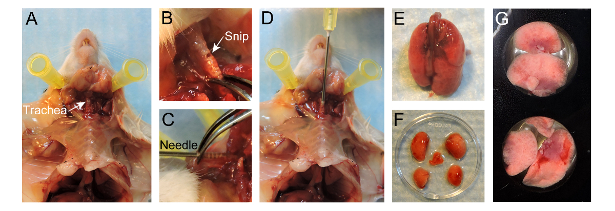
- 用手术剪刀切开皮肤至气管在胸腔,但留下胸腔完好。独立于胸腔皮肤。通过去除周围结缔组织,注意不要损伤气管本身( 图1A)暴露气管。
- 剪断的小开口在平行于软骨环暴露气管直径约为1毫米,如靠近喉部尽可能(图1B)。要小心,不要通过气管完全切断。
- 取一个20G的针和轻轻插入针4-5毫米进气管没有任何反力(图1D)。针的端部应通过气管(图1C)可见。使用镊子稳定气管针。或者,缝线可以阿罗绑UND气管以保持针就位。
注:通过插入过深,隆突可能会受到创伤,或只有一个肺的一侧可能被夸大。 - 填的注射器用400μl37℃的2%低熔点温度琼脂糖(直接从恒温浴中取出)的。确保夹层板站起来,慢慢地灌输通过针进入肺内温暖琼脂糖,使用〜400微升膨胀肺部。
注:观看肺部胸腔内充气。不要过分夸大,因为它会破裂的肺。 - 一旦肺充气,充〜胸廓的⅔,拆下注射器和保持气管内的针,以防止任何琼脂糖泄漏。
- 倒在充气肺部大约50ml 20℃的PBS,以允许肺部内的琼脂糖设置和固化。慢慢用镊子取出针并关闭气管,以防止任何非固化琼脂糖泄漏。
- 通过执行胸骨切开术暴露肺部并随后切除肺。对于肺切除,保住气管,同时通过气管切割彻底。轻轻拉动气管上,切去结缔组织和食道同时拉动肺出胸腔直到肺部从小鼠(图1E)分离。
- 沉浸在温暖的RPMI-1640肺部洗掉过量的血液,并通过使用剪刀和镊子切断裂片'主茎支气管在脐(图1F)的裂片轻轻分开。
- 放置裂片,与扁平表面向下最大化成像表面,在24孔成像板(图1G)的孔中。添加在叶前100微升37℃RPMI-1640。放置在叶片的顶部几15毫米圆形显微镜盖玻片以防止其浮动。
- 倾温PBS到周围井,以防止从EVAP所述的RPMI-1640培养基orating。插入24孔板进平衡气候室并保持在37℃的肺叶与空气和5%的CO 2。插入气候室上的共焦显微镜的阶段。
注意:其他气体的混合物(例如,5%O 2,5%CO 2的N 2以检查缺氧/低氧气的条件下,细胞的行为)也可以考虑。

图1.协议的准备肺部实时成像的。(A)准备鼠标后气管暴露。在平行于软骨环暴露气管制成(B)的小剪断。 (C),20G的针头插入4-5毫米的进气管。 (D)的400微升的2%低熔点温度琼脂糖到肺部的灌输。 (E)INFLated肺部鼠标分开。充气后(F)裂片分离。 (G)叶放置在一个24孔成像板的好。 请点击此处查看该图的放大版本。
6.采集和图像分析
注:图像可以与各种纺丝通过各种软件程序支持的磁盘共焦显微镜的获取。在这个协议中,无论是与一个特制的旋转盘共聚焦显微镜或禅与市售的旋转盘聚焦显微镜μManager用于图像采集,而了Imaris用于电影编辑和分析。
- 获取使用μManager图像。收购使用μManager软件映像的详细分步协议是前面所述18。
要么 - 获取图像使用图像分析软件,如禅( 见图S1)。
- 点击"查找"选项卡,并选择在"光路" 工具 (图S1A,红色框)目标(10倍20倍或)。随后,点击"眼睛- DAPI'看看通过目镜(图S1A,蓝盒)CFP通道。本地化样品使用显微镜手动。点击"所有Off'after组织是视野的中心。
- 点击"捕获"选项卡,设置图像采集的所有参数。
- 在"渠道"工具,单击"+"按钮(图S1B,红色框)。一个弹出菜单显示和搜索染料(S)目前在"染料数据库"(图S1B)的样本。选择染料,然后单击"添加"。
注:该计划将设置为优化所有过滤器。染料可以删除通过选择依次点击垃圾桶按钮 (图S1B,黄框)。 - 在"采集模式"菜单中,将"像素合并"到5x5的。在渠道上菜单上ECFP双击将其选中。降低激光功率到20%,使样品不会同时进行图像采集设置的参数进行漂白。
- 选中"瓷砖"框在"实验管理"部分,瓷砖工具出现在"多维收购"工具组( 图S1C)英寸点击"高级设置"按钮,从摄像机查看实时图像。在"位置"部分,点击"添加"按钮,4〜6个位置添加到实验。要删除的位置,选择位置,单击垃圾箱按钮。
- 在"采集参数"工具组中,打开了"聚焦战略"工具,然后选择"绝对E固定从下拉列表中Z位置"。
- 检查的Z-Stack框在"实验管理"部分和的Z-Stack工具出现在"多维收购"工具组( 图S1D)英寸上的一个位置双击在"位置"部分,然后按'活'。手动先设置和设置的摄像视场的最后位置。于4微米的设置间隔。
注意:程序将确定切片为选定的范围和时间间隔的数目。理想的情况下,5-7片方便,以便有足够的可视化和快速的图像采集。 - 检查在"实验管理"部分的"时间序列"对话框。设置所需的"时间"和"间隔"中的"时间序列"的工具,出现在"多维收购"工具组( 图S1E)的时间。
- 在'习得化模式"菜单中,将"像素合并"2×2。在通道菜单中荧光双击选中它并增加激光功率为100%。按"现场",并调整"曝光时间"。重复此为每个荧光基团。
- 选中"启用自动保存"复选框。在文件名选择一个文件夹和类型。所有采集的图像会自动保存此文件夹中。
- 在"实验管理"部分,点击"开始试验"开始图像采集。
- 图像采集后,编译了Imaris软件的原始数据。将图像转换为.ims文件,并可以进行调节。对于文件的转换,进行调整,并使用了Imaris保存电影的详细分步协议是前面所述18。
- 当保存影片,设定每秒(fps)的"帧率"5张。
Access restricted. Please log in or start a trial to view this content.
结果
使用纺丝盘共焦显微镜,各种小鼠模型系统和注射剂,转移微环境可以可视化并随时间跟踪。使用MMTV-PyMT; ACTB-ECFP;的c-FMS-EGFP三重转基因小鼠模型,不同的细胞组分的荧光标记(图2A,电影1)。肺实质的典型结构可以在CFP通道由于所有细胞中的β肌动蛋白启动子下表达的ECFP可视化。这些表现为大部分肺组织结构中细胞的大/多肺转移是很容易解决。髓细胞可视化在...
Access restricted. Please log in or start a trial to view this content.
讨论
这份手稿描述了转移的小鼠模型体外肺转移的实时成像的详细方法。该成像协议提供了肺的微环境中的动态和空间肿瘤细胞 - 基质相互作用的直接可视化。它是一个相对容易的和快速的方法,它允许为至少4小时的肺转移的可靠的成像。从这些实验中获得的影可以用来跟踪动态过程如细胞运动和细胞相互作用。
被描述为肺转移的产生方法有两种:一种基因工程小鼠模型和...
Access restricted. Please log in or start a trial to view this content.
披露声明
The authors have no conflicts of interest to disclose. All animal experiments were conducted in accordance with IACUC approved protocols, UCSF.
致谢
We thank Nguyen H. Nguyen for her technical help and Audrey O’Neill for support with the Zeiss Cell Observer spinning-disk confocal microscope. This work was supported by a Department of Defense postdoctoral fellowship (W81XWH-11-01-0139) and the Weizmann Institute of Science-National Postdoctoral Award Program for Advancing Women in Science (to V.P.).
Access restricted. Please log in or start a trial to view this content.
材料
| Name | Company | Catalog Number | Comments |
| MMTV-PyMT/FVB mice | Jackson Laboratory | 2374 | Female mice |
| ACTB-ECFP/FVB mice | UCSF Werb lab | Female mice | |
| c-fms-EGFP/FVB mice | UCSF Werb lab | Female mice | |
| FVB mice | Jackson Laboratory | 1800 | Female mice |
| GFP+ VO-PyMT cells | UCSF Werb lab | ||
| 70,000 kDa Dextran, rhodamine-conjugated | Invitrogen | D1818 | Dilute to 4mg/ml in 1 x PBS and store at -20 °C. Use 0.4 mg per animal. |
| 10,000 kDa Dextran, Alexa Fluor 647 conjugated | Invitrogen | D22914 | Dilute to 4mg/ml in 1 x PBS and store at -20 °C. Use 0.4 mg per animal. |
| Anti-mouse Gr-1 antibody Alexa Fluor 647 | UCSF Monoclonal antibody core | Stock 1mg/ml. Use 7 ug per animal. | |
| Anesthetic | Anesthesia approved by IACUC, used for anesthesia and/or euthanesia | ||
| 1X PBS | UCSF cell culture facility | ||
| PBS, USP sterile | Amresco INC | K813-500ML | Ultra pure grade for i.v. injection |
| Styrofoam platform | Will be used as dissection board | ||
| Fine scissors sharp | Fine Science Tools | 14060-11 | |
| Forceps | Roboz Surgical Store | RS-5135 | |
| Hot bead sterilizer | Fine Science Tools | 18000-45 | Turn ON 30min before use |
| Air | UCSF | ||
| Oxygen | UCSF | ||
| Carbon dioxide | UCSF | ||
| 1 mL syringe without needle | BD | 309659 | |
| 27 G x 1/2 needle | BD | 305109 | for i.v. injection |
| 20 G x 1 needle, short bevel | BD | 305178 | |
| Low-melting-temperature agarose | Lonza | 50111 | To make 10 ml of solution, weigh 0.2 g of agarose, add to 10 ml 1 x PBS, and heat to dissolve. Agarose will solidify at room temperature, so maintain in a 37 °C water bath until used for inflation. |
| RPMI-1640 medium without phenol red | Life Technologies | 11835-030 | |
| 24 well Imaging plate | E&K scientific | EK-42892 | |
| Glass cover slides, 15 mm | Fisher Scientific | 22-031-144 | |
| Digital CO2 and temperature controller | Okolab | DGTCO2BX | http://www.oko-lab.com |
| Climate chamber | Okolab | http://www.oko-lab.com | |
| Cell Observer spinning disk confocal microscope | Zeiss | ||
| Zen software | Zeiss | ||
| Inverted microscope | Carl Zeiss Inc | Zeiss Axiovert 200M | |
| ICCD camera | Stanford Photonics | XR-Mega-10EX S-30 | |
| Spinning disk confocal scan-head | Yokogawa Corporation | CSU-10b | |
| Imaris | Bitplane | ||
| mManager | Vale lab, UCSF | Open-source software |
参考文献
- Chaffer, C. L., Weinberg, R. A. A perspective on cancer cell metastasis. Science. 331 (6024), 1559-1564 (2011).
- Plaks, V., Kong, N., Werb, Z. The cancer stem cell niche: how essential is the niche in regulating stemness of tumor cells. Cell stem cell. 16 (3), 225-238 (2015).
- Nguyen, D. X., Bos, P. D., Massague, J. Metastasis: from dissemination to organ-specific colonization. Nat Rev Cancer. 9 (4), 274-284 (2009).
- Egeblad, M., Ewald, A. J., et al. Visualizing stromal cell dynamics in different tumor microenvironments by spinning disk confocal microscopy. Dis Model Mech. 1 (2-3), 155-167 (2008).
- Ellenbroek, S. I. J., van Rheenen, J. Imaging hallmarks of cancer in living mice. Nat Rev Cancer. 14 (6), 406-418 (2014).
- Looney, M. R., Thornton, E. E., Sen, D., Lamm, W. J., Glenny, R. W., Krummel, M. F. Stabilized imaging of immune surveillance in the mouse lung. Nat Methods. 8 (1), 91-96 (2011).
- Thornton, E. E., Krummel, M. F., Looney, M. R. Live imaging of the lung. Cur Protoc Cytom. , (2012).
- Thornton, E. E., Looney, M. R., et al. Spatiotemporally separated antigen uptake by alveolar dendritic cells and airway presentation to T cells in the lung. J Exp Med. 209 (6), 1183-1199 (2012).
- Mendoza, A., Hong, S. -H., et al. Modeling metastasis biology and therapy in real time in the mouse lung. J Clin Invest. 120 (8), 2979-2988 (2010).
- Lelkes, E., Headley, M. B., Thornton, E. E., Looney, M. R., Krummel, M. F. The spatiotemporal cellular dynamics of lung immunity. Trends Immunol. 35 (8), 379-386 (2014).
- Guy, C. T., Cardiff, R. D., Muller, W. J. Induction of mammary tumors by expression of polyomavirus middle T oncogene: a transgenic mouse model for metastatic disease. Mol Cell Biol. 12 (3), 954-961 (1992).
- Hadjantonakis, A. -K., Macmaster, S., Nagy, A. Embryonic stem cells and mice expressing different GFP variants for multiple non-invasive reporter usage within a single animal. BMC Biotechnol. 2, (2002).
- Casbon, A. -J., Reynaud, D., et al. Invasive breast cancer reprograms early myeloid differentiation in the bone marrow to generate immunosuppressive neutrophils. Proc Natl Acad Sci USA. 112 (6), 566-575 (2015).
- Donovan, J., Brown, P. Parenteral injections. Curr Protoc Immunol. , (2006).
- Halpern, J., Lynch, C. C., et al. The application of a murine bone bioreactor as a model of tumor: bone interaction. Clin Exp Metastas. 23 (7-8), 345-356 (2006).
- Sasmono, R. T., Oceandy, D., et al. A macrophage colony-stimulating factor receptor-green fluorescent protein transgene is expressed throughout the mononuclear phagocyte system of the mouse. Blood. 101 (3), 1155-1163 (2003).
- Ewald, A. J., Werb, Z., Egeblad, M. Dynamic, long-term in vivo imaging of tumor-stroma interactions in mouse models of breast cancer using spinning-disk confocal microscopy. Cold Spring Harb Protoc. (2), (2011).
- Bonnans, C., Lohela, M., Werb, Z. Real-time imaging of myeloid cells dynamics in ApcMin/+ intestinal tumors by spinning disk confocal microscopy. J Vis Exp. (92), (2014).
- Nakasone, E. S., Askautrud, H. A., et al. Imaging tumor-stroma interactions during chemotherapy reveals contributions of the microenvironment to resistance. Cancer cell. 21 (4), (2012).
- Cheng, N., Lambert, D. L. Mammary transplantation of stromal cells and carcinoma cells in C57BL/6J mice. J Vis Exp. (54), (2011).
- Al-Mehdi, A. B., Tozawa, K., Fisher, A. B., Shientag, L., Lee, A., Muschel, R. J. Intravascular origin of metastasis from the proliferation of endothelium-attached tumor cells: a new model for metastasis. Nat Med. 6 (1), 100-102 (2000).
- Wong, C. W., Song, C., et al. Intravascular location of breast cancer cells after spontaneous metastasis to the lung. Am J Pathol. 161 (3), 749-753 (2002).
- Liang, C. -C., Park, A. Y., Guan, J. -L. In vitro scratch assay: a convenient and inexpensive method for analysis of cell migration in vitro. Nat protoc. 2 (2), 329-333 (2007).
- Nelson, K., Bobba, C., Ghadiali, S., Hayes, D. J., Black, S. M., Whitson, B. A. Animal models of ex vivo lung perfusion as a platform for transplantation research. World J Exp Med. 4 (2), (2014).
- Magness, S. T., Bataller, R., Yang, L., Brenner, D. A. A dual reporter gene transgenic mouse demonstrates heterogeneity in hepatic fibrogenic cell populations. Hepatology. 40 (5), 1151-1159 (2004).
- Motoike, T., Loughna, S., et al. Universal GFP reporter for the study of vascular development. Genesis. 28 (2), (2000).
- Srivastava, M. K., Andersson, A., et al. Myeloid suppressor cells and immune modulation in lung cancer. Immunotherapy. 4 (3), (2012).
- Craig, A., Mai, J., Cai, S., Jeyaseelan, S. Neutrophil recruitment to the lungs during bacterial pneumonia. Infect Immun. 77 (2), 568-575 (2009).
- Kreisel, D., Nava, R. G., et al. In vivo two-photon imaging reveals monocyte-dependent neutrophil extravasation during pulmonary inflammation. Proc Natl Acad Sci USA. 107 (42), 18073-18078 (2010).
Access restricted. Please log in or start a trial to view this content.
转载和许可
请求许可使用此 JoVE 文章的文本或图形
请求许可探索更多文章
This article has been published
Video Coming Soon
版权所属 © 2025 MyJoVE 公司版权所有,本公司不涉及任何医疗业务和医疗服务。