
需要订阅 JoVE 才能查看此. 登录或开始免费试用。
Method Article
从羊膜中提取Lumican及其储存温度的测定
摘要
本协议描述了从羊膜(AM)中提取lumican及其储存条件作为AM提取物(AME)在-20°C,4°C和室温(RT)下6,12,20和32天以量化其蛋白质和lumican浓度。
摘要
Lumican是人羊膜(AM)中一种富含亮氨酸的小蛋白聚糖,可促进角膜上皮化和胶原纤维的组织,保持角膜透明。本工作提出了一种从AM中提取蛋白质得到Lumican的方法。此外,还评估了在不同温度和时间段下储存的AM提取物(AME)中Lumican的稳定性。解冻100mgAM并机械去上皮化。将去上皮化的AM冷冻并粉碎,直至获得细粉,用2.5mL盐水缓冲液与蛋白酶抑制剂溶解并离心进行蛋白质提取。收集上清液并在-20°C、4°C和室温(RT)下保存6、12、20和32天。之后,在每个AME中量化Lumican。该技术允许从AM中提取lumican的可访问和可获取的方案。Lumican浓度受储存时间和温度条件的影响。Lumican在AME中在-20°C和4°C下储存12天明显高于其他AME。这种lumican提取可用于开发治疗方法和药物解决方案。需要进一步的研究来确定AME发光胶在再上皮化和伤口愈合过程中的用途。
引言
角膜影响最常用的治疗方法之一是羊膜移植;然而,近年来,出现了使用羊膜组织的各种成分作为替代和辅助治疗的新建议。在研究最多的AM成分中,是从AM提取物(AME)中获得的成分1,2,3,4,5,6,7。AM含有多种可溶性因子,例如抗血管生成蛋白,白细胞介素(IL),金属蛋白酶(TIMP)的组织抑制剂,由TSG-6介导的抑制中性粒细胞细胞外陷阱的抗炎蛋白,生长因子:表皮生长因子(EGF),转化生长因子(TGF)(α和β),角质形成细胞生长因子(KGF),肝细胞生长因子(HGF)和Lumican,通过调节胶原纤维生成来维持角膜透明度1,2,3,4,5,6,7,8,9.
Lumican是一种富含亮氨酸的小蛋白聚糖(SLRP),是角膜基质基质中间质胶原酶的主要细胞外成分之一,负责组织胶原纤维并保持角膜透明度4,10,11。蛋白聚糖是细胞外基质(ECM)中的分子,是进行细胞信号传导和维持细胞内稳态的主要分子12。据报道,ECM蛋白在伤口愈合过程中驱动细胞增殖,分化和迁移过程11。
证据表明Lumican可能参与角膜再上皮化过程。Saika 等人在一项研究中表明,角膜损伤后,可以在损伤后的前 8 小时至最多 3 天内在角膜角质细胞中检测到 lumican。在第二天和第三天呈现最高浓度的lumican,该蛋白聚糖随后在第七天无法检测到13。这些数据表明Lumican参与角膜再上皮化过程的激活。另一方面,在另一项研究中,据报道,没有发光胶会延迟再上皮化;有趣的是,添加Lumican可以加速再上皮化过程4,11,13。同样,最近的一项研究报告说,Lumican可以调节角膜缘成纤维细胞14的炎症功能,这表明Lumican作为炎症,抗纤维化和再上皮化反应的调节剂发挥作用。同样,lumican可以通过与Fas-FasL等信号分子相互作用来调节角膜反应。此外,在敲除Lum-/-小鼠模型中没有Lumican表明缺乏lumican信号传导会阻止充分的角膜修复15。
该方法主要旨在展示一种从AM中提取lumican的可行且平易近人的方法。与以前的研究相比,使用这种有利的lumican提取方法可以获得相似浓度的蛋白质,从而减少了处理时间,并使研究人员更方便16。此外,这种AME发光胶可用作角膜修复和再上皮化过程的佐剂。
Access restricted. Please log in or start a trial to view this content.
研究方案
所有实验程序均已获得机构审查委员会的批准(项目编号:CEI-2020/06/04)。AM是从瓦伦西亚孔德羊膜库(来自去识别的人类受试者)获得的,该数据库是按照Chávez-García等人的描述制备的17。
1.羊膜提取物的制备
- 从羊膜库获取 100 毫克 AM。
注意:根据之前的报告,50 毫克 AM 总共分泌 10 ng/mL 的卢米康14。为了获得更高浓度的lumican,请使用100mg的AM,相当于总面积为32cm2。 - 如果AM被冷冻,请在室温下解冻。
注意:在 II B 级层流罩下执行以下步骤。 - 用10mL无菌平衡盐溶液(BSS,参见 材料表)在培养皿中洗涤AM2分钟。
- 将 BSS 倒入烧杯中。
- 重复步骤3并目视确认培养皿BSS中不存在甘油培养基。
注:重复步骤 3。根据需要,直到培养皿BSS中不存在甘油培养基。
- 将AM与10mL分散酶II(1.7IU / mL,参见 材料表)在37°C,5%CO2 下孵育30分钟。
注意:分散酶II是一种中性蛋白酶,对上皮细胞具有温和的活性。该酶有效地将完整的表皮与真皮分离,并分离完整的上皮片18。 - 分散酶孵育后,用橡胶警察进行机械去上皮化14 (见 材料表)。通过显微镜可视化确认去上皮化。
注意:去上皮化过程在使用4x和20x物镜的倒置显微镜中得到证实。可视化组织以排除任何细胞层的存在。 - 用10mL BSS在培养皿中洗涤AM2分钟。将 BSS 倒入烧杯中。
- 将去上皮化的AM(dAM)放入2 mL微量离心管中。将dAM浸入液氮中40分钟。
- 在-85°C的预冷砂浆中手动研磨冷冻dAM2-3分钟,直到获得细粉。
- 在研钵中,用 2.5 mL 蛋白酶抑制剂溶液(含蛋白酶抑制剂的 BSS)溶解 dAM 粉末。
注意:每片蛋白酶抑制剂由以下酶混合物组成:胰腺提取物(0.02mg / mL),热溶酶(金属蛋白酶)(0.0005mg / mL),胰凝乳蛋白酶(0.002mg / mL),胰蛋白酶(0.02mg / mL)和木瓜蛋白酶(0.33mg / mL)(参见 材料表)。 - 用微量移液管收集混合物,并在手术刀的帮助下清洁砂浆壁。将混合物放入 5 mL 管中。
- 与涡旋充分混合30秒。
- 通过在4°C下以34×g离心20分钟,并立即在4°C下以3360×g离心20分钟,使组织混合物匀浆。
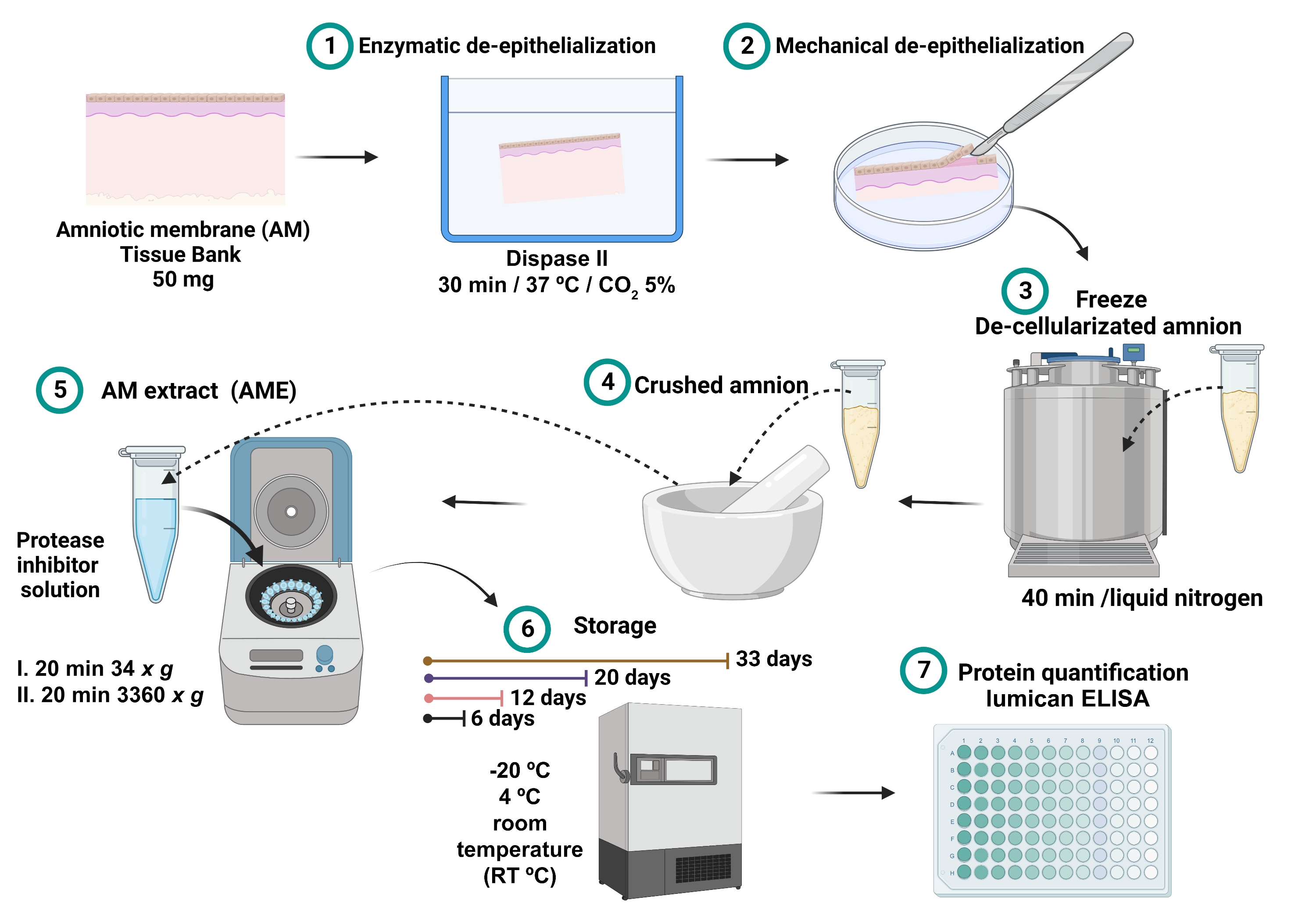
- 收集的上清液是AME(图1)。将 0.7 mL 的每个 AME 储存在不同的 2 mL 微量离心管中,在 -20 °C、4 °C 和室温 (RT) 的不同温度条件下储存 6、12、20 和 33 天。

图1:AME制备和lumican浓度测量的过程 。 将100mgAM与分散酶II在37°C孵育30分钟并机械去上皮化。将去上皮化的AM洗涤并浸入液氮中40分钟,然后粉碎直至得到细粉,用2.5mL盐水缓冲液与蛋白酶抑制剂溶解并离心。收集上清液并在-20°C,4°C和室温下储存6,12,20和32天,直到总蛋白质和Lumican定量。 请点击此处查看此图的大图。
2. AME蛋白定量
注意:AME中总蛋白的定量必须在获得后立即进行。使用Lowry蛋白测定法定量蛋白质,并遵循制造商的说明(参见 材料表)。建议将所有标准品和样品一式三份测定。
- 将 40 μL 每个 AME 样品移液到 96 孔微孔板中。
- 使用牛血清白蛋白(BSA)标准品制备到同一微孔板中的标准曲线,最终BSA浓度为0-1,500μg/ mL(0,1,5,25,250,500,750,1,000和1,500μg/ mL)。
- 将 200 μL 改良的 Lowry 试剂移液到每个孔中。立即在平板搅拌机上混合30秒。
- 用铝箔覆盖微孔板,并在室温下孵育10分钟。
- 将 20 μL 1x Folin-Ciocalteu 试剂移液到每个孔中。立即在平板搅拌机上混合30秒。
注意:要制备 1x Folin-Ciocalteu 试剂,请用超纯水 1:1 稀释 2x (2N) 试剂。在使用当天准备1x Folin-Ciocalteu试剂,因为稀释的试剂不稳定。 - 用铝箔从光上覆盖微孔板,并在室温下孵育30分钟。
- 在ELISA板光谱仪中测量660nm处样品的吸光度(参见 材料表)。
注意:可以在 650 nm 和 750 nm 之间的波长下测量颜色。 - 平均标准空白样品的660 nm吸光度值,并从标准和未知样品的其他660 nm值中减去该值。
- 用ELISA板光谱仪在端点模式下以低振荡10秒测量吸光度。
- 使用标准曲线确定每个未知样品的蛋白质浓度。
- 对于蛋白质的计算,使用Y轴上的吸光度值与每条标准BSA曲线X轴上的mg / mL浓度(以mg / mL为单位)从线性回归图中确定浓度。
- 得到线性回归方程和r值来计算蛋白质浓度。
注意:结果表示为总蛋白质相对于mg AM 的标准化相对浓度值(μg/mL 蛋白质/mg AM 组织)。
- 得到线性回归方程和r值来计算蛋白质浓度。
3. AME中Lumican的定量
注意:必须在不同储存条件和时间段储存的AME中测量Lumican的浓度。使用三明治ELISA量化Lumican并遵循制造商的说明。建议将所有标准品和样品一式两份测定。
- 将人lumican捕获抗体(参见 材料表)稀释至磷酸盐缓冲盐水(PBS)中采用的浓度。
注意:捕获抗体瓶含有120μg抗体。用 0.5 mL PBS 复溶后,在 2 μg/mL 的工作溶液中稀释捕获抗体。- 立即将每孔 100 μL 稀释的捕获抗体移液至 96 孔微孔板中。封闭板并在室温下孵育过夜。
- 吸出每个孔,并使用多通道移液器用 300 μL 洗涤缓冲液:0.05% 聚氧乙烯脱水山梨糖醇单月桂酸酯 20 在 PBS、pH 7.2-7.4(参见 材料表)中移液洗涤。重复三次。
注意:最后一次洗涤后,将盘子翻出,然后轻轻拍打纸巾,以去除任何剩余的洗涤缓冲液。 - 通过向每个孔中加入 300 μL 试剂稀释剂来封闭板:PBS 中的 1% BSA,pH 7.2-7.4,0.2 μm 过滤(参见 材料表)。在室温下孵育1小时。
- 重复步骤 2。
- 使用0-8,000 pg/mL的两倍连续稀释液将标准曲线制备到96孔微孔板中,最终浓度为125、250、500、1,000、2,000、4,000和8,000 pg/mL。ELISA夜光聚糖试剂盒包含75 ng的重组夜光聚糖标准品(见 材料表)。
- 在捕获抗体包被的 96 孔微孔板中加入 100 μL 样品和标准曲线。
- 盖上微孔板,在室温下在紧凑型摇臂中以低搅拌孵育2小时,保持速度在2-3rpm之间。
- 重复步骤 2。
- 向每个孔中加入 100 μL 生物素化检测抗体(参见 材料表)。从光中盖上,并在室温下在紧凑型摇臂中以低搅拌孵育2小时,保持速度在2-3rpm之间。
注意:生物素化检测抗体瓶含有24μg抗体。用 1.0 mL 试剂稀释剂复溶后,在 400 ng/mL 的工作溶液中稀释生物素化检测抗体。 - 重复步骤 2。
- 向每个孔中加入 100 μL 链霉亲和素-辣根过氧化物酶(HRP,参见 材料表)的工作稀释液。从光上覆盖微孔板并在室温下孵育20分钟。
注意:反应性链霉亲和素 HRP 的浓度为 40 倍。工作溶液1x链霉亲和素-HRP用试剂稀释剂制成。
注意:避免将板放在直射光下。 - 重复步骤 2。
- 最后,向每个孔中加入 100 μL 底物四甲基联苯胺(TMB,参见 材料表)溶液。
注意:用试剂盒中提供的等体积的稳定过氧化氢30%溶液制备TMB溶液。
注意:使用前立即准备溶液并将其保持在室温下。 - 在室温下在黑暗的地方孵育30分钟。
注意:避免将板放在直射光下。不要吸出TMB溶液,因为不需要进一步洗涤。 - 加入 50 μL 1N H2SO4 终止液以停止比色反应。轻轻敲击盘子以确保充分混合。
- 立即使用ELISA板光谱仪中设置为450nm的酶标仪确定每个孔的吸光度。
- 用ELISA板光谱仪在端点模式下以低振荡10秒测量吸光度。
- 平均标准空白样品的450 nm吸光度值,并从标准和未知样品的其他450 nm值中减去它。
- 使用标准曲线确定每个未知样品的lumican浓度。
- 为了计算Lumican浓度,使用Y轴上的吸光度值与每条标准lumican曲线X轴上的pg/mL浓度进行线性回归图。
- 得到线性回归方程和r值来计算发光胶浓度。
注意:卢米康的浓度相对于提取的组织毫克标准化。结果表示为lumican与mg AM(ng / mL lumican/mg AM组织)的标准化相对浓度值。
- 得到线性回归方程和r值来计算发光胶浓度。
Access restricted. Please log in or start a trial to view this content.
结果
结果报告为标准偏差 (SD) ±平均值。进行学生的 t检验和方差分析(方差分析)。 < 0.05 的 P 值被认为具有统计学意义。使用统计软件进行统计分析(见 材料表)。
AME中的总蛋白质量受时间和储存条件的影响。所有AME的基础蛋白浓度相似;总蛋白的范围为2.7±0.3μg/mL,评估的样品之间没有显着差异。然而,当样品储存12、20和32天时,观察到蛋白质浓?...
Access restricted. Please log in or start a trial to view this content.
讨论
本研究分析了AME中Lumican的存在及其在不同储存条件下的稳定性的直接相关性。有趣的是,当AME中的总蛋白质浓度被量化时,储存后蛋白质浓度增加。有证据表明,有三种机制可以改变冷冻储存中的蛋白质浓度:冷变性、溶质的冷冻浓度和冰诱导的蛋白质结构部分展开19。由于样品中液相的结晶,冷冻过程可能会影响储存样品上的蛋白质浓度。结果表明,这可能是冷冻浓度的过程,...
Access restricted. Please log in or start a trial to view this content.
披露声明
该研究由墨西哥国立自治大学研究和技术创新项目支持计划(批准号PAPIIT IN203821)和教育,科学,技术和创新部(批准号SECTEI 250/2019)资助。
致谢
作者没有相互竞争的经济利益。
Access restricted. Please log in or start a trial to view this content.
材料
| Name | Company | Catalog Number | Comments |
| 1 N H2SO4 stop solution | R&D Systems | DY994 | |
| 100 μL micropipette | Eppendorf | ||
| 1000 μL micropipette | Eppendorf | ||
| 15 mm Petri dish | Symlaboratorios | ||
| 18 G Needle (1.2 mm x 40 mm) | BD Becton Dickinson | 305211 | |
| 2 mL microcentrifuge tube | Eppendorf | Z606340 | |
| 20 mL plastic syringe | BD Becton Dickinson | 302562 | |
| 20 μL micropipette | Eppendorf | ||
| 20-200 μL micropipette | Eppendorf | ||
| 5 mL microcentrifuge tube | Eppendorf | 30119401 | |
| 96-well microplate | SARSTEDT | 821581 | |
| Aluminum foil | N/A | N/A | |
| Amniotic membrane | Instituto de Oftalmologia Conde de Valenciana Amnion Bank | 100 mg | |
| Balanced salt solution | Bausch + Lomb | BSS-403802 | |
| Beaker | N/A | N/A | |
| BioRender | BioRender | figures design | |
| Compact Rocker | BioRad | 970822DD | Mod. 5202SD-BIO |
| complete, EDTA-free, Protease inhibitor cocktail tablets | Roche | 11 873 580 001 | Protease Inhibitor |
| Daiggner vortex Genie 2 | A.Daigger & Co. , INC | 22220A | |
| Dispase II | Gibco | 17105-041 | |
| ELISA plate spectrometer | Thermo Labsystems | 35401106 | Multiscan |
| Freezer | |||
| GraphPad Prism | GraphPad Software, Inc | version 9 | statistical analysis and graphic program |
| Human lumican DuoSet ELISA kit | R&D Systems | DY2846-05 | includes human Lumican capture antibody |
| Incubator | Forma Scientific | 3326 S/N 36481-7002 | |
| Inverted light Microscope | Olympus | 6A13921 | to confirm de-epithelialization Mod.CK2 |
| Laminar flow hood | Forma Scientific | 14753-567 | Mod.1184 |
| Liquid nitrogen | N/A | N/A | |
| Mortar | N/A | N/A | |
| Multi-channel pipettor | Eppendorf | ||
| Nitrogen Tank | Thermo Scientific | Mod. Biocan 20 | |
| Paper towels | N/A | N/A | |
| Phosphate-buffered saline | R&D Systems | DY006 | |
| Pierce Modified Lowry Protein Assay Kit | Thermo Scientific | 23240 | |
| Plate sealers | R&D Systems | DY992 | |
| Reagent diluent | R&D Systems | DY995 | 1% BSA in PBS, pH 7.2-7.4, 0.2 μm filtered |
| Refrigerated centrifuge | centurion scientific Ltd | 15877 | Mod. K2015R |
| Rubber policeman cell scraper | NEST | 710001 | for mechanical de-epithelialization |
| Scalpel knife | Braun | BB521 | No. 10 or 21 |
| Streptavidin-HRP 40-fold concentrated | R&D Systems | part 893975 | |
| Substrate tetramethylbenzidine (TMB) solution | R&D Systems | DY999 | |
| Toothed tweezers | Invent Germany | 6b | inox |
| Ultrapure water | PISA | ||
| Wash buffer | R&D Systems | WA126 | 0.05% Tween 20 in PBS, pH 7.2-7.4 |
参考文献
- Jirsova, K., Jones, G. Amniotic membrane in ophthalmology: properties, preparation, storage and indications for grafting-a review. Cell and Tissue Banking. 18 (2), 193-204 (2017).
- Witherel, C., Yu, T., Concannon, M., Dampier, W., Spiller, K. Immunomodulatory effects of human cryopreserved viable amniotic membrane in a pro-inflammatory environment in vitro. Cellular and Molecular Bioengineering. 10 (5), 451-462 (2017).
- Ruiz-Cañada, C., et al. Amniotic membrane stimulates cell migration by modulating transforming growth factor-β signalling. Journal of Tissue Engineering and Regenerative Medicine. 12 (3), 808-820 (2017).
- Yeh, L., et al. Soluble lumican glycoprotein purified from human amniotic membrane promotes corneal epithelial wound healing. Investigative Opthalmology & Visual Science. 46 (2), 479(2005).
- Navas, A., et al. Anti-Inflammatory and anti-fibrotic effects of human amniotic membrane mesenchymal stem cells and their potential in corneal repair. Stem Cells Translational Medicine. 7 (12), 906-917 (2018).
- Magaña-Guerrero, F., Domínguez-López, A., Martínez-Aboytes, P., Buentello-Volante, B., Garfias, Y. Human amniotic membrane mesenchymal stem cells inhibit neutrophil extracellular traps through TSG-6. Scientific Reports. 7, 12426(2017).
- Garfias, Y., Zaga-Clavellina, V., Vadillo-Ortega, F., Osorio, M., Jimenez-Martinez, M. Amniotic membrane is an immunosuppressor of peripheral blood mononuclear cells. Immunological Investigations. 40 (2), 183-196 (2010).
- Koob, T., et al. Biological properties of dehydrated human amnion/chorion composite graft: implications for chronic wound healing. International Wound Journal. 10 (5), 493-500 (2013).
- Miyagi, H., Thomasy, S., Russell, P., Murphy, C. The role of hepatocyte growth factor in corneal wound healing. Experimental Eye Research. 166, 49-55 (2018).
- Chen, S., Mienaltowski, M., Birk, D. Regulation of corneal stroma extracellular matrix assembly. Experimental Eye Research. 133, 69-80 (2015).
- Karamanou, K., Perrot, G., Maquart, F., Brézillon, S. Lumican as a multivalent effector in wound healing. Advanced Drug Delivery Reviews. 129, 344-351 (2018).
- Theocharis, A., et al. Cell-matrix interactions: focus on proteoglycan-proteinase interplay and pharmacological targeting in cancer. FEBS Journal. 281 (22), 5023-5042 (2014).
- Saika, S., et al. Role of lumican in the corneal epithelium during wound healing. Journal of Biological Chemistry. 275 (4), 2607-2612 (2000).
- Domínguez-López, A., et al. Amniotic membrane conditioned medium (AMCM) reduces inflammatory response on human limbal myofibroblast, and the potential role of lumican. Molecular Vision. 27, 370-383 (2021).
- Vij, N., Roberts, L., Joyce, S., Chakravarti, S. Lumican regulates corneal inflammatory responses by modulating Fas-Fas Ligand signaling. Investigative Opthalmology & Visual Science. 46 (1), 88(2005).
- Mahbod, M., et al. Amniotic membrane extract preparation: What is the best method. Journal of Ophthalmic and Vision Research. 9 (3), 314-319 (2014).
- Chávez-García, C., et al. Ophthalmic indications of amniotic membrane transplantation in Mexico: an eight years Amniotic Membrane Bank experience. Cell and Tissue Banking. 17 (2), 261-268 (2015).
- Stenn, K. S., Link, R., Moellmann, G., Madri, J., Kuklinska, E. Dispase, a neutral protease from Bacillus polymyxa, is a powerful fibronectinase and type IV collagenase. Journal of Investigative Dermatology. 93 (2), 287-290 (1989).
- Bhatnagar, B. S., Bogner, R. H., Pikal, M. J. Protein stability during freezing: separation of stresses and mechanisms of protein stabilization. Pharmaceutical Development and Technology. 12 (5), 505-523 (2007).
- McClain, A. K., McCarrel, T. M. The effect of four different freezing conditions and time in frozen storage on the concentration of commonly measured growth factors and enzymes in equine platelet-rich plasma over six months. BMC Veterinary Research. 15 (1), 292(2019).
- Tamhane, A., et al. Evaluation of amniotic membrane transplantation as an adjunct to medical therapy as compared with medical therapy alone in acute ocular burns. Ophthalmology. 112 (11), 1963-1969 (2005).
- Shtein, R., et al. Autologous serum-based eye drops for treatment of ocular surface disease. Ophthalmology. 127 (1), 128-133 (2020).
- Shahriari, H., Tokhmehchi, F., Reza, M., Hashemi, N. Comparison of the effect of amniotic membrane suspension and autologous serum on alkaline corneal epithelial wound healing in the rabbit model. Cornea. 27 (10), 1148-1150 (2008).
- Schuerch, K., Baeriswyl, A., Frueh, B., Tappeiner, C. Efficacy of amniotic membrane transplantation for the treatment of corneal ulcers. Cornea. 39 (4), 479-483 (2019).
- Chen, H., et al. Amniotic membrane transplantation for persistent corneal ulcers and perforations in acute fungal keratitis. Cornea. 25 (5), 564-572 (2006).
- Guo, Q., et al. A comparison of the effectiveness between amniotic membrane homogenate and transplanted amniotic membrane in healing corneal damage in a rabbit model. Acta Ophthalmologica. 89 (4), 315-319 (2011).
- Sabater, A., Perez, V. Amniotic membrane use for management of corneal limbal stem cell deficiency. Current Opinion in Ophthalmology. 28 (4), 363-369 (2017).
- Ahmad, T., et al. Autolysis of bovine skin, its endogenous proteases, protease inhibitors and their effects on quality characteristics of extracted gelatin. Food Chemistry. 265, 1-8 (2018).
- Mullegama, S. V., et al. Nucleic acid extraction from human biological samples. Methods in Molecular Biology. 1897, 359-383 (2019).
- Skog, M., et al. The effect of enzymatic digestion on cultured epithelial autografts. Cell Transplantation. 28 (5), 638-644 (2019).
Access restricted. Please log in or start a trial to view this content.
转载和许可
请求许可使用此 JoVE 文章的文本或图形
请求许可探索更多文章
This article has been published
Video Coming Soon
版权所属 © 2025 MyJoVE 公司版权所有,本公司不涉及任何医疗业务和医疗服务。