Atención visual: fMRI Control atencional basado en la investigación del objeto
Fuente: Laboratorios de Jonas T. Kaplan y Sarah I. Gimbel, University of Southern California
El sistema visual humano es increíblemente sofisticado y capaz de procesar grandes cantidades de información muy rápidamente. Sin embargo, la capacidad del cerebro para procesar la información no es un recurso ilimitado. Atención, la capacidad para procesar selectivamente la información es relevante para los objetivos actuales y hacer caso omiso de información que no es, por lo tanto es una parte esencial de la percepción visual. Algunos aspectos de la atención son automáticas, mientras que otros están sujetos al control voluntario y consciente. En este experimento se exploran los mecanismos de control atencional voluntaria o "top-down" en el procesamiento visual.
Esta aprovecha de experimento la organización ordenada de la corteza visual para examinar cómo arriba atención selectiva puede modula el procesamiento de estímulos visuales. Ciertas regiones de la corteza visual parecen ser especializado para el procesamiento de elementos visuales específicos. Específicamente, el trabajo por Kanwisher et al. 1 ha identificado un área de la convolución del cerebro fusiforme del lóbulo temporal inferior que es significativamente más activo cuando sujetos ve caras en comparación a cuando se observan otros objetos comunes. Esta área ha llegado a ser conocido como el área fusiforme de la cara (FFA). Otra región del cerebro conocida como el área Parahippocampal del lugar (PPA), responde fuertemente a las casas y lugares, pero no a las caras. 2 dado que sabemos cómo estas regiones responden a tipos específicos de estímulos, su actividad puede ser explorada para identificar un componente clave de la atención visual de visión.
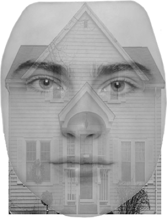
Este video muestra cómo utilizar fMRI para localizar la FFA y PPA en el cerebro y entonces examina cómo objeto-control basado en la atención modula actividad en estas áreas. El uso de un localizador funcional para restringir la prueba de hipótesis posterior es una técnica poderosa de proyección de imagen funcional. Los participantes se someterán a MRI funcional mientras se presenta con una imagen superpuesta de una cara y una casa. A pesar de una cara y una casa se presentan en cada estímulo, predecimos que los patrones de actividad en los FFA y PPA va a cambiar en base a qué artículo es ser atendido. 3
1. participante reclutamiento
- Reclutar a 20 participantes.
- Los participantes deben ser diestros y no tienen antecedentes de trastornos neurológicos o psicológicos.
- Los participantes deben tener visión normal o corregida a normal para que sean capaces de ver los indicios visuales correctamente.
- Los participantes no deben tener metal en su cuerpo. Se trata de un requisito de seguridad debido al alto campo magnético en fMRI.
- Participantes no debe sufrir de claustrofobia
En las exploraciones de localizador, FFA bilateral estaban más activas cuando temas mirar caras que cuando estaban viendo casas. Por el contrario, el PPA era más activo cuando sujetos estaban viendo casas que cuando estaban viendo caras (figura 2). Estas regiones, localizadas a través de los análisis de diseño de bloque, fueron utilizadas posteriormente como regiones de interés para extraer señales relacionadas a cambio de atención a las caras y a las casas durant...
El uso de exploraciones del localizador es una herramienta poderosa para neuroimagen cognitiva y tiene algunas ventajas distintas sobre todo cerebro. Al enfocar una hipótesis en un número pequeño de lugares específicos que han conocido las propiedades de respuesta, podemos generar predicciones muy concretas con alto poder estadístico. Todo cerebro voxel-sabio deben controlar estudios neuroimaging para las decenas de miles de pruebas estadísticas que se realizan en cada lugar en el cerebro, un proceso que reduce la ...
- Kanwisher N.G, McDermott J, Chun M.M. (1997). The fusiform face area: a module in human extrastriate cortex specialized for face perception. J. Neurosci., 17, 4302-4311.
- Epstein, R., & Kanwisher, N. (1998). A cortical representation of the local visual environment. Nature, 392, 598-601.
- Serences, J. T., Schwarzbach, J., Courtney, S. M., Golay, X., & Yantis, S. (2004). Control of Object-based Attention in Human Cortex. Cerebral Cortex, 14, 1346-1357.
Saltar a...
Vídeos de esta colección:

Now Playing
Atención visual: fMRI Control atencional basado en la investigación del objeto
Neuropsychology
40.7K Vistas

El cerebro dividido
Neuropsychology
68.1K Vistas

Mapas de motor
Neuropsychology
27.4K Vistas

Perspectivas de la neuropsicología
Neuropsychology
12.0K Vistas

Toma de decisiones y la Iowa Gambling Task
Neuropsychology
32.1K Vistas

Función ejecutiva en el trastorno del espectro autista
Neuropsychology
17.5K Vistas

Amnesia Anterógrada
Neuropsychology
30.2K Vistas

Correlatos fisiológicos de reconocimiento de la emoción
Neuropsychology
16.2K Vistas

Potenciales acontecimiento-relacionados y la tarea de Oddball
Neuropsychology
27.4K Vistas

Idioma: La N400 en incongruencia semántica
Neuropsychology
19.5K Vistas

Aprendizaje y la memoria: la tarea de recordar-sabe
Neuropsychology
17.1K Vistas

Medición de las diferencias de materia gris con Morfometría basada en Voxel: el cerebro Musical
Neuropsychology
17.2K Vistas

Descodificación de imágenes auditivas con análisis Multivoxel
Neuropsychology
6.4K Vistas

Utilizando imágenes de Tensor de difusión en la lesión cerebral traumática
Neuropsychology
16.7K Vistas

Uso de TMS para medir la excitabilidad motora durante la observación de la acción
Neuropsychology
10.1K Vistas
ACERCA DE JoVE
Copyright © 2025 MyJoVE Corporation. Todos los derechos reservados
