Un abonnement à JoVE est nécessaire pour voir ce contenu. Connectez-vous ou commencez votre essai gratuit.
Sphere Formation Assay: An In Vitro Method to Identify Pancreatic Cancer Stem Cells and Screen Therapies
Dans cet article
Overview
This video demonstrates the protocol for developing pancreatic cancer stem cell spheroids in vitro. The assay can serve as a screening tool to identify compounds that potentially target cancer stem cells.
Protocole
1.Sphere Forming Assay and Analysis
- Sphere Formation Assay and Metformin Treatment
- Take the required number of cells and add the appropriate volume of CSCs medium to prepare a cell concentration of 2,000 cells/ml. Do not keep the cell suspension on ice for no longer than 1h and mixed well prior to plating.
- Add 500 µl of 1X PBS to the first and last row of a 24-well plate for humidification in order to help minimize medium evaporation. Seed the cells into ultra-low attachment cells plates at a density of 2,000 cells per well in 1 ml of tumor sphere medium (2,000 cells/ml).
NOTE: The number of cells used for tumor sphere formation assays may vary between tumor types. - Treat at least 4 wells with vehicle (negative control) and 4 wells with each allocated treatment, e.g., 3 mM of metformin. Place the cells in an incubator set to 37 °C and supply the cells with 5% CO2 for one week.
NOTE: The medium should not be changed in order to allow the undisturbed formation of tumor spheres, but can be topped up with growth factors on a daily basis as they are not stable in culture medium. - Every other day add treatment, e.g., 3 mM of metformin or vehicle to each of the wells. Assess the number of formed tumor spheres after 5 and/or 7 days by the use of an automated cell counter that allows for counting of larger structures (Figure 1).
NOTE: Only tumor spheres with at least 40 µm should be considered and can be categorized as small (40 - 80 µm) or large (80 - 120 µm), respectively. The results can be illustrated as the percentage of tumor spheres divided by the initial number of cells seeded (e.g., 2,000 cells).
- Serial Passaging of First Generation Tumor Spheres
- After 7 days of incubation harvest the tumor spheres using a 40 µm cell strainer and centrifuge them for 5 min at 900 x g at RT.
- Dissociate the pellet of tumor spheres to single cells using trypsin, and then expand the obtained single cell cells suspension again for another 7 days.
Résultats

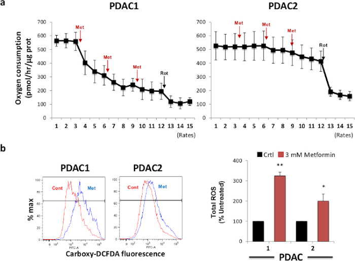
Figure 1. Metformin Treatment Inhibits Oxygen Consumption and Induces ROS Production. (A) Metformin addition inhibits oxygen consumption (OCR) of sphere-derived cells. Two different PDAC secondary spheres were treated with metformin and OCR changes were measured by extracellular flux analysis. Rotenone injection was used as a control for total mitochondrial OCR ...
Déclarations de divulgation
matériels
| Name | Company | Catalog Number | Comments |
| 24-well Ultra-low Attachment Plates | Corning | 3474 | |
| Sterile 1x Dulbecco’s Phosphate Buffered Saline | Sigma | D8537 | |
| Metformin | Sigma | D150959-5G | |
| Trypsin solution, 0.05% | Life technologies | 25300054 | |
| Incubator with CO2 input | |||
| CASY Cell Counter and Analyzer for proliferation and viability measurement | Roche Innovatis | AG CASY Model TTC 45,60,150 μm | |
| Counting Chamber/ Hemocytometer | Hausser Scientific Co | 3200 | |
| 50 ml centrifuge tubes | Corning | 430828 | |
| 15-ml polypropylene conical tube | BD Falcon | 352097 | |
| 50-ml polypropylene conical tube | BD Falcon | 352070 |
This article has been published
Video Coming Soon
Source: Lonardo, E. et al. Studying Pancreatic Cancer Stem Cell Characteristics for Developing New Treatment Strategies. J. Vis. Exp.(2015).