Un abonnement à JoVE est nécessaire pour voir ce contenu. Connectez-vous ou commencez votre essai gratuit.
Modeling Transuterine Fetal Tracheal Occlusion in Murine Model: A Surgical Procedure for Ligation of the Fetal Trachea Within a Pregnant Mouse
Dans cet article
Overview
This video demonstrates the transuterine tracheal occlusion procedure of a mouse fetus in a pregnant mouse. This model can be used to study the impact of occluded trachea on lung development.
Protocole
All procedures involving animal models have been reviewed by the local institutional animal care committee and the JoVE veterinary review board.
1. Laparotomy
- Clean the abdominal surface with alcohol and povidone-iodine. Maintain sterile conditions throughout the operation.
- Perform a vertical incision for the laparotomy of pregnant dams. Cut all layers separately.
- Identify uterine horns on each side.
- Determine the candidate fetuses for the surgery.
NOTE: Do not operate on the fetuses that are the nearest to the vagina. - Operate on two fetuses in each uterine horn if there are an even number of fetuses on each side (4 most of the time), and on 1 fetus in each uterine horn if there are an odd number of the uterus (3 most of the time).
2. Tracheal occlusion (TO)
- Use 2.5x magnification glasses for visualization.
- Position the uterine horn in a transverse fashion.
- Take the pups, facing upward, between two fingers using the eyes of the pups and the tail as a guide to position the fetus.
- Apply gentle pressure to the pup's head to allow extension of the head and therefore, visualization of the neck.
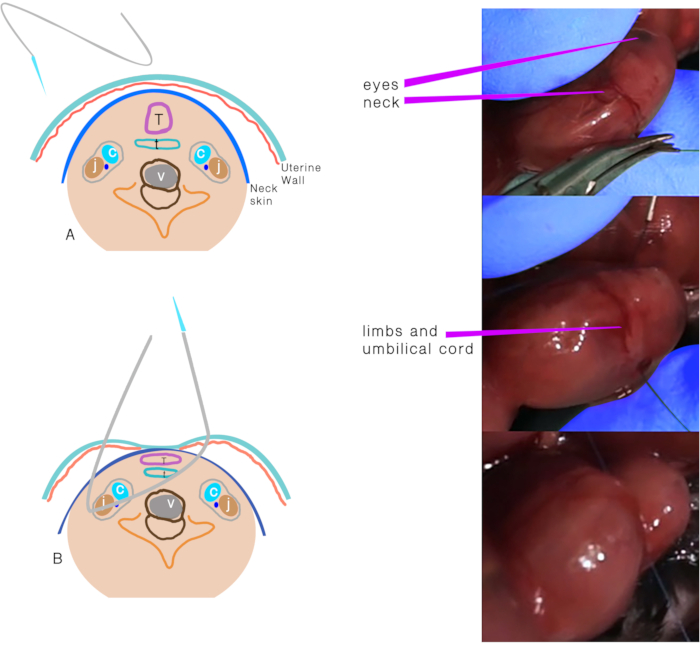
- Use a 6.0 polypropylene suture with an atraumatic needle to perform TO (Figure 1). Keep the placenta on the side and far from the entrance and exit points of the needle.
- Insert the needle transversely through the side of the uterus away from the placenta through the 1/3rd anterior part of the neck.
- Move the needle gently until the midline of the neck and direct it to the anterior part, then exit the neck between the trachea and opposite the carotid sheath and uterus.
- Knot the suture, taking care to maintain the integrity of the membranes and uterine wall, and keep the umbilical cord safe during knotting.
3. Abdominal wall closure
- Replace the uterine horn in the abdomen.
- Inject 2 mL of warm sterile saline into the peritoneal cavity before closure.
- Put a running 5/0 polyglactin suture to close the abdominal wall, and close the skin with a non-running silk suture.
- Apply 0.1 mg/kg of buprenorphine intraperitoneally for analgesia, and allow the recovery of the dam in a warm incubator.
Résultats

Figure 1. Tracheal occlusion. (A) The transuterine suture passing through the neck. (B) Schematic representation of the structures after the suture passes through and before the knot. Abbreviations: C = Carotid artery; J = Jugular vein; T =Trachea; E = Esophagus; V = Vertebra.
Déclarations de divulgation
matériels
| Name | Company | Catalog Number | Comments |
| Buprenorphine | Par Pharmaceutical | NDC 42023-179-05 | For regional anesthesia |
| Magnification glasses | USA Medical-Surgical | SLR-250LBLK | At least 2.5x |
| Silk suture | Ethicon | VCP682G | 4-0, 24 mm, cutting |
| Isoflurane | Halocarbon Life Sciences NDC | 66794-017-25 | For regional anesthesia |
| Polypropylene suture | Ethicon | Y432H | 6-0, 13 mm 1/2c Taperpoint |
This article has been published
Video Coming Soon
Source: Aydın, E. et al. Transuterine Fetal Tracheal Occlusion Model in Mice. J. Vis. Exp. (2021)