Un abonnement à JoVE est nécessaire pour voir ce contenu. Connectez-vous ou commencez votre essai gratuit.
Method Article
Outil graphique semi-automatique pour mesurer le score calcique pondéré spatialement de l’artère coronaire à partir d’images de tomodensitométrie cardiaque à grille
Dans cet article
Résumé
Cette vidéo montre l’utilisation d’un nouvel outil graphique pour mesurer le score calcique pondéré spatialement (SWCS), une alternative au score d’Agatston, pour quantifier la calcification des artères coronaires. L’outil graphique calcule le SWCS sur la base des données d’image de la tomodensitométrie cardiaque à déclenchement et des trajets définis par l’utilisateur des artères coronaires.
Résumé
La norme actuelle pour mesurer la calcification de l’artère coronaire afin de déterminer l’étendue de l’athérosclérose consiste à calculer le score d’Agatston à partir de la tomodensitométrie (TDM). Cependant, le score d’Agatston ne tient pas compte des valeurs de pixel inférieures à 130 unités de Hounsfield (HU) et des régions calciques inférieures à 1 mm2. En raison de ce seuil, le score n’est pas sensible aux petites régions de dépôt de calcium faiblement atténuées et peut ne pas détecter la micro-calcification naissante. Une mesure récemment proposée, appelée score calcique pondéré dans l’espace (SWCS), utilise également la tomodensitométrie, mais n’inclut pas de seuil pour l’HU et ne nécessite pas de signaux élevés dans des pixels contigus. Ainsi, le SWCS est sensible aux dépôts de calcium faiblement atténuants et plus petits et peut améliorer la mesure du risque de maladie coronarienne. À l’heure actuelle, le SWCS est sous-utilisé en raison de la complexité accrue des calculs. Afin de promouvoir la traduction du SWCS dans la recherche clinique et le calcul fiable et reproductible du score, l’objectif de cette étude était de développer un outil graphique semi-automatique qui calcule à la fois le SWCS et le score d’Agatston. Le programme nécessite des tomodensitogrammes cardiaques avec un fantôme d’hydroxyapatite de calcium dans le champ de vision. Le fantôme permet de dériver une fonction de pondération, à partir de laquelle le poids de chaque pixel est ajusté, ce qui permet d’atténuer les variations du signal et la variabilité entre les balayages. Avec les trois vues anatomiques visibles simultanément, l’utilisateur trace le parcours des quatre artères coronaires principales en plaçant des points ou des régions d’intérêt. Des fonctionnalités telles que le défilement pour zoomer, le double-clic pour supprimer et le réglage de la luminosité/contraste, ainsi que des conseils écrits à chaque étape, rendent le programme convivial et facile à utiliser. Une fois le traçage des artères terminé, le programme génère des rapports, qui comprennent les scores et les instantanés de tout calcium visible. Le SWCS peut révéler la présence d’une maladie subclinique, qui peut être utilisée pour une intervention précoce et des changements de mode de vie.
Introduction
La mesure de la quantité de calcium dans les artères à l’aide de la tomodensitométrie (TDM) est un moyen établi d’évaluer la gravité de l’athérosclérose coronarienne. Connaître et quantifier l’étendue de l’athérosclérose est essentiel pour déterminer le risque de maladie coronarienne future 1,2,3,4. La façon la plus courante de mesurer le calcium dans les artères coronaires est d’utiliser le score d’Agatston5. Cependant, une partie du calcul du score d’Agatston repose sur l’intensité des pixels choisis, mesurée en unités de Hounsfield (HU). Les pixels inférieurs à 130 HU ne sont pas pris en compte dans le calcul. De même, les calcifications dont la surface est inférieure à 1 mm2 ne sont pas prises en compte. En raison de ces seuils, le score d’Agatston n’est pas sensible aux petits foyers de calcification faiblement atténuants, qui peuvent encore être importants pour révéler la présence d’une maladie subclinique6.
Une mesure décrite précédemment appelée score calcique pondéré spatialement (SWCS) a été proposée pour évaluer le risque de plaque d’athérosclérose chez les patients présentant de faibles niveaux de calcification7. Contrairement au score d’Agatston, le SWCS n’utilise pas de seuillage du signal pour réduire l’impact du bruit de l’image. Au lieu de cela, il utilise un fantôme - un objet avec des concentrations connues d’hydroxyapatite de calcium (CHA) placé sur le participant de manière à ce qu’il soit dans le champ de vision du scan. Ici, un fantôme avec 0 mg/mL, 50 mg/mL, 100 mg/mL et 200 mg/mL CHA a été utilisé pendant le développement ; cependant, dans la mise en œuvre actuelle de l’outil graphique, seules les sections 0 mg/mL et 100 mg/mL sont requises. Le fantôme est utilisé pour créer une fonction de pondération spécifique à la numérisation, qui est ensuite utilisée pour peser chacun des pixels sélectionnés par l’utilisateur ainsi que ses voisins. Les pixels avec des pixels voisins qui ont un niveau d’atténuation élevé ont plus de poids que ceux qui sont entourés de pixels avec des niveaux d’atténuation plus faibles. Ce processus rend le SWCS tolérant au bruit et comparable d’un balayage à l’autre8. Le SWCS est continu et produit un score même lorsqu’il y a de faibles niveaux de calcification, ce qui permet de quantifier l’étendue de l’athérosclérose lorsque le score d’Agatston est nul. En permettant l’évaluation de la micro-calcification même lorsque le score d’Agatston est nul, le SWCS peut être important pour révéler la présence d’une maladie subclinique. Cela peut permettre une meilleure compréhension des facteurs de risque génétiques, environnementaux et autres de l’athérosclérose 9,10. Une étude antérieure, qui a examiné les personnes ayant un score d’Agatston de zéro au départ et de non zéro lors d’un suivi environ 15 ans plus tard, a observé que celles qui avaient un SWCS plus élevé au départ avaient un taux d’événements de maladie coronarienne (CHD) plus élevé. Le pouvoir prédictif du SWCS est particulièrement important dans les populations plus jeunes, où la détection et la surveillance du risque résiduel à long terme peuvent être utiles6.
Nous présentons ici un outil semi-automatique permettant de calculer le SWCS ainsi que le score d’Agatston. L’outil utilise une interface utilisateur graphique fonctionnant sur un langage de programmation compatible. L’utilisateur est en mesure d’interagir avec les images pour générer une série finale de rapports, qui incluent les deux scores calciques. Pour commencer, l’utilisateur sélectionne un cas, ou une série de fichiers DICOM (Digital Imaging and Communications in Medicine), à entrer dans le programme. Ces images doivent être des tomodensitogrammes en apnée et contrôlés par électrocardiogramme, acquises uniquement pendant la diastole pour éviter les mouvements respiratoires et cardiaques. Bien que le programme soit opérationnel avec toutes les images de tomodensitométrie cardiaque, pour produire des résultats significatifs, les images sources doivent respecter les lignes directrices minimales en matière de pointage calcique clinique11,12. À titre de référence, une épaisseur de tranche de 3 mm, une tension de crête du tube de 100 kVp, un indice de dose CT moyen de 1,19 mGy et une résolution d’image de 512 x 512 pixels sont utilisés dans l’étude ici. Toutes les images qui ne sont pas de 512 x 512 pixels sont automatiquement rééchantillonnées dans le programme pour assurer une résolution adéquate et cohérente des petites zones de calcification. Une fois les images chargées, l’utilisateur peut les voir dans les vues axiale, sagittale et coronale. On peut ensuite ajuster la luminosité et le contraste des images pour une meilleure visualisation avant de sélectionner les sections 0 mg/mL et 100 mg/mL du fantôme. Ensuite, l’utilisateur peut tracer chacune des quatre artères coronaires - l’artère antérieure descendante gauche (LAD), l’artère coronaire gauche (LCA), l’accent circonflexe gauche (LCX) et l’artère coronaire droite (RCA) - en plaçant un point, une région d’intérêt (ROI) ou une combinaison des deux pour permettre une sélection approfondie des pixels d’une artère, quelle que soit la façon dont l’artère apparaît dans le plan axial. L’utilisateur peut supprimer et remplacer ou redessiner des points et des ROI si nécessaire. Cliquez sur le bouton SWCS pour générer les rapports finaux. Les cas sont enregistrés automatiquement afin que les images, ainsi que les points et les retours sur investissement, puissent être rechargés ultérieurement. Des instructions écrites sont également disponibles à chaque étape de l’utilisation du programme, ce qui rend le programme facile à utiliser.
Protocole
Cette étude a été menée avec l’approbation du Mount Sinai Institutional Review Board (HS-20-01011), et tous les sujets ont donné leur consentement éclairé par écrit.
1. Préparation avant de commencer le protocole
- Une structure de dossiers appropriée est nécessaire pour ce programme. Commencez par créer un dossier principal pour le projet n’importe où sur l’ordinateur en cliquant avec le bouton droit de la souris et en sélectionnant l’option Nouveau dossier dans le répertoire des fichiers. Tous les fichiers DICOM d’entrée et les résultats seront stockés dans ce dossier principal.
REMARQUE : Tous les connecteurs DICOM requis par ce programme sont standard. Par conséquent, le programme est traduisible sur toutes les plates-formes de scanner. - Dans ce dossier principal, créez deux nouveaux dossiers en cliquant avec le bouton droit de la souris et en sélectionnant deux fois l’option Nouveau dossier .
- Renommez le premier de ces deux dossiers pour qu’il soit évident qu’il stocke des données brutes (par exemple, Original_Data) en cliquant dessus avec le bouton droit de la souris et en sélectionnant l’option Renommer .
- Créez un nouveau dossier pour un patient donné dans ce dossier Original_Data en cliquant avec le bouton droit de la souris et en sélectionnant l’option Nouveau dossier . Renommez-le en tant qu’identifiant de patient anonymisé en cliquant avec le bouton droit de la souris sur le dossier et en sélectionnant l’option Renommer . Importez uniquement l’ensemble complet des fichiers DICOM bruts de ce patient dans ce dossier.
- Répétez l’étape 1.3.1 pour tous les patients à analyser dans le programme.
- Renommez le deuxième de ces deux dossiers en Meta_Data pour le stockage des résultats en cliquant dessus avec le bouton droit de la souris et en sélectionnant l’option Renommer . Ce dossier sera vide jusqu’à ce que le programme soit exécuté et que les résultats soient générés.
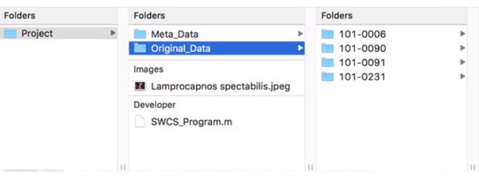
- Téléchargez le fichier du programme (fichier supplémentaire 1) et l’image de la pochette (fichier supplémentaire 2) et déplacez-les du dossier des téléchargements vers le dossier principal du projet par glisser-déposer. La configuration finale du dossier du projet doit ressembler à la figure 1.

Figure 1 : Format du dossier principal du projet. Cette figure montre comment le dossier principal du projet doit être structuré et formaté pour une utilisation correcte du programme. Veuillez cliquer ici pour voir une version agrandie de cette figure.
2. Lancement du programme
- Ouvrez le code du programme en double-cliquant sur son fichier dans le dossier principal du projet. Cela ouvrira le logiciel et affichera le code du programme.
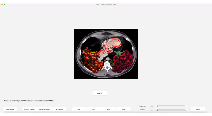
- Entrez dans la fenêtre de l’éditeur en cliquant une fois n’importe où dessus. Cliquez sur le bouton vert Exécuter situé dans le ruban supérieur de l’onglet de l’éditeur pour lancer le programme. La fenêtre initiale du programme, après ouverture, doit ressembler à l’image de la figure 2.
REMARQUE : Des instructions écrites sur les actions attendues de l’utilisateur et la progression de la génération des résultats seront affichées dans la zone inférieure gauche tout au long de l’utilisation du programme. - Cliquez sur le bouton Ouvrir DICOM dans le coin inférieur gauche. Cela ouvrira le répertoire files.
- Accédez au dossier principal du projet, au dossier contenant les données d’origine et à un patient à analyser. Cliquez une fois sur le dossier du patient pour qu’il soit mis en surbrillance, puis cliquez sur Ouvrir.
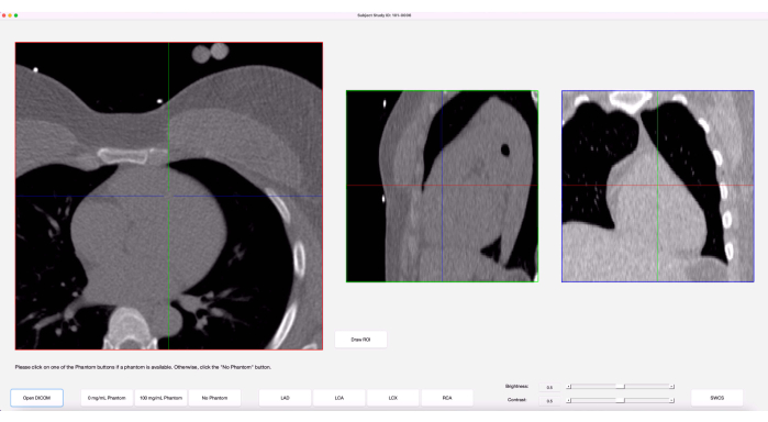
- Les images s’affichent désormais en trois vues : axiale, sagittale et coronale. Le survol et le défilement d’une vue particulière parcourent les tranches de cette vue. Le réticule de chaque vue affiche l’emplacement du pointeur à ce moment-là. Réglez la luminosité et le contraste dans la partie inférieure droite du programme en faisant glisser les barres, comme dans la Figure 3.

Figure 2 : Fenêtre initiale du programme. Le programme, lorsqu’il est lancé initialement, comporte les boutons disposés avec une image d’art. Veuillez cliquer ici pour voir une version agrandie de cette figure.

Figure 3 : Interface utilisateur graphique (GUI). Une fois les images chargées, l’interface graphique du programme affiche trois vues anatomiques des images ainsi qu’un réticule sur chaque vue, représentant le curseur. Veuillez cliquer ici pour voir une version agrandie de cette figure.
3. Analyse de la calcification de l’artère coronaire
- Lorsque vous êtes prêt à commencer l’analyse, cliquez sur le bouton Fantôme 0 mg/mL . Passez la souris sur la vue axiale et faites défiler vers le haut ou vers le bas jusqu’à ce que la section 0 mg/mL du fantôme soit visible.
- Déplacez le curseur au centre du fantôme 0 mg/mL dans la vue axiale. Maintenant, sans déplacer le curseur, observez le réticule dans les vues sagittale et coronale.
- Faites défiler lentement quelques tranches vers le haut et vers le bas jusqu’à ce que le réticule des trois vues soit au centre du fantôme à 0 mg/mL. Cliquez une fois pour placer une grille de points de 10 x 10 sur la tranche actuelle et ses deux tranches voisines. Cela provoquera l’apparition d’un groupe rouge de cercles dans la vue axiale et l’apparition d’une colonne de trois points dans les vues sagittale et coronale. Ces points seront utilisés dans le calcul de la fonction de pondération.
- Répétez les étapes 3.1 à 3.3 pour le fantôme de 100 mg/mL.
- Si un fantôme n’est pas disponible ou est inutilisable en raison de problèmes de qualité, cliquez sur le bouton Aucun fantôme pour obtenir une agrégation de 10 fantômes d’échantillons et la fonction de pondération correspondante à utiliser.
- Lorsque vous avez terminé de sélectionner les fantômes, commencez à tracer chacune des quatre artères coronaires en cliquant sur l’un des boutons de l’artère - LAD, LCA, LCX ou RCA.
- Passez la souris sur la vue axiale et faites défiler l’écran pour accéder à l’extrémité proximale ou distale de l’artère choisie. Observez la forme de l’artère dans cette tranche.
- Si la forme de l’artère est circulaire dans la vue axiale de cette tranche et qu’elle a un diamètre maximal de 5 mm, suivez ces sous-étapes. S’il n’est pas circulaire, passez à l’étape 3.9.
- Placez un point sur l’artère en cliquant une fois sur le centre de l’artère dans la vue axiale. Le point placé sera affiché avec un cercle de 5 mm de diamètre. Si l’artère ne rentre pas dans le cercle, supprimez-la en double-cliquant dessus et passez à l’étape 3.9 pour dessiner un ROI. Le point apparaîtra également dans les vues sagittales et coronales.
- Si l’artère est plus facile à visualiser dans la vue sagittale ou coronale, placez-y un point à la place. Assurez-vous qu’il est aligné avec le centre de l’artère dans la vue axiale.
- Si un point placé est accidentel ou n’est pas optimal, il peut être supprimé en double-cliquant rapidement dessus dans l’une des trois vues. Le texte dans la zone en bas à gauche vous avertira que le point a été supprimé.
- Si la forme de l’artère n’est pas circulaire dans le plan axial, suivez les étapes ci-dessous.
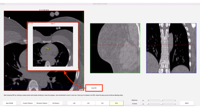
- Tout en vous assurant que la coupe axiale souhaitée est visible, cliquez sur le bouton Dessiner le retour sur investissement. Dans la fenêtre contextuelle, faites défiler l’écran pour effectuer un zoom avant/arrière et commencez à cliquer une fois autour de l’artère pour la tracer, comme dans la figure 4.
REMARQUE : Contrairement aux points, les ROI ne peuvent être placés que dans la vue axiale. - Tant que le retour sur investissement est toujours ouvert, la touche de retour arrière peut être utilisée pour supprimer le point précédent placé dans le tracé de l’artère. Pour fermer le ROI, double-cliquez à l’endroit où le dernier point doit être placé ou double-cliquez sur le premier point placé.
- Ajustez davantage le retour sur investissement fermé et affinez-le en faisant glisser les points de périmètre du retour sur investissement ou en double-cliquant sur le périmètre d’un retour sur investissement pour ajouter un point.
- Une fois le ROI fermé, deux boutons apparaîtront en bas de la fenêtre pop-up : Verrouiller et Redessiner le ROI. Si le retour sur investissement doit être redessiné, cliquez sur le bouton Redessiner le retour sur investissement pour effacer le retour sur investissement actuel et en dessiner un nouveau.
- Lorsque vous êtes satisfait du retour sur investissement actuel, cliquez sur le bouton Verrouiller et fermez la fenêtre contextuelle en cliquant sur le bouton rouge dans le coin supérieur gauche (Mac) ou sur le bouton X dans le coin supérieur droit (PC) de la fenêtre contextuelle.
- Un retour sur investissement peut être supprimé même une fois qu’il est verrouillé en double-cliquant sur l’un des points de périmètre dans la vue axiale de la fenêtre principale du programme (et non sur la fenêtre contextuelle).
- Tout en vous assurant que la coupe axiale souhaitée est visible, cliquez sur le bouton Dessiner le retour sur investissement. Dans la fenêtre contextuelle, faites défiler l’écran pour effectuer un zoom avant/arrière et commencez à cliquer une fois autour de l’artère pour la tracer, comme dans la figure 4.

Figure 4 : Fonction de retour sur investissement de l’écran. Lorsque l’option Dessiner le retour sur investissement est sélectionnée, une fenêtre contextuelle de la tranche axiale actuelle s’affiche. Le jaune indique un retour sur investissement qui a été précédemment dessiné sur cette tranche. Veuillez cliquer ici pour voir une version agrandie de cette figure.
- Faites défiler vers le haut ou vers le bas une tranche dans la vue axiale et répétez les étapes 3.8-3.9 jusqu’à ce que l’extrémité de l’artère choisie soit atteinte. Lorsque vous avez terminé, cliquez à nouveau sur le bouton de l’artère finie pour examiner le tracé de l’artère et vous assurer qu’aucun point accidentel n’a été placé.
REMARQUE : Il est possible de placer plus d’un point ou d’un ROI sur une tranche axiale donnée. Les lignes supplémentaires reliant deux ROI sur une tranche peuvent être ignorées. Si des coupes d’une artère donnée ne sont pas étiquetées, un message d’erreur apparaîtra. Fermez le message et étiquetez la ou les tranches manquées. - Passez à la suivante des quatre artères et répétez les étapes 3.6 à 3.10.
- Lorsque les quatre artères ont été tracées, cliquez sur le bouton SWCS dans le coin inférieur droit pour générer les résultats. La zone en bas à gauche affichera la progression et affichera « Traitement terminé » lorsque vous aurez terminé. Fermez la fenêtre du programme en cliquant sur le bouton rouge dans le coin supérieur gauche (Mac) ou sur le bouton X dans le coin supérieur droit (PC).
4. Accès aux résultats
- Pour accéder aux résultats du cas qui vient d’être analysé, ouvrez le répertoire des fichiers et accédez au dossier principal du projet. Allez dans le dossier Meta_Data et remarquez qu’un nouveau dossier portant le même nom que le dossier de données d’origine de ce sujet est apparu.
- Dans ce dossier, il y aura trois types de documents : CSV, PNG et PDF. Examinez les PDF pour obtenir le score final SWCS et Agatston pour le cas, ainsi que la fonction de pondération utilisée.
NOTE : Les CSV stockent les coordonnées des différents points/ROI placés lors de l’analyse de ce sujet. Le fait d’avoir ces CSV permettra aux images de ce sujet d’être rouvertes dans le programme plus tard et de faire apparaître automatiquement les points/ROI précédents. Toute modification apportée au dossier, lors de sa réouverture, sera automatiquement répercutée dans les fichiers CSV.
Résultats
Les résultats représentatifs présentés dans cette section montrent ce qu’implique une utilisation réussie du programme. Ici, un patient avec un score d’Agatston supérieur à zéro est utilisé à titre d’exemple. Comme nous l’avons vu précédemment, les résultats contenus dans le dossier de métadonnées d’un patient contiendront des feuilles de calcul sous forme de fichiers CSV, des images sous forme de fichiers PNG et des rapports sous forme de fichiers PDF, comme le montre la fig...
Discussion
Bien que le protocole de ce programme soit relativement facile à suivre, il y a quelques étapes critiques qui sont nécessaires pour une utilisation réussie et des résultats fiables. Avant de commencer, il est important de s’assurer que les données des patients qui seront utilisées dans le cadre de ce programme sont anonymisées afin d’assurer la confidentialité des patients. La mise en forme et le nommage initiaux du dossier principal du projet doivent être corrects pour que le programme puisse reconnaître ...
Déclarations de divulgation
Les auteurs déclarent qu’ils n’ont aucun conflit d’intérêts à divulguer.
Remerciements
Ce travail a été soutenu par R01ES029967 subvention du NIH.
matériels
| Name | Company | Catalog Number | Comments |
| Calcium Hydroxyapatite | Sigma-Aldrich | 289396-100G | Suspended in EpoxAcast 690 resin for phantom creation |
| Clinical Cardiac CT Scanner | Siemens | SOMATOM Force Dual Source CT | Used for the source images; Any cardiac CT will be sufficient |
| EpoxAcast 690 | Smooth-On | 03641 | Used for phantom creation |
| MATLAB | Mathworks | R2019a | Requires Image Processing Toolbox and Statistics and Machine Learning Toolbox; Any version compatible with and able to run version R2019a scripts is sufficient |
| Standard Computer | N/A | N/A | macOS or Windows operating system |
| syngo.via | Siemens | VB60A_HF04 | Commercial software used for computing Agatston score for validation study |
Références
- O'Malley, P. G., Taylor, A. J., Jackson, J. L., Doherty, T. M., Detrano, R. C. Prognostic value of coronary electron-bean computed tomography for coronary heart disease events in asymptomatic populations. The American Journal of Cardiology. 85 (8), 945-948 (2000).
- Budoff, M. J., et al. Assessment of coronary artery disease by cardiac computed tomography. Circulation. 114 (16), 1761-1791 (2006).
- Rumberger, J. A., Simons, D. B., Fitzpatrick, L. A., Sheedy, P. F., Schwartz, R. S. Coronary artery calcium area by electron-beam computed tomography and coronary atherosclerotic plaque area. Circulation. 92 (8), 2157-2162 (1995).
- Mautner, G. C., et al. Coronary artery calcification: assessment with electron beam CT and histomorphometric correlation. Radiology. 192 (3), 619-623 (1994).
- Agatston, A. S., et al. Quantification of coronary artery calcium using ultrafast computed tomography. Journal of the American College of Cardiology. 15 (4), 827-832 (1990).
- Shea, S., et al. Spatially weighted coronary artery calcium score and coronary heart disease events in the multi-ethnic study of atherosclerosis. Circulation: Cardiovascular Imaging. 14 (1), e011981 (2021).
- Liang, C. J., Budoff, M. J., Kaufman, J. D., Kronmal, R. A., Brown, E. R. An alternative method for quantifying coronary artery calcification: the multi-ethnic study of atherosclerosis (MESA). BMC Medical Imaging. 12, 14 (2012).
- McCollough, C. H., et al. Coronary artery calcium: a multi-institutional, multimanufacturer international standard for quantification at cardiac CT. Radiology. 243 (2), 527-538 (2007).
- Detrano, R., et al. Coronary calcium as a predictor of coronary events in four racial or ethnic groups. The New England Journal of Medicine. 358 (13), 1336-1345 (2008).
- Budoff, M., et al. Cardiovascular events with absent or minimal coronary calcification: The Multi-Ethnic Study of Atherosclerosis (MESA). American Heart Journal. 158 (4), 554-561 (2009).
- Hecht, H. S., et al. 2016 SCCT/STR guidelines for coronary artery calcium scoring of noncontrast noncardiac chest CT scans: A report of the Society of Cardiovascular Computed Tomography and Society of Thoracic Radiology. Journal of Cardiovascular Computed Tomography. 11 (1), 74-84 (2017).
- American College of Radiology. ACR-NASCI-SPR practice parameter for the performance and interpretation of cardiac computed tomography (CT). American College of Radiology. , (2021).
Réimpressions et Autorisations
Demande d’autorisation pour utiliser le texte ou les figures de cet article JoVE
Demande d’autorisationExplorer plus d’articles
This article has been published
Video Coming Soon