Method Article
Cryoconservation des cellules germinales primordiales et renaissance des souches de drosophile
* Ces auteurs ont contribué à parts égales
Dans cet article
Résumé
Il est hautement souhaitable de mettre au point une méthode de conservation à long terme des souches de drosophile comme solution de rechange au transfert fréquent de mouches adultes dans des flacons d’aliments frais. Ce protocole décrit la cryoconservation des cellules germinales primordiales de la drosophile et la renaissance de la souche via leur transplantation sur des embryons hôtes agamétiques.
Résumé
Les souches de drosophiles doivent être maintenues par le transfert fréquent de mouches adultes dans de nouveaux flacons. Cela comporte un risque de détérioration mutationnelle et de changements phénotypiques. Il est donc impératif de mettre au point une méthode alternative de conservation à long terme sans de tels changements. Malgré des tentatives antérieures réussies, la cryoconservation des embryons de drosophile n’est toujours pas d’une utilité pratique en raison de sa faible reproductibilité. Nous décrivons ici un protocole de cryoconservation des cellules germinales primordiales (PGC) et de reconstitution de souches par transplantation de PGC cryoconservés dans des embryons hôtes agamétiques de Drosophila melanogaster (D. melanogaster). Les PGC sont très perméables aux agents cryoprotecteurs (CPA), et les variations développementales et morphologiques entre les souches sont moins problématiques que dans la cryoconservation des embryons. Dans cette méthode, les PGC sont prélevés sur environ 30 embryons de donneurs, chargés dans une aiguille après le traitement CPA, puis cryoconservés dans de l’azote liquide. Pour produire des gamètes dérivés d’un donneur, les PGC cryoconservés dans une aiguille sont décongelés, puis déposés dans environ 15 embryons hôtes agamétiques. Une fréquence d’au moins 15 % de mouches fertiles a été atteinte avec ce protocole, et le nombre de descendants par couple fertile était toujours plus que suffisant pour faire revivre la souche originale (le nombre moyen de descendants étant de 77,2 ± 7,1), ce qui indique la capacité des PGC cryoconservés à devenir des cellules souches germinales. Le nombre moyen de mouches fertiles par aiguille était de 1,1 ± 0,2, et 9 aiguilles sur 26 ont produit deux ou plusieurs descendants fertiles. Il a été constaté que 11 aiguilles suffisent pour produire 6 descendants ou plus, dans lesquels au moins une femelle et un mâle sont probablement inclus. L’hôte agametique permet de faire revivre la souche rapidement en croisant simplement des mouches femelles et mâles nouvellement émergées. De plus, les PGC ont le potentiel d’être utilisés dans des applications de génie génétique, telles que l’édition du génome.
Introduction
Le maintien des souches de drosophile par le transfert de mouches adultes dans de nouveaux flacons de nourriture entraîne inévitablement l’accumulation de mutations et de changements épigénétiques au fil du temps. Il est impératif de mettre au point une méthode alternative pour le maintien à long terme des souches de drosophile sans de tels changements, en particulier pour les souches de référence dans lesquelles l’ensemble du génome doit être maintenu. Plusieurs tentatives réussies de cryoconservation d’embryons ou d’ovaires de drosophiles ont été décrites 1,2,3. Malheureusement, ils ne sont toujours pas d’une utilité pratique en raison de leur faible reproductibilité. En effet, les embryons à un stade précoce ont un faible taux de survie après cryoconservation en raison de leur forte teneur en vitellus, ce qui empêche la perméation et la diffusion de l’agent cryoprotecteur (CPA) 2,3. La perméabilité au CPA est également sévèrement limitée par les couches cireuses des embryons à un stade avancé. Il est difficile et prend beaucoup de temps de trouver une période spécifique à la souche au cours de laquelle les embryons ont un taux de survie élevé et une couche de cire plus fine. Récemment, Zhan et al.4 ont amélioré les méthodes de perméabilisation des embryons, de chargement de CPA et de vitrification et ont cryoconservé avec succès des embryons de plusieurs souches. Cependant, les méthodes ne sont pas faciles à appliquer car la viabilité des embryons après perméabilisation a tendance à être faible. Par conséquent, il est encore nécessaire d’améliorer et de développer des approches alternatives. Les méthodes de cryoconservation des cellules germinales primordiales (PGC) constituent une approche alternative pour le maintien à long terme des souches de drosophile.
La transplantation de PGC (également appelée cellule polaire) a été utilisée pour générer des chimères germinales, en particulier des femelles, afin d’étudier des processus tels que les effets maternels des mutations létales zygotiques et la détermination du sexe des cellules germinales 5,6,7,8,9,10,11,12 . Les PGC sont beaucoup plus petits que les embryons et sont susceptibles d’être très perméables à la plupart des cryoprotecteurs. De plus, la variation développementale et morphologique entre les souches est moins problématique, et un hôte agametique permet une restauration rapide de génomes entiers. Nous avons récemment mis au point une nouvelle méthode de cryoconservation PGC13, qui empêche les changements génétiques et épigénétiques autrement inévitables dans les souches de drosophile. Nous vous présentons ici le protocole détaillé.
Cette méthode de cryoconservation nécessite une expertise spécifique dans la manipulation et l’instrumentation des PGC. Bien qu’une approche étape par étape puisse être une solution efficace pour ceux qui ne la connaissent pas, elle peut ne pas convenir aux petits laboratoires en raison des exigences en matière d’instrumentation. Ce protocole de cryoconservation PGC peut être plus facilement adapté pour être utilisé avec différentes espèces de drosophiles et différentes espèces d’insectes que les protocoles de cryoconservation d’embryons en raison de différences de développement et morphologiques plus faibles. Les PGC peuvent également être utilisés dans des applications de génie génétique, telles que l’édition du génome 14,15,16. En résumé, cette méthode peut être utilisée dans les centres de stockage et autres laboratoires pour maintenir les souches de mouches et d’autres insectes pendant des périodes prolongées sans changements.
Protocole
1. Préparation de l’équipement
- Système de micromanipulateur : Assembler un système de micromanipulateur pour prélever et transplanter des cellules (figure 1A).
- Lames de verre de collection PGC (Figure 2A)
- Pour préparer la colle à l’heptane, coupez du ruban adhésif double face d’environ 30 cm de long et faites-le tremper toute la nuit dans 7 ml de solution d’heptane de qualité technique (ordinaire).
- Tracez deux lignes de référence parallèles pour l’alignement de l’embryon au dos d’une lame de verre.
- Étalez des gouttes de la colle heptane ci-dessus sur la lame de verre (sur le côté sans les lignes) à l’aide d’une pipette Pasteur. Séchez à l’air libre la surface de la lame jusqu’à ce qu’elle devienne blanche.
- Répétez l’ajout et l’étalement de gouttes de colle heptane et séchez à nouveau la lame.
REMARQUE : La colle empêche les solutions liquides de se répandre sur la surface plane et facilite le chargement des solutions aqueuses dans une aiguille. - Pour fabriquer des cadres de piscine d’embryons, collez trois couches de ruban vinyle standard de 0,2 mm d’épaisseur, comme du ruban isolant, sur une planche à découper. Découpez le ruban en rectangles de 1,5 cm de large. Ensuite, découpez les trois couches de ruban adhésif en laissant un cadre de 2 à 3 mm.
REMARQUE : Un cadre de pool d’embryons est apposé après l’alignement des embryons, pour former un pool d’embryons.
- Aiguilles de transplantation
REMARQUE : Toutes les aiguilles disponibles dans le commerce au moment de cette étude étaient trop étroites ou trop larges pour la cryoconservation des PGC.- Fabriquez une aiguille à l’aide d’un capillaire en verre et d’un extracteur. Nous utilisons un extracteur NARISHIGE PN-31 avec le niveau de chauffage à 85,0-98,4, le niveau principal de l’aimant à 57,8 et le sous-niveau de l’aimant à 45,0.
- Pour fabriquer une aiguille d’une épaisseur de paroi d’environ 1 μm et d’une pointe d’environ 200 μm de longueur avec un diamètre intérieur de 10-12 μm, polissez la pointe de l’aiguille en trois étapes (Figure 3). Tout d’abord, meulez la pointe de l’aiguille à un angle de 30° à une vitesse de 780 tr/min jusqu’à ce que la pointe ait un diamètre intérieur de 10-12 μm. Cette première étape de broyage dure environ 1 h.
REMARQUE : Pour éviter de casser la pointe de l’aiguille, tournez d’abord la meule, puis déplacez doucement l’aiguille vers le bas sur la meule. - Tracez une ligne sur le dessus de l’aiguille pour suivre l’angle souhaité. Tournez l’aiguille dans le sens inverse des aiguilles d’une montre à 90° et polissez-la à nouveau à une vitesse de 180 tr/min. Cela prend environ 5 min.
- Tournez l’aiguille de 45° dans le sens des aiguilles d’une montre et polissez-la à une vitesse de 180 tr/min pendant une seconde.
- Placez une lame de verre de collecte avec une goutte de mélange d’acide chromique (ATTENTION : toxique) sur la platine du microscope. Fixez l’aiguille au support capillaire (Figure 1D) à un angle de 10° à 13° par rapport à la surface de la lame, déplacez délicatement l’aiguille vers le bas et plongez la pointe dans le mélange d’acide chromique.
- En tirant et en poussant le piston (Figure 1B), chargez et déchargez mécaniquement la solution de l’aiguille plusieurs fois pour éliminer les débris de verre dans l’aiguille. Assurez-vous également de nettoyer le mur extérieur.
- Lavez l’intérieur et l’extérieur de l’aiguille deux fois avec de l’eau distillée pour éliminer complètement l’acide chromique.
2. Collecte et cryoconservation des PGC
- Collecte d’embryons
- Transférez un nombre approprié de mouches de la souche donneuse d’intérêt (environ 450 pour chaque sexe pour le gobelet de prélèvement d’embryons) dans un gobelet de prélèvement d’embryons muni d’une plaque de prélèvement d’embryons (figure 1E) et incubez-les à 25 °C. Nous utilisons généralement des mouches parentales âgées de 3 à 5 jours qui sont élevées dans des conditions moins denses à température ambiante (23-25 °C).
- Effectuez deux prélèvements de 30 minutes et jetez les œufs pondus. Étant donné que les femelles peuvent conserver les œufs fécondés qui se développent dans l’oviducte, cette étape est nécessaire pour synchroniser la ponte à l’étape 2.1.3 (figure 4).
- Après les deux prélèvements, prélever les embryons pendant 50 min, puis incuber les embryons collectés dans une chambre humidifiée à 25 °C pour permettre aux embryons de se développer jusqu’au stade blastoderme (stade précoce 517). Le temps d’incubation est généralement de 100 min, mais peut être prolongé jusqu’à 120 min, selon la souche (Figure 4).
REMARQUE : Une chambre humidifiée est fabriquée en plaçant une serviette en papier humide au fond d’une boîte en plastique et en la vaporisant avec un brouillard d’eau avant utilisation. Dans les embryons de stade précoce 5, la formation des PGC est complète, mais la cellularisation somatique ne l’est pas. Le stade exact d’un embryon est déterminé au microscope composé à l’étape 2.4.
- Embryons déchorionnants
- Déposez une goutte d’eau distillée sur une crépine à mailles en acier inoxydable (150 mailles, ouverture de 109 μm, diamètre de fil de 60 μm ; Graphique 1F). À l’aide d’une pince, prélevez les embryons de la plaque de prélèvement d’embryons et mettez-les dans la goutte d’eau.
- Pressez du papier de soie contre la passoire par le dessous pour absorber l’eau. Ajouter des gouttelettes de solution fraîche d’hypochlorite de sodium à 5 % (sous forme de Cl) aux embryons et tapoter continuellement la passoire pendant 10 s.
- Lavez les embryons en les aspergeant directement d’eau distillée et pressez du papier de soie contre la passoire par le dessous pour absorber l’eau. Répétez cette étape 3 fois.
- Alignement d’embryons déchorionisés
- À l’aide d’un microscope stéréoscopique, utilisez une pince pour transférer les embryons. Alignez les embryons déchorionnés sur deux rangées sur une lame de verre de prélèvement PGC le long des deux lignes de référence (Figure 2A). Les embryons sont orientés avec leur face antérieure vers la droite (le côté à manipuler) et la face ventrale vers le haut.
REMARQUE : Cette étape devrait être terminée en 20 minutes, au cours desquelles nous alignons généralement environ 40 embryons. - Fixez un cadre de piscine d’embryons autour des embryons sur la lame en verre de prélèvement PGC. Déposez 1 μL de solution de CPA (1 solution d’Ephrussi-Beadle Ringer, EBR, contenant 20 % d’éthylène glycol et 1 M de saccharose ; 1 μL de liquide liquide : 130 mM de NaCl, 5 mM de KCl, 2 mM de CaCl2 et 10 mM de Hepes à un pH de 6,9) à deux endroits distincts dans la zone délimitée par le cadre et remplissez le bassin d’huile de silicone pour éviter que les embryons ne se dessèchent (figure 2A).
REMARQUE : Pour préparer la solution de CPA, dissoudre complètement 10,26 g de saccharose dans environ 20 mL de H2O distillé contenant 3 mL de solution 10 x EBR. Ajouter 6 mL d’éthylène glycol, puis ajouter le H2O distillé jusqu’à 30 mL. Après un mélange complet, filtrez la solution à travers une membrane jetable de 0,22 mm.
- À l’aide d’un microscope stéréoscopique, utilisez une pince pour transférer les embryons. Alignez les embryons déchorionnés sur deux rangées sur une lame de verre de prélèvement PGC le long des deux lignes de référence (Figure 2A). Les embryons sont orientés avec leur face antérieure vers la droite (le côté à manipuler) et la face ventrale vers le haut.
- Collecte de PGC
- Placez la lame de verre de collecte PGC de l’étape 2.3.2 sur la platine d’un microscope équipé d’un système de micromanipulateur. Fixez l’aiguille au support capillaire et amenez le premier embryon de la rangée de gauche et la pointe de l’aiguille dans le même plan focal. Chargez l’huile de silicone dans l’aiguille pendant 2-3 s.
- Commencez le prélèvement de PGC à partir d’embryons dans la rangée de gauche. À l’aide d’une lentille d’objectif 20x, déplacez doucement la pointe de l’aiguille vers la surface de l’extrémité antérieure de l’embryon et pénétrez l’embryon vers l’extrémité postérieure, non pas en déplaçant l’aiguille mais en déplaçant la platine du microscope.
- Lorsque la pointe de l’aiguille atteint l’extrémité postérieure, rétractez légèrement l’aiguille et déchargez complètement le jaune de l’aiguille juste à l’intérieur de la couche de cellules somatiques.
- Tout en maintenant la pression constante dans l’aiguille, déplacez la pointe de l’aiguille vers les PGC juste à l’intérieur du pôle postérieur et chargez doucement, mais sans prendre beaucoup de temps, les PGC.
- Retirez rapidement l’aiguille de l’embryon et déchargez le jaune et d’autres contaminants de l’aiguille dans le pool d’huile de silicone, en gardant les PGC dans l’aiguille. Ensuite, chargez de l’huile de silicone propre de la piscine.
- Répétez les étapes 2.4.2 à 2.4.5 pour les autres embryons de la rangée de gauche. Avant de prélever des PGC à partir d’un nouvel embryon, déposez autant que possible l’huile de silicone chargée à l’étape 2.4.5 à l’intérieur de la couche de cellules somatiques tout en gardant les PGC chargées dans l’aiguille. Cela permet de s’assurer que les PGC nouvellement chargés sont adjacents aux PGC précédemment collectés sans qu’aucun matériau intermédiaire ne s’interpose entre eux.
- Après avoir terminé le prélèvement des PGC à partir des embryons de la rangée de gauche, séparez autant que possible les PGC du jaune et des autres contaminants. Pour ce faire, déposez tous les PGC dans l’aiguille à la surface d’un embryon et retirez tout jaune ou autre contaminant vers un autre embryon voisin.
- Ensuite, collectez les PGC des embryons de la rangée de droite. Combinez les PGC collectés dans les rangées de droite et de gauche.
- Application d’un agent cryoprotecteur (CPA) sur les PGC
- Après avoir lavé l’aiguille avec du CPA en une goutte, chargez du CPA frais dans une autre goutte dans l’aiguille et ajoutez du CPA aux PGC déposés sur l’embryon. Le volume de CPA doit être équivalent à celui des PGC.
- Retirez autant de CPA que possible du groupe de PGC 1-2 s après l’ajout de CPA. Les PGC rétrécissent légèrement et deviennent de forme carrée immédiatement après l’ajout de CPA.
- Videz l’aiguille puis chargez de l’huile de silicone pendant 5 s ou plus. Chargez tous les PGC collectés, puis chargez à nouveau l’huile de silicone pendant 5 s ou plus. Les PGC sont maintenant pris en sandwich entre deux couches d’huile de silicone (Figure 2B).
REMARQUE : Il est important d’éliminer autant de jaune d’œuf, de CPA et d’autres contaminants que possible.
- Cryoconservation des PGC
- Ouvrez le robinet d’arrêt à trois voies (Figure 1C), puis détachez l’aiguille du micromanipulateur. Épongez l’huile de la surface de l’aiguille avec du papier de soie doux. Ne touchez pas directement la pointe de l’aiguille avec le mouchoir.
- Fixez l’aiguille à un porte-aiguille et verrouillez-la en position à la base à l’aide de ruban adhésif en vinyle (Figure 1H). Collez une étiquette sur le tube de support.
- Congelez le support avec l’aiguille pointant vers le bas en l’immergeant dans de l’azote liquide. Ne relâchez pas le support tant que le liquide n’a pas cessé de pétiller hors de la grille.
- Stockez le support dans un réservoir de stockage d’azote liquide dans la zone de la phase liquide, et non dans la zone de la phase vapeur.
3. Décongélation et repiquage des PGC
- Collecte, déchorionisation et alignement d’embryons de mouches hôtes agamétiques
- Prélever et déchorionner les embryons de mouches hôtes agamétiques en suivant l’étape 2.
- Alignez les embryons hôtes agamétiques de stade 5 sur une lame de verre de transplantation. Cependant, cette fois-ci, orientez le postérieur vers la droite (le côté à manipuler) et ventral vers le haut (Figure 5). Alignez environ 30 embryons sur deux rangées en 20 min.
- Pendant l’alignement des embryons, faites fonctionner un humidificateur pendant 2 à 10 minutes si l’humidité ambiante l’exige (tableau 1). L’humidité idéale est de 30 % à 40 %, mais elle peut varier en fonction des conditions thermiques.
- Décongélation et transplantation de PGC dans des embryons hôtes
- Pour décongeler rapidement les PGC cryoconservés, glissez le support contenant l’aiguille dans la solution 1x EBR à température ambiante avec l’aiguille pointant vers le bas et maintenez-le immergé pendant 10 s.
- Placez la lame de verre de transplantation sur la platine d’un microscope. Fixez l’aiguille lyophilisée au support capillaire et amenez le premier embryon de la rangée de gauche et la pointe de l’aiguille dans le même plan focal.
- À l’aide d’une lentille d’objectif 20x, déplacez doucement la pointe de l’aiguille vers la surface de l’extrémité postérieure de l’embryon.
- Poussez doucement l’extérieur de chaque embryon et assurez-vous qu’ils reprennent lentement leur forme d’origine. L’aiguillon confirmera que la pression interne de l’embryon n’est ni trop élevée ni trop basse.
- Déplacez doucement l’aiguille et pénétrez un embryon à partir du pôle postérieur.
- Déposez doucement environ 10 à 20 PGC juste à l’intérieur du pôle postérieur, précisément entre la membrane vitelline et la couche de cellules somatiques de l’embryon. Évitez de les déposer dans la couche de cellules somatiques. Si le liquide périvitelline s’écoule de l’embryon, aspirez le liquide qui s’écoule dans l’aiguille et retirez-le.
- Retirez l’aiguille de l’embryon. Répétez les étapes 3.2.5 et 3.2.6 pour les embryons subséquents.
4. Incubation d’embryons et restauration des souches de donneurs
- Retirer tous les embryons qui n’ont pas reçu de PGC transplantés et incuber les embryons restants dans une chambre humidifiée (Figure 1G) à 25 °C.
- Au moins 24 h après la transplantation et dès que possible après l’éclosion, utiliser des pinces pour ramasser et transférer les larves écloses dans des flacons de nourriture standard pour drosophiles et incuber à 25 °C.
- Pour raviver la souche, croisez des femelles et des mâles nouvellement émergés (figure 6).
NOTE : Les hôtes agametiques permettent de restaurer l’ensemble du génome en une seule fois sans croiser les souches de chromosomes équilibrants. La coexistence de mâles agamétiques dans une fiole n’aura pas d’importance parce que les femelles, même si elles sont accouplées avec eux, ne montrent pas de réponses post-accouplement à long terme, y compris une diminution de la réceptivité à la régénération18,19.
Résultats
L’efficacité de la transplantation de PGC cryoconservés a été rapportée par Asaoka et al.13 et est donnée dans le tableau 2 pour la transplantation de PGC cryoconservés pendant 1 jour ou plus dans de l’azote liquide. Le taux d’éclosion était de 168/208 embryons transplantés (80,8 %) et la viabilité de l’embryon à l’adulte était de 87/208 (41,8 %). La fréquence des mouches fertiles était de 28/87 (32,2%). Cette fréquence ne différait pas entre les PGC cryoconservés pendant 8 à 30 jours et ceux cryoconservés pendant 31 à 150 jours (20/57 vs 8/30, G' = 0,63, p >0,1, d.f. = 1). Le nombre moyen de descendants par couple était de 77,2 ± 7,1 (n = 18, 28-122), ce qui indique la capacité des PGC cryoconservés à devenir des cellules souches germinales. Sur les 26 aiguilles, 10 n’ont produit aucune descendance fertile, 7 aiguilles ont produit 1 descendance fertile, 7 aiguilles ont produit 2 descendances fertiles et 2 aiguilles ont produit 3 ou 4 descendances fertiles. Le nombre moyen de mouches fertiles par aiguille était de 1,1 ± 0,2. Sur la base de ces données, avec un degré de confiance de 95 %, 11 aiguilles suffisent pour produire 6 descendants ou plus, dans lesquels au moins une femelle et un mâle sont probablement inclus.
Dans les expériences ci-dessus, nous avons utilisé des embryons exprimant l’ARNm ovo-A dans des PGC (nanos>ovo-A, OvoA_OE embryons) comme hôte agamétique. Sur 669 femelles F1 et 720 mâles F1 issus de couples nanos>ovo-A transplantés, il n’y avait pas d’échappatoire dérivé des PGC hôtes. Plusieurs mutants oskar (osk) sont également des agametiques sensibles à la température20,21. Parce qu’un mutant osk avec une viabilité homozygote élevée et le phénotype agamétique n’est plus disponible, nous avons recréé le mutant faux-sens osk[8] 20 par édition génomique assistée par CRISPR/Cas9. Ces mouches étaient complètement agamétiques (0 évasion sur 230 femelles et 192 mâles) à 25 °C, mais quelques échappées ont émergé à 23 °C (1 sur 248 femelles et 1 sur 290 mâles). Les nanos>ovo-A sont donc recommandés comme embryons hôtes agamétiques. Les stocks UASp-ovo-A et nanos-Gal4 13 seront bientôt disponibles auprès du Centre de stockage de la drosophile de KYOTO.
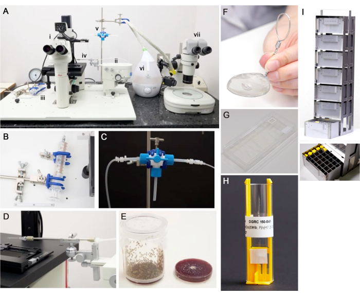
Figure 1 : Matériel requis. (A) Un système de micromanipulateur pour la collecte et la transplantation de cellules. i) microscope inversé, ii) micromanipulateur mécanique, iii) seringue, iv) support capillaire, v) robinet d’arrêt à trois voies, vi) humidificateur et vii) stéréomicroscope. (B) Une seringue. (C) Un robinet d’arrêt à trois voies et des tubes en silicone relient une seringue et un support capillaire. (D) Une aiguille et un support capillaire sont attachés à un micromanipulateur. (E) Un gobelet de prélèvement d’embryons avec une plaque de prélèvement d’embryons (6 cm de diamètre, 7,7 cm de hauteur). (F) Une crépine à mailles en acier inoxydable. (G) Un récipient utilisé comme chambre humide avec une lame de verre. Pour maintenir l’humidité, placez du papier humide sur le fond et fermez le couvercle. (H) Un porte-aiguille avec une aiguille pour la cryoconservation. (I) Un rack de stockage pour la cryoconservation et une boîte avec des aiguilles. Veuillez cliquer ici pour voir une version agrandie de cette figure.
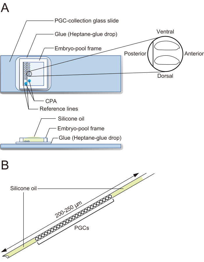
Figure 2 : Une lame de verre de prélèvement de PGC et une aiguille de cryoconservation. (A) Une lame de verre de collecte de cellules germinales primordiales (PGC) enduite de colle. Les embryons déchorionnés sont alignés sur deux rangées et orientés avec leur face antérieure vers la droite (le côté à manipuler) et la face ventrale vers le haut. Un cadre de piscine d’embryons est fixé, deux gouttes de solution d’agents cryoprotecteurs (CPA) sont déposées et la piscine est remplie d’huile de silicone. (B) Une aiguille doit contenir une quantité aussi faible que possible de jaune d’œuf et d’autres contaminants. Les PGC sont pris en sandwich entre deux couches d’huile de silicone lorsqu’ils sont cryoconservés dans de l’azote liquide. Veuillez cliquer ici pour voir une version agrandie de cette figure.
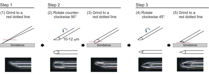
Figure 3 : Fabrication de l’aiguille. Méthode de polissage de la pointe en trois étapes pour fabriquer une aiguille avec une taille de trou appropriée et une pointe pointue. Veuillez cliquer ici pour voir une version agrandie de cette figure.
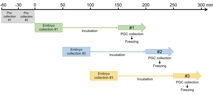
Figure 4 : Schéma de collecte d’embryons. Après deux pré-collectes, nous collectons généralement trois ou quatre fois par jour. Veuillez cliquer ici pour voir une version agrandie de cette figure.
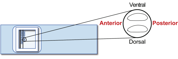
Figure 5 : Alignement de l’embryon de l’hôte. Alignement des embryons de l’hôte sur une lame de verre. Veuillez cliquer ici pour voir une version agrandie de cette figure.
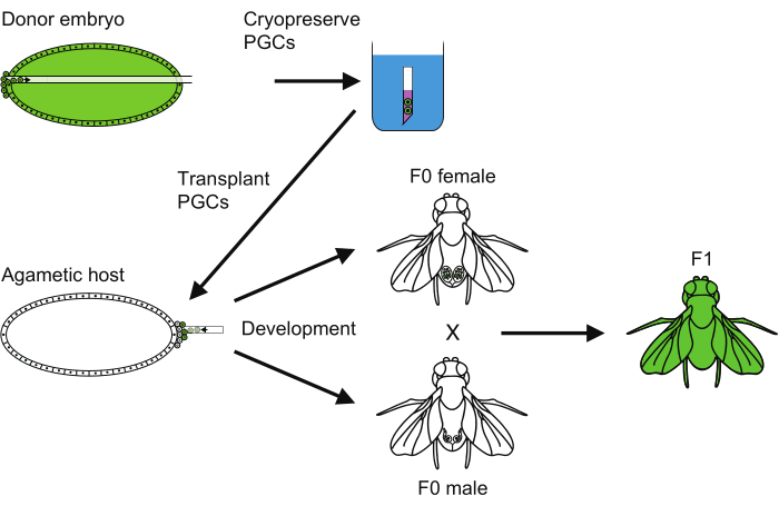
Figure 6 : Vue d’ensemble de la méthode de cryoconservation PGC. Tour d’horizon de toutes les étapes suivies pour réaliser la cryoconservation des cellules germinales primordiales (PGC). Veuillez cliquer ici pour voir une version agrandie de cette figure.
| Humidité ambiante | |||
| < 30 % | ~ 30% | > 30 % | |
| Aligner les embryons de l’hôte (~20 min) | Utilisez un humidificateur pendant 2 à 10 minutes | Utilisez un humidificateur par intermittence pendant 1 min | N’utilisez pas d’humidificateur |
| Décongélation des PGC de donneurs | Sans objet | Sans objet | Sans objet |
| Réglettes à séchage à l’air libre | Omettre cette étape | Omettre cette étape | 5 min (en anglais) |
| Appliquer de l’huile de silicone | Sans objet | Sans objet | Sans objet |
| Greffer des PGC | Sans objet | Sans objet | Sans objet |
| Toutes ces étapes devraient être terminées en 50 min. | |||
Tableau 1 : Séchage des embryons lors de l’alignement embryonnaire et de la décongélation des PGC.
| Souche donneuse | Période de cryoconservation | Nombre d’embryons transplantés (A) | Nombre de larves écloses (B) (éclosion, B/A) | Nombre d’adultes enfermés (C) (viabilité de l’œuf à l’adulte, C/A) | Nombre d’adultes fertiles (D) (fréquence des mouches fertiles, D/C) |
| M17 (en anglais seulement) | 8 - 30 jours | 134 | 108 (80.6%) | 57 (42.5%) | 20 (35.1%) |
| M17 (en anglais seulement) | 31 - 150 jours | 74 | 60 (81.1%) | 30 (40.5%) | 8 (26.7%) |
| M17 : yw ; TM6B, P{Dfd-GMR-nvYFP}4, Sb[1] Tb[1] ca[1]/ Pri[1] | |||||
Tableau 2 : Efficacité de la transplantation de PGC cryoconservés. Ce tableau est modifié à partir de13. Toutes les données proviennent d’hôtes agamétiques.
Discussion
Un facteur essentiel pour le succès de la cryoconservation et de la renaissance des PGC est d’utiliser de bons embryons. Les jeunes femelles (p. ex., âgées de 3 à 5 jours) doivent être utilisées pour le prélèvement d’embryons. Les embryons du donneur et de l’hôte sont évalués par inspection microscopique, et seuls ceux au stade du blastoderme (stade 5) sont utilisés12. Pour le prélèvement de PGC, nous alignons généralement environ 40 embryons de donneurs sur une période de 20 minutes et recueillons des PGC d’environ 30 embryons au stade précoce 5 ; Les embryons plus âgés et défectueux ne sont pas utilisés. Après la cryoconservation et la décongélation, les PGC doivent conserver leur forme ; Les PGC se rompent en cas d’échec de conservation. Les embryons hôtes doivent également être au stade 5 et avoir une pression interne modérée ; Les embryons doivent reprendre lentement leur forme d’origine après une légère poussée. Les embryons trop et insuffisamment séchés ne se développeront pas normalement après la transplantation. Étant donné que la transplantation hétérosexuelle de PGC ne produit pas de gamètes chez la drosophile 5,10, la transplantation de PGC à partir d’embryons de donneurs multiples dans des embryons hôtes est plus susceptible de donner des adultes fertiles. À cette fin, nous recueillons généralement des PGC à partir d’environ 30 embryons par aiguille.
En tant que cryoprotecteurs, nous avons essayé l’éthylène glycol, le diméthylsulfoxyde et le glycérol avec du saccharose à différentes concentrations. Nous avons déterminé que l’EBR contenant 20 % d’éthylène glycol et 1 M de saccharose était le meilleur13 ; cependant, l’utilisation de différents cryoprotecteurs peut améliorer la conservation des PGC22.
Cette méthode de cryoconservation nécessite des compétences spécialisées dans la manipulation des PGC, et environ 6 semaines de formation sont nécessaires pour collecter et transplanter confortablement les PGC. Afin d’évaluer et d’améliorer les compétences en compétences, celle-ci peut être divisée en six étapes de formation : 1) aligner les embryons sur une lame de verre, 2) contrôler un manipulateur, 3) transplanter des PGC d’un embryon dans un autre embryon sans cryoconservation, 4) transplanter des PGC de 10 embryons ou plus dans 5 à 10 embryons, 5) transplanter des PGC après application de CPA, et 6) transplanter des PGC après congélation-décongélation. Chaque étape peut prendre 1 semaine. Les objectifs à court terme de l’étape 3 sont un taux d’éclosion de 40 %, une viabilité de l’embryon à l’adulte de 10 % à 20 % et une fréquence de mouches fertiles de 20 %.
La cryoconservation PGC nécessite des instruments coûteux et un personnel hautement qualifié. Par conséquent, cette méthode peut ne pas être adoptée par de nombreux laboratoires. Cependant, la méthode PGC actuelle présente plusieurs aspects importants. Tout d’abord, les PGC sont beaucoup plus petits que les embryons et sont très perméables aux cryoprotecteurs. En revanche, la perméabilité aux cryoprotecteurs est sévèrement limitée par les couches cireuses des embryons de drosophile, ce qui constitue le problème le plus grave de la cryoconservation des embryons. En effet, des études antérieures ont fait de grands efforts pour trouver une fenêtre temporelle dans laquelle les embryons ont un taux de survie élevé et une couche de cire plus fine. Le second s’intéresse aux variations développementales et morphologiques entre les souches. Les PGC sont prélevés sur des embryons de stade précoce 5 (2 h 30 min-3 h 20 min après la ponte), tandis que la cryoconservation embryonnaire est effectuée sur des embryons de stade 16 (14-22 h après la ponte). Les embryons sont donc beaucoup plus âgés et présentent une variation de souche beaucoup plus importante dans la fenêtre temporelle optimale pour la cryoconservation par rapport à la cryoconservation PGC. En effet, la fréquence des hôtes produisant une descendance dérivée d’un donneur ne variait pas entre les cinq souches étudiées par Asaoka et al.13, bien que les hôtes ne soient pas agamétiques. De plus, les PGC ont le potentiel d’être utilisés dans des applications de génie génétique, telles que l’édition du génome 14,15,16.
Déclarations de divulgation
Les auteurs n’ont aucun conflit d’intérêts à déclarer.
Remerciements
Nous remercions le KYOTO Drosophila Stock Center pour les souches de mouches. Nous remercions également Mme Wanda Miyata pour l’édition en anglais du manuscrit et le Dr Jeremy Allen d’Edanz (https://jp.edanz.com/ac) pour l’édition d’une ébauche de ce manuscrit. Ce travail a été soutenu par des subventions (JP16km0210072, JP17km0210146, JP18km0210146) de l’Agence japonaise pour la recherche et le développement médicaux (AMED) à T.T.-S.-K., des subventions (JP16km0210073, JP17km0210147, JP18km0210145) d’AMED à S.K., une subvention (JP20km0210172) d’AMED à T.T.-S.-K. et S.K., une subvention d’aide à la recherche scientifique (C) (JP19K06780) de la Société japonaise pour la promotion de la science (JSPS) à T.T.-S.-K., et une subvention d’aide à la recherche scientifique dans des domaines innovants (JP18H05552) de JSPS à S.K.
matériels
| Name | Company | Catalog Number | Comments |
| Acetic acid | FUJIFILM Wako Pure Chemical Corporation | 017-00256 | For embryo collection |
| Agar powder | FUJIFILM Wako Pure Chemical Corporation | 010-08725 | For embryo collection |
| Calcium chloride | FUJIFILM Wako Pure Chemical Corporation | 038-24985 | For EBR solution |
| Capillary | Sutter Instrument | B100-75-10-PT | BOROSILICATE GLASS; O.D: 1.0mm, I.D: 0.75mm , length: 10cm, 225Pcs |
| Capillary holder | Eppendorf | 5196 081.005 | Capillary holder 4; for micromanipulation |
| Chromic acid mixture | FUJIFILM Wako Pure Chemical Corporation | 037-05415 | For needle washing |
| CPA solution | 1x EBR containing 20% ethylene glycol and 1M sucrose | ||
| Double-sided tape | 3M | Scotch w-12 | For glue extracting |
| Ephrussi–Beadle Ringer solution (EBR) | 130 mM NaCl, 5 mM KCl, 2 mM CaCl2, and 10 mM Hepes at pH 6.9 | ||
| Ethanol (99.5) | FUJIFILM Wako Pure Chemical Corporation | 057-00451 | For embryo collection |
| Ethylene glycol | FUJIFILM Wako Pure Chemical Corporation | 054-00983 | For CPA solution |
| Falcon 50 mm x 9 mm bacteriological petri dish | Corning Inc. | 351006 | For embryo collection |
| Forceps | Vigor | Type5 Titan | For embryo handling |
| Grape juice | Asahi Soft Drinks Co., LTD. | Welch's Grape 100 | For embryo collection |
| Grape juice agar plate | 50% grape juice, 2% agar, 1% ethanol, 1% acetic acid | ||
| Heptane | FUJIFILM Wako Pure Chemical Corporation | 084-08105 | For glue extracting |
| Humidifier | APIX INTERNATIONAL CO., LTD. | FSWD2201-WH | For embryo preparation |
| Inverted microscope | Leica Microsystems GmbH | Leica DM IL LED | For micromanipulation |
| Luer-lock glass syringe | Tokyo Garasu Kikai Co., Ltd. | 0550 14 71 08 | Coat a plunger with silicon oil (FL-100-450CS);for micromanipulation |
| Mechanical micromanipulator | Leica Microsystems GmbH | For micromanipulation | |
| Micro slide glass | Matsunami Glass Ind., Ltd. | S-2441 | For embryo aligning |
| Microgrinder | NARISHIGE Group | Custom order | EG-401-S combined EG-401 and MF2 (with ocular lens MF2-LE15 ); for needle preparation |
| Microscope camera | Leica Microsystems GmbH | Leica MC170 HD | For micromanipulation |
| Needle holder | Merck KGaA | Eppendorf TransferTip (ES) | For cryopreservation |
| Potassium chloride | Nacalai Tesque, Inc. | 28514-75 | For EBR solution |
| Puller | NARISHIGE Group | PN-31 | For needle preparation; the heater level is set to 85.0-98.4, the magnet main level to 57.8, and the magnet sub level to 45.0. |
| PVC adhesive tape for electric insulation | Nitto Denko Corporation | J2515 | For embryo-pool frame |
| Silicon oil | Shin-Etsu Chemical, Co, Ltd. | FL-100-450CS | For embryo handling |
| Sodium chloride | Nacalai Tesque, Inc. | 31320-05 | For EBR solution |
| Sodium hypochlorite solution | FUJIFILM Wako Pure Chemical Corporation | 197-02206 | Undiluted and freshly prepared; for embryo breaching |
| Sucrose | Nacalai Tesque, Inc. | 30404-45 | For CPA solution |
Références
- Brüschweiler, W., Gehring, W. A method for freezing living ovaries of Drosophila melanogaster larvae and its application to the storage of mutant stocks. Experientia. 29, 134-135 (1973).
- Steponkus, P. L., et al. Cryopreservation of Drosophila melanogaster embryos. Nature. 345, 170-172 (1990).
- Mazur, P., Cole, K. W., Hall, J. W., Schreuders, P. D., Mahowald, A. P. Cryobiological preservation of Drosophila embryos. Science. 258 (5090), 1932-1935 (1992).
- Zhan, L., Li, M. G., Hays, T., Bischof, J. Cryopreservation method for Drosophila melanogaster embryos. Nat Comm. 12, 2412 (2021).
- Van Deusen, E. B. Sex determination in germ line chimeras of Drosophila melanogaster. Development. 37 (1), 173-185 (1977).
- Breen, T. R., Duncan, I. M. Maternal expression of genes that regulate the bithorax complex of Drosophila melanogaster. Dev Biol. 118, 442-456 (1986).
- Schupbach, T., Wieschaus, E. Germline autonomy of maternal-effect mutations altering the embryonic body pattern of Drosophila. Dev Biol. 113, 443-448 (1986).
- Irish, V., Lehmann, R., Akam, M. The Drosophila posterior-group gene nanos functions by repressing hunchback activity. Nature. 338, 646-648 (1989).
- Hülskamp, M., Schröder, C., Pfeifle, C., Jäckle, H., Tautz, D. Posterior segmentation of the Drosophila embryo in the absence of a maternal posterior organizer gene. Nature. 338, 629-632 (1989).
- Steinmann-Zwicky, M., Schmid, H., Nöthiger, R. Cell-autonomous and inductive signals can determine the sex of the germ line of Drosophila by regulating the gene Sxl. Cell. 57 (1), 157-166 (1989).
- Stein, D., Roth, S., Vogelsang, E., Nüsslein-Volhard, C. The polarity of the dorsoventral axis in the drosophila embryo is defined by an extracellular signal. Cell. 65 (5), 725-735 (1991).
- Kobayashi, S., Yamada, M., Asaoka, M., Kitamura, T. Essential role of the posterior morphogen nanos for germline development in Drosophila. Nature. 380, 708-711 (1996).
- Asaoka, M., et al. Offspring production from cryopreserved primordial germ cells in Drosophila. Comm Biol. 4 (1), 1159 (2021).
- Blitz, I. L., Fish, M. B., Cho, K. W. Y. Leapfrogging: primordial germ cell transplantation permits recovery of CRISPR/Cas9-induced mutations in essential genes. Development. 143 (15), 2868-2875 (2016).
- Koslová, A., et al. Precise CRISPR/Cas9 editing of the NHE1 gene renders chickens resistant to the J subgroup of avian leukosis virus. Proc Natl Acad Sci U S A. 117 (4), 2108-2112 (2020).
- Zhang, F. Efficient generation of zebrafish maternal-zygotic mutants through transplantation of ectopically induced and Cas9/gRNA targeted primordial germ cells. J Genet Genom. 47 (1), 37-47 (2020).
- Campos-Ortega, J. A., Hartenstein, V. Stages of Drosophila Embryogenesis. The Embryonic Development of Drosophila. , (1997).
- Manning, A. A sperm factor affecting the receptivity of Drosophila melanogaster females. Nature. 194, 252-253 (1962).
- Kubli, E. Sex-peptides: seminal peptides of the Drosophila male. Cell Mol Life Sci. 60, 1689-1704 (2003).
- Lehmann, R., Nüsslein-Volhard, C. Abdominal segmentation, pole cell formation, and embryonic polarity require the localized activity of oskar, a maternal gene in drosophila. Cell. 47 (1), 141-152 (1986).
- Kiger, A. A., Gigliotti, S., Fuller, M. T. Developmental genetics of the essential Drosophila Nucleoporin nup154: allelic differences due to an outward-directed promoter in the P-element 3′ end. Genetics. 153 (2), 799-812 (1999).
- Rienzi, L. F., et al. Perspectives in gamete and embryo cryopreservation. Semin Reprod Med. 36 (5), 253-264 (2018).
Réimpressions et Autorisations
Demande d’autorisation pour utiliser le texte ou les figures de cet article JoVE
Demande d’autorisationExplorer plus d’articles
This article has been published
Video Coming Soon