JoVE Logo
Faculty Resource Center

Sign In

Charité Universitätsmedizin Berlin

6 ARTICLES PUBLISHED IN JoVE

image

Immunology and Infection

Highly Resolved Intravital Striped-illumination Microscopy of Germinal Centers
Zoltan Cseresnyes *1,2, Laura Oehme *3, Volker Andresen 4, Anje Sporbert 2, Anja E. Hauser *3,5, Raluca Niesner *1
1Biophysical Analytics, German Rheumatism Research Center, Leibniz Institute, 2Microscopy Core Facility, Max-Delbrück Center for Molecular Medicine, 3Immunodynamics, German Rheumatism Research Center, Leibniz Institute, 4LaVision Biotec GmbH, 5Immunodynamics and Intravital Imaging, Charité - University of Medicine

High-resolution intravital imaging with enhanced contrast up to 120 µm depth in lymph nodes of adult mice is achieved by spatially modulating the excitation pattern of a multi-focal two-photon microscope. In 100 µm depth we measured resolutions of 487 nm (lateral) and 551 nm (axial), thus circumventing scattering and diffraction limits.

image

Developmental Biology

Automated Quantification of Hematopoietic Cell – Stromal Cell Interactions in Histological Images of Undecalcified Bone
Sandra Zehentmeier 1, Zoltan Cseresnyes 2,3, Juan Escribano Navarro 4, Raluca A. Niesner 2, Anja E. Hauser 1,5
1Immunodynamics, German Rheumatism Research Center, a Leibniz Institute, 2Biophysical Analytics, German Rheumatism Research Center, a Leibniz Institute, 3Max-Delbrück Center for Molecular Medicine, 4Wimasis GmbH, 5Immunodynamics and Intravital Imaging, Charité - University of Medicine

A strategy to quantitatively analyze histological data in the bone marrow is presented. Confocal microscopy of fluorescently labeled cells in tissue sections results in 2-dimensional images, which are automatically analyzed. Co-localization analyses of different cell types are compared to data from simulated images, giving quantitative information about cellular interactions.

image

JoVE Core

Morphology-Based Distinction Between Healthy and Pathological Cells Utilizing Fourier Transforms and Self-Organizing Maps
Fabian L. Kriegel 1,2, Ralf Köhler 2, Jannike Bayat-Sarmadi 2, Simon Bayerl 3, Anja E. Hauser 2,3, Raluca Niesner 2, Andreas Luch *1, Zoltan Cseresnyes *4
1Department of Chemical and Product Safety, German Federal Institute for Risk Assessment (BfR), 2Deutsches Rheuma-Forschungszentrum (DRFZ) Berlin, a Leibniz Institute, 3Charité Universitätsmedizin Berlin, 4Applied Systems Biology, Leibniz Institute for Natural Product Research and Infection Biology Hans Knöll Institute

Here, we provide a workflow that allows the identification of healthy and pathological cells based on their 3-dimensional shape. We describe the process of using 2D projection outlines based on the 3D surfaces to train a Self-Organizing Map that will provide objective clustering of the investigated cell populations.

image

Education

Digital Home-Monitoring of Patients after Kidney Transplantation: The MACCS Platform
Wiebke Duettmann 1, Marcel G. Naik 1, Danilo Schmidt 2, Matthias Pfefferkorn 1, Matthias Kurz 1, Verena Graf 1, Andrea Kreichgauer 3, Severin Hoegl 4, Max Haenska 4, Tim Gielsdorf 1, Tillmann Breitenstein 1, Bilgin Osmanodja 1, Petra Glander 1, Jannis Bakker 1, Manuel Mayrdorfer 1, Carolin J. Gethmann 1, Friederike Bachmann 1, Mira Choi 1, Eva Schrezenmeier 1, Bianca Zukunft 1, Fabian Halleck 1, Klemens Budde 1
1Department of Nephrology and internal intensive Care, Charité Universitätsmedizin Berlin, 2IT Department, Charité Universitätsmedizin Berlin, 3Department of Neurology, Charité Universitätsmedizin Berlin, 4comjoodoc business solutions GmbH

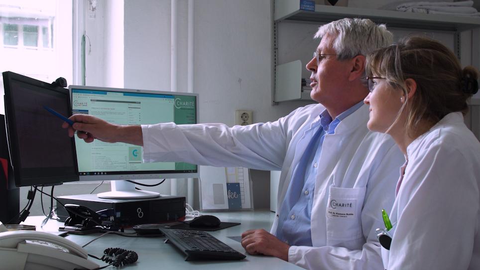
The MACCS platform is a comprehensive telemedicine concept aiming at better outcomes after kidney transplantation by sharing key medical information between patients and physicians. A telemedicine team reviews incoming data to detect potential complications and to improve adherence in kidney transplant recipients to achieve better long-term outcomes.

image

Education

TBase - an Integrated Electronic Health Record and Research Database for Kidney Transplant Recipients
Danilo Schmidt 1, Bilgin Osmanodja 2, Matthias Pfefferkorn 2, Verena Graf 2, Dirk Raschke 1, Wiebke Duettmann 2, Marcel G. Naik 2, Carolin J. Gethmann 2, Manuel Mayrdorfer 2, Fabian Halleck 2, Lutz Liefeldt 2, Petra Glander 2, Oliver Staeck 2, Michael Mallach 1, Martin Peuker 1, Klemens Budde 2
1Division IT, Charité - Universitätsmedizin Berlin, 2Department of Nephrology and Medical Intensive Care, Charité - Universitätsmedizin Berlin

TBase combines an electronic health record with an innovative research database for kidney transplant recipients. TBase is built upon an in-memory database platform, connected to different hospital systems, and used for regular outpatient care. It automatically integrates all relevant clinical data including transplantation-specific data creating a unique research database.

image

Neuroscience

Quantification of Microglial Engulfment of Synaptic Material Using Flow Cytometry
Bilge Ugursu 1, Susanne A. Wolf 1,2
1Psychoneuroimmunology, Max-Delbrück-Center for Molecular Medicine in the Helmholtz Association, 2Experimental Ophthalmology, Charité Universitätsmedizin Berlin

Here we present two protocols to quantify microglial engulfment of vGLUT1-positive synapses and pHRodo Red-labeled crude synaptosomes using flow cytometry.

JoVE Logo

Privacy

Terms of Use

Policies

Research

Education

ABOUT JoVE

Copyright © 2024 MyJoVE Corporation. All rights reserved