純粋な培養物と縞めっき:混合サンプルからの単一細菌コロニーの単一の分離
概要
ソース: ティルデ・アンダーソン1, ロルフ・ルード1
1臨床科学ルンド, 感染医学の部門, ルンド大学生物医学センター, 221 00 ルンド, スウェーデン
微生物の生物多様性を特定することは不可能に見えるが、推定1兆種の共存種(1,2)と本当に驚異的である。特に過酷な気候は、ヒト胃の酸性環境(3)または南極の氷河湖(4)のような、特定の種によって支配されてもよいが、細菌は典型的に混合培養に見出される。各株は、別の(5)の成長に影響を与える可能性があるため、コロニーを分離し、栽培する能力(1つのタイプのみで構成される)コロニーは、同様に臨床および学術的な設定で不可欠となっています。純粋な培養は、さらなる遺伝的(6)およびプロテオミクス検査(7)、サンプル純度の分析、およびおそらくより注目すべき、臨床サンプルからの感染剤の同定および特徴付けを可能にする。
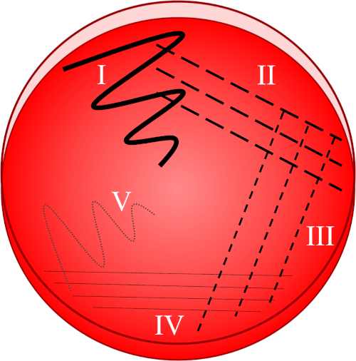
細菌は、成長要件の広い範囲を持っており、要求の厳しい種と断固たる種(8)の両方を維持するように設計された栄養培地の多くの種類があります。成長培地は、液体形態(スープとして)または典型的な寒天系(赤藻類由来のゲル化剤)固体形態のいずれかで調製することができる。スープへの直接接種は、遺伝的に多様な、あるいは混合細菌集団を生成するリスクを伴うのに対し、めっきと再ストリークは、各細胞が非常に類似した遺伝的構成を有する純粋な文化を作成します。ストリークプレート技術は、サンプルの進行性希釈に基づいており(図1)、個々の細胞を互いに分離することを目的としています。培養物および指定環境によって持続される任意の生存細胞(以下、コロニー形成部、CFUと称される)は、その後、バイナリ核分裂を介して娘細胞の単離コロニーを見出すことができる。細菌群内の急速な突然変異率にもかかわらず、この細胞群は一般的にクローンとみなされている。その結果、この集団を収穫し、再ストリークすることで、その後の作業は1つの細菌タイプのみを含むことが保証されます。

図1:ストリークプレートは、元のサンプルの進行性希釈に基づいています。I)接種液は、最初はジグザグ運動を用いて分散し、比較的密な細菌集団を持つ領域を作成する。II-IV)ストリークは、4番目の象限に達するまで、毎回無菌接種ループを使用して、前の領域から引き出されます。V)プレートの中央に向かう最終的なジグザグ運動は、接種が著しく希釈された領域を形成し、コロニーが互いに別々に現れることを可能にする。
ストリークプレート技術は、選択的および/または差動媒体の使用と組み合わせることもできます。選択的培地は特定の生物の増殖を阻害する(例えば、抗生物質の添加を通じて)、微分培地は単に別のものと区別するのに役立ちます(例えば、血液寒天プレートの血化を通じて)。
微生物学のすべての仕事の根底にあるのは無菌(無菌)技術の使用である。危険な菌株の意図しない成長、エアロゾル形成、機器/人員の汚染のリスクがあるので、すべての細菌培養物は潜在的に病原性と見なされるべきです。これらのリスクを最小限に抑えるために、すべてのメディア、プラスチック、金属、ガラス製品は、通常、使用前後のオートクレーブによって殺菌され、残留細胞を効果的に拭き取る121°C付近の高圧飽和蒸気にさらされます。ワークスペースは、一般に、使用前および使用後の両方のエタノールを使用して消毒されます。ラボのコートと手袋は、常に感染性薬剤との作業中に着用されます。
手順
1. 設定
- すべての微生物は、それらが危険であるかのように扱われるべきです。常にラボのコートと手袋を着用し、長い髪を結び、傷が特によく保護されていることを確認してください。
- 70%エタノールを使用して殺菌して作業スペースを準備します。
- 寒天プレート、サンプル溶液、および殺菌前のプラスチック接種ループまたは金属ループとブンゼンの炎の箱が近くにあることを確認します。使い捨て可能な、プラスチックループは通常前殺菌される。金属ループは70%エタノールに浸し、ブンセン炎の青い領域の近くに保持し、熱くなるまで加熱する必要があります。プレートの蓋を上げて(汚染を防ぐためにわずかに)、固化媒体に対してそれをタップして、ワイヤーを冷却することができます。
- 作業スペースの繰り返し殺菌と手と手首の徹底的な洗浄/殺菌で各手順を終了します。
2. プロトコル
- メディアの準備
- 利用される細菌種/株を維持する固体培地(
結果
申請書と概要
参考文献
- The Human Microbiome Project C. Structure, Function and Diversity of the Healthy Human Microbiome. Nature. 486:207-214. (2012)
- Locey KJ, Lennon JT. Scaling laws predict global microbial diversity. Proceedings of the National Academy of Sciences. 113 (21) 5970-5975 (2016)
- Skouloubris S, Thiberge JM, Labigne A, De Reuse H. The Helicobacter pylori UreI protein is not involved in urease activity but is essential for bacterial survival in vivo. Infection and Immunity. 66:4517-21. (1998)
- Mikucki JA, Auken E, Tulaczyk S, Virginia RA, Schamper C, Sørensen KI, Doran PT, Dugan H, Foley N. Deep groundwater and potential subsurface habitats beneath an Antarctic dry valley. Nature Communications. 6:6831. (2015)
- Mullineaux-Sanders C, Suez J, Elinav E, Frankel G. Sieving through gut models of colonization resistance. Nature Microbiology. 3:132-140. (2018)
- Fournier PE, Drancourt M, Raoult D. Bacterial genome sequencing and its use in infectious diseases. Lancet Infectious Diseases. 7:711-23 (2007)
- Yao Z, Li W, Lin Y, Wu Q, Yu F, Lin W, Lin X. Proteomic Analysis Reveals That Metabolic Flows Affect the Susceptibility of Aeromonas hydrophila to Antibiotics. Scientific Reports. 6:39413 (2016)
- Medina D, Walke JB, Gajewski Z, Becker MH, Swartwout MC, Belden LK. Culture Media and Individual Hosts Affect the Recovery of Culturable Bacterial Diversity from Amphibian Skin. Frontiers in Microbiology. 8:1574 (2017)
タグ
スキップ先...
このコレクションのビデオ:

Now Playing
純粋な培養物と縞めっき:混合サンプルからの単一細菌コロニーの単一の分離
Microbiology
166.2K 閲覧数

ウィノグラツキーカラムの作成:堆積物サンプル中の微生物種を濃縮する方法
Microbiology
129.5K 閲覧数

シリアル希釈とめっき:微生物列挙
Microbiology
316.3K 閲覧数

エンリッチメント培養:選択的および差動媒体における好気性微生物と嫌気性微生物の培養
Microbiology
132.1K 閲覧数

16S rRNAシーケンシング:細菌種を同定するPCRベースの技術
Microbiology
189.1K 閲覧数

成長曲線:コロニー形成単位と光学密度測定を用いて成長曲線を生成する
Microbiology
296.5K 閲覧数

抗生物質感受性試験:2つの抗生物質のMIC値を決定し、抗生物質の相乗効果を評価するエプシロメーター試験
Microbiology
93.8K 閲覧数

顕微鏡検査と染色:グラム、カプセル、内胞染色
Microbiology
363.5K 閲覧数

プラークアッセイ:プラーク形成単位(PFU)としてウイルス定数を決定する方法
Microbiology
186.3K 閲覧数

適応塩化カルシウム手順を用いて大腸菌細胞の形質転換
Microbiology
86.9K 閲覧数

結合:アンピシリン耐性をドナーからレシピエント大腸菌に移す方法
Microbiology
38.3K 閲覧数

ファージトランスダクション:アンピシリン耐性をドナーからレシピエント大腸菌に伝達する方法
Microbiology
29.1K 閲覧数
Copyright © 2023 MyJoVE Corporation. All rights reserved