JoVE 비디오를 활용하시려면 도서관을 통한 기관 구독이 필요합니다. 전체 비디오를 보시려면 로그인하거나 무료 트라이얼을 시작하세요.
In Vivo Electroporation: A Site-Specific Method to Transfect Zebrafish
Overview
This video describes in vivo electroporation, which is a technique for DNA delivery into ependymoglial cells in adult zebrafish.
프로토콜
1. Electroporation
- Remove the fish from the injection set-up while still holding it in the sponge.
- Immerse the inner side of the tip of the electrodes in the ultrasound gel.
- Cover the fish telencephalon with a small amount of ultrasound gel.
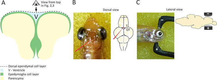
- Position the fish head between the electrodes, placing the positive electrode at the ventral side of the fish’s head and the negative electrode on the dorsal side (Figure 1C), while still holding the fish’s body in the sponge. This sets the direction of the flow of the current necessary to electroporate ependymoglia positioned at subventricular zone.
- Press the electrodes gently and precisely against the telencephalon (Figure 1 C). Administer the current with the foot pedal. Hold the electrodes in place until all five pulses are finished.
결과

Figure 1: Schematic representation of coronal section of the everted zebrafish telencephalon. (A) Scheme of a coronal section of zebrafish telencephalon, highlighting the position of ependymoglial cells, which are lining ventricular surface and building the ventral ventricular wall. Dorsal ependymal layer is bridging the two hemispheres and covering the ventricle (V), located in between two cell...
자료
| Name | Company | Catalog Number | Comments |
| Ultrasound gel | SignaGel, Parker laboratories INC | 15-60 | Electrode Gel |
| BTX Tweezertrodes Electrodes | Platinum Tweezertrode, BTX Harvard Apparatus | 45-0486 | 1mm diameter |
| Electroporation device | BTX ECM830 Square Wave Electroporation System, BTX Harvard Apparatus | 45-0662 |
This article has been published
Video Coming Soon
Source: Durovic, T., et al. Electroporation Method for In Vivo Delivery of Plasmid DNA in the Adult Zebrafish Telencephalon. J. Vis. Exp. (2019).
Copyright © 2025 MyJoVE Corporation. 판권 소유