JoVE 비디오를 활용하시려면 도서관을 통한 기관 구독이 필요합니다. 전체 비디오를 보시려면 로그인하거나 무료 트라이얼을 시작하세요.
Method Article
Zebrafish 배아 눈 조직의 Microdissection
요약
이 문서는 망막 안료 상피 하나에서 세 일 postfertilization의 배아에게 부착과 않고 zebrafish의 망막 microdissect하는 방법을 설명합니다.
초록
Zebrafish는 때문에 급속한의 눈 개발에 연구에 인기있는 동물 모델입니다
프로토콜
1 부 : microdissection 전 준비
- 솔루션
- E3 매체 4 (5 MM NaCl, 0.17 MM KCl, 0.33 MM CaCl 2, 0.33 MM MgSO 4).
- 링어의 솔루션 1, 5 (116 MM NaCl, 2.9 KCl MM 1.8 MM CaCl 2, 5 MM HEPES, pH7.2), 필터 살균.
- 5N NaOH.
- 텅스텐 바늘의 화학 에칭
- 점토하여 페트리 접시에 5N NaOH를 포함하는 작은 비커를 확보.
- 그것은 NaOH 용액과 접촉할 수 있도록 비커의 측면에 종이 클립을 부착합니다.
- 종이 클립과 텅스텐 와이어의 짧은 조각 (~ 2cm)의 권력을 긍정적인 전극에 DC 전원 공급 장치의 음극을 연결합니다.
- 작은 점토 공과 텅스텐 와이어의 다른 쪽 끝을 싸서 NaOH 용액에이 끝부분을 찍어.
- 약 3V로 전압을 증가시키고 좋은 에칭 속도가 접수 때까지 NaOH 용액에 철사를 찍어 내려. 좋은 품질의 바늘은 보통 10 분 이내에 에칭 수 있습니다.
- 솔루션에 노출 텅스텐 와이어 점차 얇은 될 것입니다. 흙 공이 꺼져 방울 때, 여전히 긍정적인 전극에 연결된 전선의 다른 한쪽은 날카로운 바늘 모양을 것입니다.
- 소금 예금을 제거하는 링어의 솔루션 또는 E3와 날카로운 바늘을 씻어.
- 테이프로 나무 작은 주걱이나 쉽게 처리할 수있는 바늘 홀더에 바늘을 연결합니다.
- 문화 플레이트
- 팰컨 60 X 15mm의 폴리스티렌 문화 번호판은 망막 microdissection에 사용할 준비가 된 것입니다.
- RPE 연결된 망막을 해부를위한 30 분 해부하기 전에 표면의 접착력을 없애 링어의 솔루션을 포함하는 문화 판에서 완전히이 배아를 부술. 곧 절개 전에 신선한 링어의 솔루션을 광범위하게 접시를 씻으십시오.
- 배아의 수집 및 준비
- 성인 zebrafish 4, 5 설명된대로 유지됩니다.
- 구분선에 의해 번식 전에 밤에 pairwise 정적 사육 탱크에있는 부모 물고기를 분리합니다.
- 객실 표시등이 다음날 아침에 켜져 후 분할기를 제거합니다. 부모가 매 30 10 분 간격으로 시간을 통과하도록 허용합니다. 각 교차로 후 생선을 분리.
- 28 별도로 E3 매체의 각 교차로에서 수집한 배아를 유지 ° C.
- 설립 기준 1, 6에 따라 10~12시간 포스트 수정 (HPF)와 restage 즉시 특정 시간 지점에서 해부하기 전 무대 배아.
2 부 : 해부 - 뇌의 제거 및 안구 노출 1
- 이 절에서 설명하는 절차는 망막 (부품 3A)와 RPE 연결된 망막 (파트 3B) 해부 일반적인 수 있습니다.
- 모든 미세 해부는 더몬트 집게의 끝부분에 의해 행해지며 조직을 위치에 대한 더몬트 포셉 또 한 쌍의에 의해 지원됩니다. 화학적 - 새겨져 텅스텐 바늘은 미세한 조작 필요한 경우 사용할 수 있습니다.
- 모든 미세 해부는 1X 목적 또는 동급을 갖춘 올림푸스 SZX16 현미경 ~ 5 배속 배율에서 수행됩니다.
- 28 벤치 최고 인큐베이터에서 E3 중간에 배아를 유지 ° C 해부하는 동안 쉽게 배아 액세스에 대한 현미경 옆에.
- 현미경 단계는 유전자 발현에 대한 온도 변동의 영향을 최소화하기 위해 온도 플레이트에 의해 28 ° C로 예열합니다.
- 제 1 부 # 3에서 설명한대로 팰컨 60 X 15mm 문화 플레이트에 링어의 솔루션에 대한 특정 발달 단계에서 배아를 전송 준비.
- 시체에서 빠르게 앞쪽에 트렁크의 일부로 머리를 잘라.
- 지원 포셉과 문화 판에서이 조직의 뒷부분 끝을 핀.
- 앞쪽에 이마에서 시작 등의 측면에 머리를 엽니다.
- 눈의 중간 측면이 노출되고 더 조작에 대한 상향 직면되도록 머리를 제거합니다.
부품 3A : 망막 절개 1
- 로 제 2에 설명되어있는 배아를 준비합니다.
- 조심스럽게 망막에있는 작은 오프닝을 볼 때까지 포셉의 일각에 의해 위쪽으로 직면하고있는 안구 공의 중간 측면에 노출된 RPE를 브러시.
- 빗질과 망막의 중간 측면은 거의 모든 노출 때까지 작업을 필링을 계속합니다. 긁힘 및 노출된 망막을 펀칭을 피한다.
- 이제 아래 직면하고있는 망막, 접근의 측면 RPE를 제거하려면 해당과 문화 플레이트 표면에서 약 45 °의 각도로 칫솔.
- C로 RPE의 분리 부분을 달아하여 오라 세라타 상대적으로 단단히 연결되어 RPE를 제거다음 ulture의 해부에 대한 판하고 망막을 부드럽게 리프트.
- 잔류 RPE를 정리하기 위해 문화 접시의 바닥에있는 망막을 롤.
- 우리는 배아가 깔린되면이 특정 팰컨 폴리스티렌 문화 플레이트가 약 20 분 RPE의 우선 준수를 가지고 것으로 나타났습니다. 이 속성은 RPE 잔존물의 제거를 위해 활용됩니다. 그러나, 망막 세포는 높은 배율에 따라 밀접하게 검사 수있는 낮은 정도로 표면에 막대기를 않습니다. 하나는 RPE 제거 및 망막 무결성의 완성도간에 균형을 공격한다.
- 렌즈는 종종 문화 판을 준수하고 압연 과정에서 망막에서 분리합니다. 때때로, 그것은 렌즈 표면에 소용돌이 치는 동작과 함께 새겨져 텅스텐 바늘에 의해 렌즈를 분리하는 것이 필요합니다.
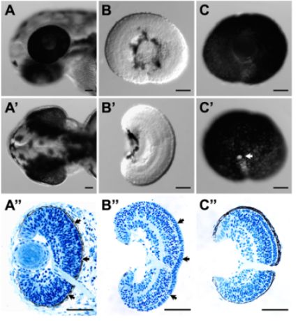
- 이러한 절차를 성공적으로 조직의 무결성을 손상시키지 않고 망막의 RPE를 제거할 수 있습니다. 이것은 좋은 전반적인 형태 (그림 1B와 B ') 및 조직학 (그림 1B ")로 표시되는 화살표 특히, photoreceptor 레이어와 RPE 사이에있는 세포외 기질의 보존 (그림 1B 매우 좋습니다."; 전체 배아 (그림 1A "))에서 조직학 비교.
부품 3B : RPE 연결된 망막 절개 3
- 로 제 2에 설명되어있는 배아를 준비합니다.
- 보조 포셉하여 문화 판에 머리를 핀. 뒷부분 측면 측면에서 부드럽게 눈을 들어 앞쪽에쪽으로 롤.
- RPE 계층의 외부에 추정 맥락막과 scleral 조직은 피부에 상대적으로 긴밀하게 연결되어 있으며, 주로 앞쪽에쪽으로 조심스럽게 눈을 압연에 의해 해제 벗겨 수 있습니다.
- 눈 측면 측면이 노출되고 안구가 계속 피부에 신경을 쓰는 경우 후 텅스텐 바늘로 렌즈를 제거합니다.
- 이러한 절차를 성공적으로 망막을 가지고있는 전체의 RPE 계층을 보존할 수 있습니다. 추정 choroids과 scleral 조직은 크게 (그림 1C, C '와 C') 제거할 수 있습니다.
4 부 : RNA 작업에 조직 샘플 컬렉션
- 해부 표본으로 1 일 이전에 설명한 스트림 RNA 특성화를위한 RNase 무료 microfuge 튜브에 TRIzol 수집하실 수 있습니다.
대표 결과

그림 1. (A) 래터럴과 절개 전 52 HPF에서 zebrafish 애벌레 머리 (A ') 등의 볼 수 있습니다. (A) & (A '). (B) 래터럴과 (B'54 HPF에서 해부 망막의) 지느러미 참조하십시오. 망막의 표면 측면 모두에서 손상되지 않았어요에서 애벌레 머리 ( ") 해당 histological 섹션 그리고 지느러미 전망. (B) & (B ')에서 해부하는 망막의 (B ") 해당 histological 섹션을 참조하십시오. 망막과 망막 라미네이션의 구조는 손상되었다. 특히, photoreceptor 계층 및 RPE (A 사이의 세포외 기질은 "(화살표, 화살표) 잘 해부 망막 B)에 보존되었다." (C) 래터럴 52 HPF에서 해부 RPE 연결된 망막의 (C ') 중간 볼 수 있습니다. RPE 계층도를 해부 조직의 histological 섹션 (C ')로 표시되었던, 손상 및 지속되었다. C'의 하얀 부분은 시신경 (화살표)입니다. 조직학 들어, 조직 샘플은 4 %의 수집 및 고정되었습니다 3 설명된대로 paraformaldehyde. 삽입 이러한 샘플 sectioning 플라스틱이 수행되었다. 스케일 막대 = 50 μm의이.
토론
zebrafish 아이 조직의 Microdissection 효과적으로 그대로 망막 및 RPE 연결된 망막을 얻을 수 있습니다. 이것은 실질적으로 특정 안구 조직 (예 : 망막 또는 RPE)에 관한 표현 연구를 지원합니다. 사실, 우리는 성공적으로 전체 망막 1 RPE 3 RNA 발현 프로파일을 얻기 위해이 절차를 이용합니다. 해당 프로필의 유틸리티는 강력 망막 차별 돌연변이에 불안정하게된다 는걸 알수 경로와 ...
공개
감사의 말
이 작품은 퍼듀 대학에서 생물학의학과에서 시작 기금에 의해 지원됩니다.
자료
| Name | Company | Catalog Number | Comments |
| Cordless pestle motor | VWR international | 47747-370 | |
| DC power supply | Lascar | PSU130 | Any DC supply would work. The specific voltage of a different machine will need further optimization. |
| Disposable pestle & microtube, 1.5 mL (DNase, RNase and pyrogen-free) | VWR international | 47747-366 | These are used for tissue collection in TRIzol for expression analysis. |
| Dumont #5 forceps, Tips: 0.05 x 0.01mm, Inox | World Precision Instruments, Inc. | 500341 | Fine tip dimension is desirable but is not inflexible, as one may need to sharpen the tip from time to time. |
| Dumont #5SF forceps, Tips: 0.025 x 0.005mm, Inox | Fine Science Tools | 11252-00 | Fine tip dimension is desirable but is not inflexible, as one may need to sharpen the tip from time to time. |
| Falcon polystyrene culture plates, 60 X 15 mm | BD Biosciences | 351007 | These plates are used as dissection plates. |
| Olympus SZX16 Stereomicroscope | Olympus Corporation | SZX16 | Any stereomicroscope would work. We used Leica stereomicroscope in previous studies1-3 without any issues. We also use the 1X objective exclusively for the dissection even though we have a 2X objective installed. |
| Sharpening stone | Fine Science Tools | 29008-01 | Use this to sharpen the tip of the forceps if necessary |
| Thermo plate | Tokai Hit | MATS-U55SZX2B | This is used to maintain the temperature of the tissue throughout dissection and minimize the influence of temperature fluctuation on gene expression. We also put the whole microscope in an environmentally controlled room at 28°C during dissection in previous studies1-3 with good success. |
| Trizol, 100 mL | Invitrogen | 15596-026 | |
| tungsten wire, 0.015 inch diameter | World Precision Instruments, Inc. | TGW1510 | |
| Wooden Applicator | Puritan | 807 | This is used for holding the chemically-etched tungsten needle. |
참고문헌
- Leung, Y. F., Dowling, J. E. Gene expression profiling of zebrafish embryonic retina. Zebrafish. 2, 269-283 (2005).
- Leung, Y. F., Ma, P., Link, B. A., Dowling, J. E. Factorial microarray analysis of zebrafish retinal development. Proc Natl Acad Sci U S A. 105, 12909-12914 (2008).
- Leung, Y. F., Ma, P., Dowling, J. E. Gene expression profiling of zebrafish embryonic retinal pigment epithelium in vivo. Invest Ophthalmol Vis Sci. 48, 881-890 (2007).
- Nusslein-Volhard, C., Dahm, R. . Zebrafish : a practical approach. , (2002).
- Westerfield, M. The zebrafish book : a guide for the laboratory use of zebrafish (Danio rerio). , (2000).
- Kimmel, C. B., Ballard, W. W., Kimmel, S. R., Ullmann, B., Schilling, T. F. Stages of embryonic development of the zebrafish. Dev Dyn. 203, 253-310 (1995).
- Fadool, J. M., Dowling, J. E. Zebrafish: a model system for the study of eye genetics. Prog Retin Eye Res. 27, 89-110 (2008).
재인쇄 및 허가
JoVE'article의 텍스트 или 그림을 다시 사용하시려면 허가 살펴보기
허가 살펴보기더 많은 기사 탐색
This article has been published
Video Coming Soon
Copyright © 2025 MyJoVE Corporation. 판권 소유